Podcast
Questions and Answers
Which of the following imaging techniques provides a dynamic, real-time view of brain activity?
Which of the following imaging techniques provides a dynamic, real-time view of brain activity?
- Computed Tomography (CT)
- Electroencephalogram (EEG) (correct)
- Magnetic Resonance Imaging (MRI)
- All of the above
Computed Tomography (CT) scans use strong radio waves to produce images of the brain.
Computed Tomography (CT) scans use strong radio waves to produce images of the brain.
False (B)
What is a primary use of Functional Magnetic Resonance Imaging (fMRI) in the context of brain tumors?
What is a primary use of Functional Magnetic Resonance Imaging (fMRI) in the context of brain tumors?
Plan for tumor removal
During an MRI, magnetic field lines up ______ in hydrogen atoms.
During an MRI, magnetic field lines up ______ in hydrogen atoms.
Match the imaging technique with its primary advantage:
Match the imaging technique with its primary advantage:
Which imaging technique is LEAST suitable for patients with metal implants?
Which imaging technique is LEAST suitable for patients with metal implants?
EEG is known for its high spatial resolution, providing a clear and precise location of neural activity.
EEG is known for its high spatial resolution, providing a clear and precise location of neural activity.
Besides diagnosing brain tumors, what other condition can MRI help assess in the brain?
Besides diagnosing brain tumors, what other condition can MRI help assess in the brain?
In CT scans, a rotating ______ beam moves 360 degrees around the head.
In CT scans, a rotating ______ beam moves 360 degrees around the head.
A researcher aims to study the millisecond-level changes in neuronal activity immediately following a visual stimulus. Which neuroimaging technique would be MOST appropriate for recording these rapid brain processes?
A researcher aims to study the millisecond-level changes in neuronal activity immediately following a visual stimulus. Which neuroimaging technique would be MOST appropriate for recording these rapid brain processes?
Flashcards
Electroencephalogram (EEG)
Electroencephalogram (EEG)
Dynamic imaging showing brain electrical activity via scalp electrodes; high temporal resolution.
Computed Tomography (CT)
Computed Tomography (CT)
Static images made by rotating X-ray around the head, creating a 3D model.
Magnetic Resonance Imaging (MRI)
Magnetic Resonance Imaging (MRI)
Static images using radio and magnetic waves to align hydrogen protons; no ionizing radiation.
Functional MRI (fMRI)
Functional MRI (fMRI)
Signup and view all the flashcards
High temporal resolution (EEG)
High temporal resolution (EEG)
Signup and view all the flashcards
Strengths of EEG
Strengths of EEG
Signup and view all the flashcards
Weaknesses of EEG
Weaknesses of EEG
Signup and view all the flashcards
Uses of CT
Uses of CT
Signup and view all the flashcards
Uses of MRI
Uses of MRI
Signup and view all the flashcards
Uses of fMRI
Uses of fMRI
Signup and view all the flashcards
Study Notes
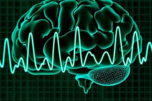
- Electroencephalogram (EEG) shows dynamic image changes in real time, reflecting brain activity via electrodes placed on the scalp to detect brain wave patterns.
Strengths
- High temporal resolution, allowing detection of rapid changes in brain waves.
- Safe and non-invasive.
Weaknesses
- Low spatial resolution, which makes precise locations of neural activity unclear.
- Requires gel on electrodes which can be messy.
Uses
- Diagnosing epilepsy.
- Sleep research.
- Determining brain death.
- Computed Tomography (CT) produces static pictures.
- Rotating x-ray beam moves 360 degrees around the head, taking multiple x-ray photos, and computer pieces these together to make a 3D model.
Uses
- Checking for skull fractures.
- Diagnosing brain tumors.
- Measuring tumor size.
- Helps assess injury from brain trauma.
- Magnetic Resonance Imaging (MRI) produces still pictures, functioning with radio and magnetic waves.
- Magnetic fields align protons in hydrogen atoms, then radiation tips the protons out of alignment.
Strengths
- More detailed pictures.
- Doesn't expose patients to ionising radiation.
- Safe for pregnant women.
Weaknesses
- Not safe for patients with magnetic metal in their body.
- Loud banging noises can disturb patients.
Uses
- Diagnosing brain tumors.
- Assessing strokes.
- Assessing brain injury from trauma.
- Functional Magnetic Resonance (fMRI) is a type of dynamic, functioning neuroimaging that produces dynamic pictures.
- Creates a 3D map of brain neurons that communicate through electrical impulses and neurotransmitters.
Strengths
- Can determine location of neural activity with accuracy.
- No iodising radiation.
Weaknesses
- No metal on or in the body.
- Takes longer to detect changes in neural activity.
Uses
- Plan for tumor removal.
- Evaluation the effects of a stroke.
Studying That Suits You
Use AI to generate personalized quizzes and flashcards to suit your learning preferences.