Podcast
Questions and Answers
What is the frequency range for beta waves in EEG analysis?
What is the frequency range for beta waves in EEG analysis?
- 0.1 - 3.5 Hz
- 14.0 - 30.0 Hz (correct)
- 4.0 - 7.5 Hz
- 8.0 - 13 Hz
Which of the following statements about the N400 ERP is accurate?
Which of the following statements about the N400 ERP is accurate?
- It reflects the relatedness of words rather than sentence understanding. (correct)
- It requires invasive procedures for accurate measurement.
- It is exclusively linked to sentence comprehension.
- It occurs primarily in the delta frequency range.
What is a key advantage of using EEG over behavioral paradigms?
What is a key advantage of using EEG over behavioral paradigms?
- Invasiveness
- Better spatial resolution than MEG
- Excellent temporal resolution (correct)
- Higher signal-to-noise ratio
Which of the following is a limitation of EEG?
Which of the following is a limitation of EEG?
What technology is utilized in magnetoencephalography (MEG) to detect magnetic fields generated by neuronal activity?
What technology is utilized in magnetoencephalography (MEG) to detect magnetic fields generated by neuronal activity?
What does electroencephalography (EEG) primarily measure?
What does electroencephalography (EEG) primarily measure?
Which of the following is NOT a technique for artifact rejection in EEG pre-processing?
Which of the following is NOT a technique for artifact rejection in EEG pre-processing?
What is the primary purpose of the Event-Related Potential (ERP) technique?
What is the primary purpose of the Event-Related Potential (ERP) technique?
How does EEG primarily differ from MEG in terms of measurement focus?
How does EEG primarily differ from MEG in terms of measurement focus?
In EEG data analysis, what does PCA/ICA stand for?
In EEG data analysis, what does PCA/ICA stand for?
What is a common artifact that may affect EEG readings?
What is a common artifact that may affect EEG readings?
Which aspect of EEG makes it useful for assessing conditions such as epilepsy?
Which aspect of EEG makes it useful for assessing conditions such as epilepsy?
What is indicated by a paroxysmal spike and wave pattern in an EEG recording?
What is indicated by a paroxysmal spike and wave pattern in an EEG recording?
What is a primary limitation of fMRI in measuring neuronal activity?
What is a primary limitation of fMRI in measuring neuronal activity?
What is an important aspect of Prediction Error (PE) in behavioral learning?
What is an important aspect of Prediction Error (PE) in behavioral learning?
What do ERP studies in EEG focus on?
What do ERP studies in EEG focus on?
Which of the following does NOT improve fMRI research quality?
Which of the following does NOT improve fMRI research quality?
Which statement best describes the relationship between EEG and spatial resolution?
Which statement best describes the relationship between EEG and spatial resolution?
What does the activation pattern from passive face viewing tasks illustrate about neuroimaging?
What does the activation pattern from passive face viewing tasks illustrate about neuroimaging?
What is the main advantage of combining methods in neuroscience research?
What is the main advantage of combining methods in neuroscience research?
Which method is best suited for understanding when a neural response occurs?
Which method is best suited for understanding when a neural response occurs?
Flashcards
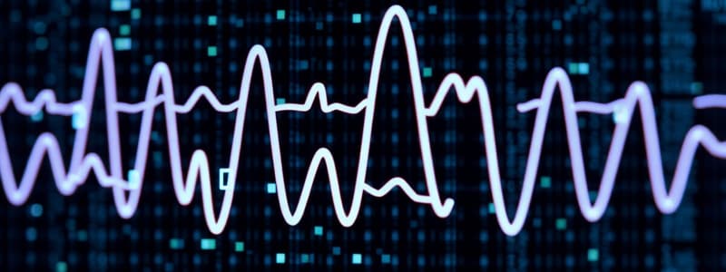
Frequency analysis of EEG
Frequency analysis of EEG
Analyzing EEG data based on the frequency of neuronal oscillations, such as 10 Hz representing 10 cycles per second.
Time-frequency analysis
Time-frequency analysis
Examining how the amplitude (power) of brain waves changes over time.
EEG Frequency Bands
EEG Frequency Bands
Different ranges of brain wave frequencies, each associated with different brain states or activities (e.g., delta, theta, alpha, beta, gamma).
N400 ERP
N400 ERP
Signup and view all the flashcards
MEG (Magnetoencephalography)
MEG (Magnetoencephalography)
Signup and view all the flashcards
What does EEG measure?
What does EEG measure?
Signup and view all the flashcards
EEG and behavioral state
EEG and behavioral state
Signup and view all the flashcards
EEG artifact
EEG artifact
Signup and view all the flashcards
EEG pre-processing
EEG pre-processing
Signup and view all the flashcards
ERP (Event-Related Potential)
ERP (Event-Related Potential)
Signup and view all the flashcards
ERP averaging
ERP averaging
Signup and view all the flashcards
Why is ERP important?
Why is ERP important?
Signup and view all the flashcards
EEG: Temporal vs Spatial
EEG: Temporal vs Spatial
Signup and view all the flashcards
False Positives (Type 1 Error)
False Positives (Type 1 Error)
Signup and view all the flashcards
False Negatives (Type 2 Error)
False Negatives (Type 2 Error)
Signup and view all the flashcards
fMRI Measures What?
fMRI Measures What?
Signup and view all the flashcards
Voxel Limitations
Voxel Limitations
Signup and view all the flashcards
fMRI Temporal Resolution
fMRI Temporal Resolution
Signup and view all the flashcards
Reward Prediction Error (PE)
Reward Prediction Error (PE)
Signup and view all the flashcards
PE & Dopamine
PE & Dopamine
Signup and view all the flashcards
PE & Striatum
PE & Striatum
Signup and view all the flashcards
Study Notes
Neuroscience Methods & Issues
- This lecture covers neuroscience methods for studying cognition, including EEG, MEG, fMRI (and PET)
- Methods are focused on measuring brain activity related to cognitive function
- Four main areas of focus include neural function measurements (EEG, MEG), combined function/structure measurements (fMRI, PET), general issues with neuro methods, and the future of combined methods
Functional Neuroimaging Methods: Temporal and Spatial Resolution
- Graph shows temporal and spatial resolution differences between MEG/EEG, fMRI, and PET
- MEG/EEG have excellent temporal resolution, measuring millisecond-level activity changes. However, their spatial resolution is limited
- fMRI has good spatial resolution, allowing accurate location of activity, though its temporal resolution is lower than EEG/MEG (seconds)
- PET also has good spatial resolution, but has the poorest temporal resolution compared to the other methods (days).
Neural Function (EEG, MEG)
- EEG measures large populations of electrical neuronal activity.
- EEG data is continuous and varies by behavioural states, which can suggest cognitive function
- EEG is used for diagnosing issues like epilepsy
- MEG detects magnetic fields produced by neuronal activity.
- MEG signals are more localised than EEG information which helps pinpoint brain regions related to activity
- MEG analysis involves positioning SQUIDs around the head, detecting auditory responses to a stimulus, generating contour maps and localising the stimulus-response in the brain
EEG Pre-processing
- Essential steps include filtering, epoch segmentation, artifact rejection (blinks or movements) and baseline correction
- Data cleaning is crucial to prevent data distortion and accurately reflect the response to a cognitive task
Event-Related Potentials (ERPs)
- Using data from many trials (e.g. responses to stimuli), researchers can average out noise to identify neural responses to stimuli that may be unique
- Data is time-locked to events of interest, often stimuli and behaviours in a task to isolate event-related signals
- ERPs are important neuro signals that can be used to understand cognitive events in the brain
Frequency Analysis of EEG Data
- EEG activity is characterized by frequency, or oscillations
- The different oscillations include various states such as theta, alpha, beta, and gamma rhythms, which can be tracked over time
- Time-frequency analysis is used to measure changes in the amplitude of the wave across time
Example: The N400 ERP
- The N400 is an ERP component related to semantic processing
- An example sentence (like 'He spread his toast with…socks') can be used to illustrate this
- The amplitude of the N400 component in the brain shows how relatable words are in a sentence
However Interpretation Can Be More Difficult
- The N400 component may not be unique to sentence processing but also due to semantic priming from other context words
- Priming describes how previous information can change following responses to a stimulus (related words).
EEG Issues and Considerations
- Advantages are excellent temporal resolution, non-invasiveness, and low cost
- Disadvantages include limited spatial resolution, long setup time, and low signal-to-noise ratio (SNR)
Magnetoencephalography (MEG)
- MEG records magnetic fields produced by neuronal activity, offering better spatial localization than EEG
- Detecting these signals needs super-conducting sensors and amplifiers (SQUIDs) which collect and interpret the signals
- The data is then overlaid onto images of the brain from another imaging technique (e.g. MRI) to provide a visual localisation of the signals
MEG Analysis
- Data processing often consists of positioning SQUIDs around the head, introducing a stimulus that generates a behaviour (like a tone that elicits an auditory response), creating contour maps showing areas of activity, and identifying the dipole signal related to the response in the brain
MEG Issues and Considerations
- Advantages include good temporal resolution and an ability to identify the activity of neuronal sources in the brain
- Disadvantages include small signals and the inverse problem which determines the exact location of electrical activity within the brain
Structural Analysis: Magnetic Resonance Imaging (MRI)
- MRI uses magnetic field properties within the brain tissue using hydrogen atoms to record an image of the brain
- These signals can be used to generate images of brain structures
- fMRI uses this magnetic property to understand brain activity related to cognitive tasks through oxygenated measurements.
Functional Magnetic Resonance Imaging (fMRI)
- fMRI monitors the ratio of oxygenated and deoxygenated haemoglobin in the blood in the brain, inferring which areas of the brain are active
- Active regions of the brain will take up more oxygen to supply brain cells
- It's an indirect measure of brain activity but has great spatial resolution
fMRI Data
- fMRI data is composed of voxels (3D cubes) that identify brain activity
- Each voxel can contain a great number of neurons, and so the signal reflects the population activity within the voxel
fMRI Sources of Noise
- fMRI data has several sources of noise, in addition to physical movement issues like the subject moving, external sounds and changes in brain activity over time
fMRI Preprocessing
- Steps in preprocessing can include motion compensation (prevent inaccurate data due to movement), slice timing correction (adjust for differing times during image acquisition), spatial filtering (improving the quality of the signal), and temporal filtering (removing high-frequency fluctuations of the data)
Co-Registration and Normalisation
- Co-registration refers to aligning fMRI data with individual and structural anatomical images, like MRI
- Normalisation refers to adjusting multiple subjects' fMRI data, making it comparable, by aligning their data to a standard brain model via warping their images
fMRI Example Experiment (Smoking)
- A study examined attentional bias and neuro-activity in smokers with exposure to smoking and/or neutral images
Conclusion and Future Directions
- EEG and MEG results should be considered to understand when neural events happen, and how to combine results from these techniques across different cognitive tasks
- Combining multiple methods helps understanding neurological processes better than each single technique
- There is a potential to identify future treatment options especially when these results are considered from a meta-analysis.
Meta-Analysis of Prediction Error (PE) Literature
- PE literature studies have been categorized into valence (positive/negative value), magnitude (size of the event), and signed PE groups (combination of valence and magnitude)
- Studies have shown separate networks within the brain are correlated with individual PE categories.
- The striatum is one area of the brain that appears to correlate across different PE classifications.
Prediction Error (PE)
- Prediction Error (PE) is the difference between the expected reward and the outcome of a cognitive task. PE data can be used to update future expectations and behaviours
- Dopamine neurons, among others, are used to identify this Prediction Error
- The striatum is an important brain region involved in this prediction error pathway
Concurrent fMRI and EEG
- EEG studies have revealed different temporal networks to valence, magnitude and PE in studies combining fMRI and EEG
- Researchers used EEG informed analysis to highlight distinct brain regions that fMRI alone could not find
Sample Sizes
- 50 participants is considered a large sample size
- It is crucial that researchers consider relevant factors of sample size because effects might be smaller even if the sample size is large, and that bigger studies will not always be meaningful
Bringing It All Together
- Formulating a clear research question based on literature is critical to ensure the research is based on a good foundation
- Developing a proper research question needs careful consideration of all the relevant background research to help build on existing results
Reversal Learning Task
- A behavioural paradigm or task used to study decision-making in the brain which demonstrates the transition in decision-making processes.
- Stimuli and feedback are displayed and responses analysed in different conditions to measure how brain regions change in responses
- This helps understand learning related to cognitive processes.
Combining Methods
- Combining different methods can offer insights beyond what a single method can provide
- Combining different methods might enhance understanding of the human brain more fundamentally but it could also introduce issues that need to be carefully considered
PET Issues and Considerations
- PET is exposed to radiation, making short-term tasks the only use case for these scans
- PET is an expensive technique, making it limited to research purposes rather than clinical and routine applications
- PET can measure blood flow and metabolic rates in the brain, which are crucial measures for understanding cognitive processes.
General Limitations of fMRI
- fMRI data reflects blood flow in a brain region and not neural activity directly, so results have limitations in interpreting specific neural activity
- A large number of neurons might be represented by each voxel, making it difficult to isolate the specific neural activity in that region, and the data is only measured coarsely via time measurements
- fMRI responds very slowly compared to neural signalling time, so it cannot accurately measure changes in neural activity
EEG and fMRI Issues and Considerations
- The different considerations regarding EEG and fMRI can be summarised as their respective strengths (e.g., EEG has good temporal but poor spatial resolution) and weaknesses (spatial or temporal limitations)
Studying That Suits You
Use AI to generate personalized quizzes and flashcards to suit your learning preferences.