Podcast
Questions and Answers
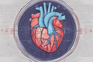
What is echocardiography?
What is echocardiography?
The use of ultrasound in order to study the function and morphology of the heart without being invasive
What equipment is needed for echocardiography?
What equipment is needed for echocardiography?
Echocardiology table, ultrasound gel or alcohol
When performing an echocardiogram on a dog, where should the probe be placed on the right side?
When performing an echocardiogram on a dog, where should the probe be placed on the right side?
- 2nd-4th intercostal spaces
- 4th-6th intercostal spaces (correct)
- 3rd-5th intercostal spaces
- 5th-7th intercostal spaces
When performing an echocardiogram on a dog, where should the probe be placed on the left side?
When performing an echocardiogram on a dog, where should the probe be placed on the left side?
How does the scanning location for echocardiography in cats differ from that in dogs?
How does the scanning location for echocardiography in cats differ from that in dogs?
Which scanning position allows visualization of all four heart chambers, the mitral valve, and the tricuspid valve?
Which scanning position allows visualization of all four heart chambers, the mitral valve, and the tricuspid valve?
Which echocardiographic view allows visualization of all four heart chambers, the aorta, the mitral valve, and the tricuspid valve?
Which echocardiographic view allows visualization of all four heart chambers, the aorta, the mitral valve, and the tricuspid valve?
Which echocardiographic view is best for assessing the left ventricle, papillary muscles, intraventricular septum, and right ventricle?
Which echocardiographic view is best for assessing the left ventricle, papillary muscles, intraventricular septum, and right ventricle?
How can the mitral valve be brought into view when using the right parasternal transverse short-axis view?
How can the mitral valve be brought into view when using the right parasternal transverse short-axis view?
Which echocardiographic view is best used to measure blood flow velocity as it moves from the atria to the ventricles?
Which echocardiographic view is best used to measure blood flow velocity as it moves from the atria to the ventricles?
What is the primary purpose of the left parasternal apical 5-chamber view?
What is the primary purpose of the left parasternal apical 5-chamber view?
What is the primary purpose of M-mode in echocardiography?
What is the primary purpose of M-mode in echocardiography?
In an M-mode echocardiogram, what does the x-axis represent?
In an M-mode echocardiogram, what does the x-axis represent?
In an M-mode echocardiogram, what does the y-axis represent?
In an M-mode echocardiogram, what does the y-axis represent?
Which of the following best describes the advantage of using M-mode in echocardiography?
Which of the following best describes the advantage of using M-mode in echocardiography?
What is fractional shortening?
What is fractional shortening?
What is the normal Fractional shortening for the dog?
What is the normal Fractional shortening for the dog?
Flashcards
Echocardiography
Echocardiography
The use of ultrasound to study heart function and structure non-invasively.
Echocardiography equipment
Echocardiography equipment
Necessary items include an echocardiology table, ultrasound gel or alcohol.
Probe placement on a dog (right side)
Probe placement on a dog (right side)
The probe should be placed between the 4th and 6th intercostal spaces.
Probe placement on a dog (left side)
Probe placement on a dog (left side)
Signup and view all the flashcards
Echocardiography in cats
Echocardiography in cats
Signup and view all the flashcards
Right parasternal long-axis view
Right parasternal long-axis view
Signup and view all the flashcards