Podcast
Questions and Answers
What does the P wave in an ECG represent?
What does the P wave in an ECG represent?
- AV nodal depolarisation
- Ventricular repolarisation
- Atrial depolarisation (correct)
- Ventricular depolarisation
Which lead is formed from the difference between the left arm and right arm electrodes?
Which lead is formed from the difference between the left arm and right arm electrodes?
- Lead aVR
- Lead II
- Lead I (correct)
- Lead III
Which of the following accurately describes the role of the right leg electrode?
Which of the following accurately describes the role of the right leg electrode?
- It is primarily used as an earth electrode.
- It must be positioned directly above the heart.
- It can significantly impact the ECG readings.
- It is placed to reduce electrical interference. (correct)
How are the augmented unipolar limb leads represented in standard ECG?
How are the augmented unipolar limb leads represented in standard ECG?
What is the relationship between all limb leads in an ECG?
What is the relationship between all limb leads in an ECG?
Which segment in the ECG corresponds to ventricular repolarisation?
Which segment in the ECG corresponds to ventricular repolarisation?
What does the hexaxial reference system help with?
What does the hexaxial reference system help with?
What does the QRS complex in an ECG indicate?
What does the QRS complex in an ECG indicate?
What is the position of electrode V1?
What is the position of electrode V1?
Where is electrode V4 placed?
Where is electrode V4 placed?
What is the function of Wilson’s central terminal in ECG recording?
What is the function of Wilson’s central terminal in ECG recording?
Which of the following statements about chest electrodes is correct?
Which of the following statements about chest electrodes is correct?
What is the primary orientation plane that chest leads record electrical activity in?
What is the primary orientation plane that chest leads record electrical activity in?
Which electrode is placed horizontally with V4?
Which electrode is placed horizontally with V4?
What role do limb electrodes play in relation to the chest leads?
What role do limb electrodes play in relation to the chest leads?
Which lead is correctly positioned at the left sternal edge in the 4th intercostal space?
Which lead is correctly positioned at the left sternal edge in the 4th intercostal space?
Flashcards are hidden until you start studying
Study Notes
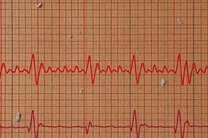
ECG Fundamentals
- The cardiac conduction system orchestrates heart rhythms through a sequence of electrical events: atrial depolarization, AV nodal depolarization, ventricular depolarization, and ventricular repolarization.
- Corresponding ECG waves include the P wave (atrial depolarization), PR segment (AV nodal depolarization), QRS complex (ventricular depolarization), and T wave (ventricular repolarization).
Limb Electrode Positions
- Standard limb electrodes:
- Red: Right arm (below elbow)
- Yellow: Left arm (below elbow)
- Green: Left leg (below knee)
- Black: Right leg (below ankle)
- Limb electrodes form an equilateral triangle with the heart at its center known as Einthoven's triangle.
Standard Limb Leads
- Standard limb leads include I, II, and III, which can be calculated mathematically from each other:
- Lead I = VLA – VRA
- Lead II = VLL – VRA
- Lead III = VLL - VLA
Augmented Unipolar Limb Leads
- Augmented leads include aVR, aVL, and aVF, with specific orientations and roles in ECG interpretation.
Hexaxial Reference System
- All limb leads are mathematically interrelated; knowing any two leads allows calculation of the third.
- The orientation of each limb lead affects its ECG appearances based on the direction of electrical current flow.
Right Leg Electrode
- The right leg electrode minimizes interference but is not an earth electrode; it can be placed anywhere.
Frontal Plane vs. Horizontal Plane
- Limb leads capture electrical activity in the frontal plane (right/left, up/down).
- Chest leads are required for assessing electrical activity in the horizontal (transverse) plane.
Standard Chest Electrode Positions
- V1: 4th intercostal space, right sternal edge
- V2: 4th intercostal space, left sternal edge
- V3: Midway between V2 and V4
- V4: 5th intercostal space, mid-clavicular line
- V5: Horizontal with V4, anterior axillary line
- V6: Horizontal with V4, mid-axillary line
Wilson’s Central Terminal
- Functions as the negative pole for chest leads by electrically joining the three limb electrodes.
Chest Leads Configuration
- All chest electrodes act as positive poles.
- Common layout includes: aVR, aVL, V1, V2, V3, V4, V5, V6, II, III, and aVF.
Typical 12-Lead ECG Format
- Represents standard arrangement of all limb and chest leads, essential for comprehensive cardiac assessment.
Studying That Suits You
Use AI to generate personalized quizzes and flashcards to suit your learning preferences.