Podcast
Questions and Answers
Which sensory function allows the detection of multiple points of contact on the skin?
Which sensory function allows the detection of multiple points of contact on the skin?
- Discriminative Touch (correct)
- Proprioception
- Pressure Sensation
- Vibration Detection
Pacinian corpuscles are responsible for detecting fine touch and superficial pressure.
Pacinian corpuscles are responsible for detecting fine touch and superficial pressure.
False (B)
What is the primary function of muscle spindles?
What is the primary function of muscle spindles?
Detecting stretch and proprioception.
The _____ pathway is essential for sensations related to touch, proprioception, and vibration.
The _____ pathway is essential for sensations related to touch, proprioception, and vibration.
Match the sensory receptors with their primary function:
Match the sensory receptors with their primary function:
Which type of nerve fibers are considered the fastest in conduction speed?
Which type of nerve fibers are considered the fastest in conduction speed?
C fibers are well myelinated and transmit signals quickly.
C fibers are well myelinated and transmit signals quickly.
What is the function of Ruffini corpuscles?
What is the function of Ruffini corpuscles?
The group of axons that transmits sensory information upward is called an _____ tract.
The group of axons that transmits sensory information upward is called an _____ tract.
Which receptors are associated with detecting hair movement?
Which receptors are associated with detecting hair movement?
What type of sensory information does the Fasciculus Gracilis primarily carry?
What type of sensory information does the Fasciculus Gracilis primarily carry?
The dorsal root ganglion contains cell bodies of motor neurons.
The dorsal root ganglion contains cell bodies of motor neurons.
Which part of the thalamus serves as a relay station for sensory information to the cortex?
Which part of the thalamus serves as a relay station for sensory information to the cortex?
The second-order neurons in the sensory pathway cross over via __________.
The second-order neurons in the sensory pathway cross over via __________.
Match the sensory fiber types with their descriptions:
Match the sensory fiber types with their descriptions:
What is the primary role of the dorsal column medial lemniscus pathway?
What is the primary role of the dorsal column medial lemniscus pathway?
The primary somatosensory cortex is located in the frontal lobe.
The primary somatosensory cortex is located in the frontal lobe.
What is the significance of the somatosensory homunculus?
What is the significance of the somatosensory homunculus?
Sensory pathways are organized __________ to ensure specific body areas are accurately represented in the CNS.
Sensory pathways are organized __________ to ensure specific body areas are accurately represented in the CNS.
Which of the following fibers are responsible for connecting different regions within the same hemisphere?
Which of the following fibers are responsible for connecting different regions within the same hemisphere?
Which receptor is primarily responsible for detecting fine discriminative touch?
Which receptor is primarily responsible for detecting fine discriminative touch?
Type A fibers are associated with the slow transmission of pain signals.
Type A fibers are associated with the slow transmission of pain signals.
What is the function of Golgi tendon organs?
What is the function of Golgi tendon organs?
_______ is the awareness of joint, muscle, and limb positions without visual input.
_______ is the awareness of joint, muscle, and limb positions without visual input.
Match the following sensory receptors with their primary function:
Match the following sensory receptors with their primary function:
What speed does Aβ fibers typically transmit at?
What speed does Aβ fibers typically transmit at?
The dorsal column medial lemniscus pathway is involved in transmitting motor information to the muscles.
The dorsal column medial lemniscus pathway is involved in transmitting motor information to the muscles.
Where do first-order neurons reside in the dorsal column medial lemniscus pathway?
Where do first-order neurons reside in the dorsal column medial lemniscus pathway?
The dorsal column medial lemniscus pathway is considered an __________ tract.
The dorsal column medial lemniscus pathway is considered an __________ tract.
Which type of receptor responds primarily to deep pressure and vibration?
Which type of receptor responds primarily to deep pressure and vibration?
What does the Fasciculus gracilis primarily carry?
What does the Fasciculus gracilis primarily carry?
The first-order neurons in the sensory pathways originate in the spinal cord.
The first-order neurons in the sensory pathways originate in the spinal cord.
What is the role of the medial lemniscus?
What is the role of the medial lemniscus?
The sensory fibers that enter the spinal cord are organized from __________ to __________ from sacral to cervical.
The sensory fibers that enter the spinal cord are organized from __________ to __________ from sacral to cervical.
Which structure serves as a relay point for sensory information in the thalamus?
Which structure serves as a relay point for sensory information in the thalamus?
Lesions in the spinal cord can affect specific regions due to somatotopic organization.
Lesions in the spinal cord can affect specific regions due to somatotopic organization.
Match the following structures with their corresponding roles:
Match the following structures with their corresponding roles:
What is the significance of proprioception in the nervous system?
What is the significance of proprioception in the nervous system?
The fibers that cross over to the contralateral side at the dorsal part of the medulla are known as __________.
The fibers that cross over to the contralateral side at the dorsal part of the medulla are known as __________.
What happens when there is an infarction of the lenticular striate arteries?
What happens when there is an infarction of the lenticular striate arteries?
Which type of receptor is primarily responsible for detecting stretch in muscles?
Which type of receptor is primarily responsible for detecting stretch in muscles?
Type A fibers are the slowest in conduction speed among nerve fibers.
Type A fibers are the slowest in conduction speed among nerve fibers.
What is the primary function of Meissner's corpuscles?
What is the primary function of Meissner's corpuscles?
The ability to identify two separate points of contact on the skin is referred to as __________.
The ability to identify two separate points of contact on the skin is referred to as __________.
Match the following receptors with their primary functions:
Match the following receptors with their primary functions:
Which type of fibers is associated with touch, pressure, and vibration sensations?
Which type of fibers is associated with touch, pressure, and vibration sensations?
Proprioception involves the awareness of body part positions without visual input.
Proprioception involves the awareness of body part positions without visual input.
Where do first-order neurons reside in the dorsal column medial lemniscus pathway?
Where do first-order neurons reside in the dorsal column medial lemniscus pathway?
The bundle of axons in the central nervous system is known as a __________.
The bundle of axons in the central nervous system is known as a __________.
Which receptors are found deeper in the dermis and hypodermis?
Which receptors are found deeper in the dermis and hypodermis?
What type of sensory information does the Fasciculus cuneatus primarily carry?
What type of sensory information does the Fasciculus cuneatus primarily carry?
First-order neurons in the sensory pathways originate in the spinal cord.
First-order neurons in the sensory pathways originate in the spinal cord.
What is the primary role of the medial lemniscus in sensory pathways?
What is the primary role of the medial lemniscus in sensory pathways?
The _____ is the area in the brain that serves as a relay point for sensory information before it reaches the cortex.
The _____ is the area in the brain that serves as a relay point for sensory information before it reaches the cortex.
Match the following spinal structures with their functions:
Match the following spinal structures with their functions:
Which of the following best describes the term 'somatotopic organization'?
Which of the following best describes the term 'somatotopic organization'?
The internal capsule is responsible for connecting the left and right hemispheres of the brain.
The internal capsule is responsible for connecting the left and right hemispheres of the brain.
In which part of the brain do the crossing fibers of the sensory pathway occur?
In which part of the brain do the crossing fibers of the sensory pathway occur?
Sensory fibers entering the spinal cord are organized from _____ to _____ from sacral to cervical.
Sensory fibers entering the spinal cord are organized from _____ to _____ from sacral to cervical.
What is a key clinical implication of infarction in the lenticular striate arteries?
What is a key clinical implication of infarction in the lenticular striate arteries?
What sensation does the dorsal column medial lemniscus pathway primarily convey?
What sensation does the dorsal column medial lemniscus pathway primarily convey?
Aδ fibers are the fastest type of nerve fibers.
Aδ fibers are the fastest type of nerve fibers.
What type of receptor is responsible for detecting deep pressure and vibration?
What type of receptor is responsible for detecting deep pressure and vibration?
The awareness of joint, muscle, and limb positions without visual input is known as __________.
The awareness of joint, muscle, and limb positions without visual input is known as __________.
Match the following sensory receptors with their primary function:
Match the following sensory receptors with their primary function:
Which type of nerve fibers are involved in touch, pressure, and vibration sensations?
Which type of nerve fibers are involved in touch, pressure, and vibration sensations?
Meissner's corpuscles respond to deep pressure and vibration.
Meissner's corpuscles respond to deep pressure and vibration.
Where do first-order neurons reside in the dorsal column medial lemniscus pathway?
Where do first-order neurons reside in the dorsal column medial lemniscus pathway?
The dorsal column medial lemniscus pathway transmits information from the peripheral nervous system to the __________.
The dorsal column medial lemniscus pathway transmits information from the peripheral nervous system to the __________.
Which receptors are involved in monitoring hair movement?
Which receptors are involved in monitoring hair movement?
What type of sensory information does the Fasciculus cuneatus primarily carry?
What type of sensory information does the Fasciculus cuneatus primarily carry?
First-order neurons in the dorsal column medial lemniscus pathway cross to the contralateral side in the spinal cord.
First-order neurons in the dorsal column medial lemniscus pathway cross to the contralateral side in the spinal cord.
Name the structures that contain second-order neurons in the medulla.
Name the structures that contain second-order neurons in the medulla.
The medial lemniscus is a fiber bundle that carries sensory information upwards to the __________.
The medial lemniscus is a fiber bundle that carries sensory information upwards to the __________.
Match the following spinal cord pathways with their primary functions:
Match the following spinal cord pathways with their primary functions:
Which structure serves as a relay point in the thalamus for sensory information?
Which structure serves as a relay point in the thalamus for sensory information?
Proprioception is not important for motor coordination.
Proprioception is not important for motor coordination.
What is the significance of the lenticular striate arteries?
What is the significance of the lenticular striate arteries?
Sensory fibers entering the spinal cord are organized __________ from sacral to cervical.
Sensory fibers entering the spinal cord are organized __________ from sacral to cervical.
Which type of fibers are responsible for connecting the two hemispheres of the brain?
Which type of fibers are responsible for connecting the two hemispheres of the brain?
Which pathway is responsible for carrying sensory information from the lower limbs and trunk?
Which pathway is responsible for carrying sensory information from the lower limbs and trunk?
The nuclei gracilis and cuneatus are located in the spinal cord.
The nuclei gracilis and cuneatus are located in the spinal cord.
What is the primary role of the ventral posterior lateral nucleus (VPL) in the thalamus?
What is the primary role of the ventral posterior lateral nucleus (VPL) in the thalamus?
The internal arcuate fibers cross over to the __________ side at the dorsal part of the medulla.
The internal arcuate fibers cross over to the __________ side at the dorsal part of the medulla.
Match the following spinal cord structures with their functions:
Match the following spinal cord structures with their functions:
Which of the following accurately describes the organization of sensory fibers entering the spinal cord?
Which of the following accurately describes the organization of sensory fibers entering the spinal cord?
First-order neurons originate in the dorsal root ganglion and provide direct connections to the cortex.
First-order neurons originate in the dorsal root ganglion and provide direct connections to the cortex.
What important role does proprioception play in the nervous system?
What important role does proprioception play in the nervous system?
Infarction of the lenticular striate arteries can lead to potential loss of __________.
Infarction of the lenticular striate arteries can lead to potential loss of __________.
Match the following pathways with their associated functions:
Match the following pathways with their associated functions:
Which type of receptor is primarily responsible for detecting deep pressure and vibration?
Which type of receptor is primarily responsible for detecting deep pressure and vibration?
The dorsal column medial lemniscus pathway is responsible for transmitting motor information to the muscles.
The dorsal column medial lemniscus pathway is responsible for transmitting motor information to the muscles.
What is the awareness of joint, muscle, and limb positions without visual input called?
What is the awareness of joint, muscle, and limb positions without visual input called?
The __________ is an essential pathway for touch and proprioceptive sensations.
The __________ is an essential pathway for touch and proprioceptive sensations.
Match the following types of nerve fibers with their characteristics:
Match the following types of nerve fibers with their characteristics:
Which type of receptors are responsible for detecting hair movement?
Which type of receptors are responsible for detecting hair movement?
Type A fibers are associated with the slow transmission of pain signals.
Type A fibers are associated with the slow transmission of pain signals.
Name one function of Ruffini corpuscles.
Name one function of Ruffini corpuscles.
The dorsal root ganglion contains cell bodies of __________ neurons.
The dorsal root ganglion contains cell bodies of __________ neurons.
Which receptors are primarily located in the dermal papillae and responsible for fine touch?
Which receptors are primarily located in the dermal papillae and responsible for fine touch?
Flashcards are hidden until you start studying
Study Notes
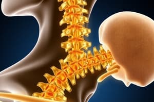
Dorsal Column Medial Lemniscus Pathway Overview
- Vital pathway for sensations related to touch, proprioception, pressure, vibration, and stretch.
- Facilitates discriminative touch, also known as two-point touch, important for identifying multiple points of contact on the skin.
Key Sensory Functions
- Discriminative Touch: Ability to detect fine touch and differentiate between two points touching the skin.
- Pressure and Stretch Sensation: Monitors pressure applied to the skin and stretching of the dermis.
- Vibration Detection: Specialized receptors sense vibrations.
- Proprioception: Awareness of body position and movement in space, vital for coordination and balance.
Ascending Tract Definition
- An ascending tract is a bundle of axons in the central nervous system that transmits sensory information upward.
Sensory Receptors Involved
- Meissner's Corpuscles: Located in dermal papillae, responsive to fine touch and discriminative touch.
- Merkel's Discs: Found in the stratum basale, sensitive to fine touch and superficial pressure.
- Pacinian Corpuscles: Located deeper in the dermis, respond to deep pressure and vibrations.
- Ruffini Corpuscles: Detect stretch of the skin and sustained pressure.
- Perechial Nerve Endings: Associated with hair follicles, aiding in detecting hair movement.
- Muscle Spindles: Comprised of intrafusal fibers detecting stretch, contains nuclear chain and nuclear bag fibers.
- Golgi Tendon Organs: Monitor tendon stretch, protecting against excessive tension.
Nerve Fiber Classification
- Nerve fibers are classified by myelination:
- A fibers: Highly myelinated, fastest conduction (up to 120 m/s).
- B fibers: Moderately myelinated.
- C fibers: Poorly myelinated or unmyelinated, slowest conduction.
- Type I A fibers: Carry proprioceptive signals from muscle spindles.
- Type I B fibers: Transmit signals from Golgi tendon organs.
- A Beta fibers: Relay touch, pressure, and vibration sensations from various receptors.
Spinal Cord Pathway Organization
- Dorsal root ganglion contains cell bodies of sensory neurons; first-order neurons enter the spinal cord.
- Information is processed in the posterior gray horn and travels through dorsal white columns.
- Fasciculus Gracilis: Carries sensory information from below T6, mainly lower body and legs.
- Fasciculus Cuneatus: Transmits sensory information from above T6, covering the upper body and arms.
- Organization of sensory pathways ensures specific areas of the body are represented accurately in the CNS through somatotopic arrangement.
Clinical Relevance
- Understanding the upper and lower body representation is crucial for diagnosing lesions.
- Lesions can affect specific regions based on the organization of fibers, impacting sensory perception.
Summary
- The dorsal column medial lemniscus pathway is essential for processing key sensory modalities through various specialized receptors, with tight relevance to overall body awareness, coordination, and sensory discrimination. The pathway's organization in the spinal cord aids in accurate representation and efficient sensory information processing.### Spinal Cord Pathways
- Fibers entering the spinal cord remain ipsilateral (same side) at the spinal cord level, critical for clinical understanding.
- First-order neurons originate in the dorsal root ganglion with peripheral processes connecting to receptors and central processes entering the spinal cord.
Fasciculus Cuneatus and Fasciculus Gracilis
- Fasciculus cuneatus carries information from upper body sensations (above T6); Fasciculus gracilis carries information from lower body sensations (below T6).
- Both fasciculi project to second-order neurons located in the medulla, specifically in the nucleus gracilis and nucleus cuneatus.
Crossing Over and Medial Lemniscus Formation
- Second-order neurons cross over via internal arcuate fibers after processing sensations in the medulla.
- The medial lemniscus is formed as these fibers ascend through the brainstem, transitioning to contralateral pathways.
Thalamic Relay
- Fibers converge on the ventral posterior lateral nucleus (VPL) of the thalamus, which serves as a relay station for sensory information to the cortex.
- The VPL sends projections through the posterior limb of the internal capsule to the primary somatosensory cortex.
Internal Capsule and Sensory Information
- The posterior limb of the internal capsule carries sensory fibers; infarcts in supplying arteries can lead to sensory loss.
- Internal capsule and corona radiata consist of projection fibers, facilitating the transmission of sensory information to higher cortical centers.
Somatosensory Areas in Cortex
- The primary somatosensory cortex, located near the central sulcus, receives organized sensory input mapped to specific body areas (sensory homunculus).
- The secondary somatosensory cortex complements the primary area, with distinct locations for sensations from various body parts.
Somatic Topographic Organization
- Somatosensory information is organized topographically in the cortex, allowing precise localization of sensations (e.g., touch from the foot, hand, face).
- Sensory homunculus provides a visual representation of the body's sensory distribution in the cortex.
White Matter Fiber Types
- Projection fibers connect different brain regions, transporting information upwards (sensory) and downwards (motor pathways).
- Commissural fibers, like the corpus callosum, interconnect the two hemispheres, while association fibers connect regions within the same hemisphere (e.g., arcuate fasciculus).
Summary of Pathways and Fiber Types
- Understanding the paths and connections of sensory information is essential for grasping the functioning of the nervous system.
- Differentiating between types of fibers is crucial for studying neural communication within the central nervous system.
Dorsal Column Medial Lemniscus Pathway Overview
- Crucial pathway for sensory modalities such as touch, proprioception, pressure, vibration, and stretch.
- Enables discriminative touch, allowing the detection of multiple skin contact points.
Key Sensory Functions
- Discriminative Touch: Fine touch sensitivity; can identify two distinct points on the skin.
- Pressure and Stretch Sensation: Monitors skin pressure and dermal stretching.
- Vibration Detection: Specialized receptors respond to vibratory stimuli.
- Proprioception: Body position and movement awareness, essential for balance and coordination.
Ascending Tract Definition
- Bundle of axons in the central nervous system that carries sensory information upwards.
Sensory Receptors Involved
- Meissner's Corpuscles: Located in dermal papillae, responsive to fine and discriminative touch.
- Merkel's Discs: Provide sensitivity to fine touch and superficial pressure.
- Pacinian Corpuscles: Detect deep pressure and vibrations, located in deeper dermis.
- Ruffini Corpuscles: Sensitive to skin stretch and sustained pressure.
- Perechial Nerve Endings: Associated with hair follicles to detect hair movement.
- Muscle Spindles: Detect stretch via intrafusal fibers, including nuclear chain and nuclear bag fibers.
- Golgi Tendon Organs: Monitor tendon stretch to prevent excessive tension.
Nerve Fiber Classification
- A fibers: Highly myelinated; fastest conduction speed (up to 120 m/s).
- B fibers: Moderately myelinated.
- C fibers: Poorly myelinated or unmyelinated; slowest conduction.
- Type I A fibers: Proprioceptive signals from muscle spindles.
- Type I B fibers: Signals from Golgi tendon organs.
- A Beta fibers: Relay touch, pressure, and vibration sensations.
Spinal Cord Pathway Organization
- Dorsal root ganglion contains sensory neuron cell bodies; first-order neurons enter the spinal cord.
- Information is processed in the posterior gray horn and ascends through dorsal white columns.
- Fasciculus Gracilis: Carries sensory info from lower body (below T6).
- Fasciculus Cuneatus: Transmits sensory info from upper body (above T6).
- Somatotopic organization ensures accurate representation of body areas in the CNS.
Clinical Relevance
- Understanding body representation helps diagnose spinal cord lesions.
- Lesions can impact sensory perception based on fiber organization.
Spinal Cord Pathways
- Fibers entering the spinal cord remain ipsilateral, critical for clinical assessments.
- Dorsal root ganglion neurons connect to receptors and enter the spinal cord for processing.
Fasciculus Cuneatus and Fasciculus Gracilis
- Fasciculus cuneatus: Upper body sensations (above T6).
- Fasciculus gracilis: Lower body sensations (below T6).
- Project to second-order neurons in the medulla (nucleus gracilis and nucleus cuneatus).
Crossing Over and Medial Lemniscus Formation
- Second-order neurons cross over via internal arcuate fibers in the medulla.
- Medial lemniscus formed as these fibers ascend in the brainstem, shifting to contralateral pathways.
Thalamic Relay
- Fibers converge at the ventral posterior lateral nucleus (VPL) of the thalamus, acting as a relay for sensory info to the cortex.
- The VPL transmits projections through the internal capsule to the primary somatosensory cortex.
Internal Capsule and Sensory Information
- Posterior limb of the internal capsule carries sensory fibers; artery infarcts can cause sensory losses.
- Contains projection fibers facilitating the transmission of sensory information to cortical regions.
Somatosensory Areas in Cortex
- The primary somatosensory cortex is near the central sulcus, mapping specific body area sensations (sensory homunculus).
- Secondary somatosensory cortex complements the primary area, dedicated to sensations from various parts.
Somatic Topographic Organization
- Sensory information organized topographically for precise localization (e.g., touch sensations from specific body parts).
- Sensory homunculus visually represents the distribution of sensory modalities in the cortex.
White Matter Fiber Types
- Projection fibers: Connect different brain regions, transport sensory and motor information.
- Commissural fibers: Link two hemispheres (e.g., corpus callosum).
- Association fibers: Connect regions within the same hemisphere (e.g., arcuate fasciculus).
Summary of Pathways and Fiber Types
- Comprehending sensory pathways is crucial for understanding the nervous system's function.
- Distinguishing between fiber types aids in studying neural communication in the CNS.
Dorsal Column Medial Lemniscus Pathway
- Transmits touch, proprioception, pressure, vibration, and stretch sensations to the brain.
- Enables discriminative touch, allowing perception of fine touch and distinguishing between two points of contact.
Key Sensations
- Two-point touch: Ability to detect two distinct points on the skin.
- Pressure and stretch detection enhances tactile perception.
- Proprioception: Recognition of limb and joint positions without visual cues.
Ascending Tract Overview
- The pathway is classified as an ascending tract for sensory relay to the brain.
- Composed of bundles of axons in the central nervous system.
Types of Receptors Involved
- Meissner’s corpuscles: Sensitive to fine touch, located in dermal papillae.
- Merkel’s disks: Detect fine touch and superficial pressure above dermal papillae.
- Pacinian corpuscles: Respond to deep pressure and vibration, found deeper within the dermis.
- Ruffini corpuscles: Sensitive to skin stretch and vibrations, located in dermis and joint capsules.
- Peritrichial nerve endings: Monitor hair movement around hair follicles.
- Muscle spindles: Detect muscle stretch via intrafusal fibers.
- Golgi tendon organs: Monitor tension within tendons during muscle contractions.
Nerve Fiber Types
- Type A fibers: Myelinated, categorized into subtypes Aα, Aβ, and Aδ.
- Aα fibers: Fastest type, conducting up to 120 m/s, linked with muscle spindles and Golgi tendon organs.
- Aβ fibers: Moderate speed, around 30 m/s, associated with touch, pressure, and vibration.
- Aδ fibers: Slower conduction, relaying fast pain and crude touch sensations.
Pathway into the CNS
- Sensory information enters the dorsal root ganglion where first-order neurons are located.
- Central processes extend to the dorsal gray horn of the spinal cord, then ascend through the dorsal white column.
- Fasciculus gracilis: Carries sensory data from lower limbs and trunk (below T6).
- Fasciculus cuneatus: Transmits sensory data from upper limbs and upper body trunk (above T6).
Somatotopic Organization
- Sensory fibers are organized medially to laterally in the spinal cord from sacral to cervical regions.
- Medial fibers represent sacral and lumbar regions; lateral fibers correspond to thoracic and cervical regions.
Functional Importance
- Proprioception is vital for motor coordination; relied upon by the cerebellum and cerebral cortex.
- Rapid signal transmission aids in reflexive actions and quick responses to stimuli.
Spinal Cord Pathways
- Sensory fibers retain ipsilateral positioning as they enter the spinal cord, significant for clinical purposes.
- First-order neurons from the dorsal root ganglion connect to receptors and enter the spinal cord via fasciculus cuneatus and gracilis.
Medulla and Nucleus Organization
- Second-order neurons found in the medulla at the nuclei gracilis and cuneatus, involved in processing sensory information.
Crossing Mechanism
- Internal arcuate fibers cross to the contralateral side in the dorsal part of the medulla after reaching the nuclei.
- Post-crossover, fibers ascend through the brainstem as the medial lemniscus.
Thalamic Relay and Internal Capsule
- The medial lemniscus converges at the ventral posterior lateral nucleus (VPL) in the thalamus, acting as a sensory relay center.
- Axons from VPL proceed through the internal capsule to the somatosensory cortex.
Blood Supply Implications
- The lenticular striate arteries supply the internal capsule; infarcts here may lead to sensation loss, emphasizing the pathway's clinical significance.
Somatosensory Pathways
- Primary (S1) and secondary (S2) somatosensory cortices represent sensory information from specific body regions, organized as a sensory homunculus.
Fiber Types and Brain Connections
- Projection fibers in the internal capsule and corona radiata facilitate inter-area brain communication.
- Commissural fibers link hemispheres (e.g., corpus callosum), while association fibers connect regions within a hemisphere (e.g., arcuate fasciculus between Wernicke’s and Broca’s areas).
Summary of Important Structures
- Dorsal root ganglion: Location of first-order neurons.
- Nuclei gracilis and cuneatus: Houses second-order neurons in the medulla.
- Internal arcuate fibers: Crosses to the opposite side for sensory transmission.
- Medial lemniscus: Ascending fiber bundle for sensory information.
- Ventral posterior lateral nucleus: Thalamic relay for transmitting sensory data.
- Internal capsule: Major projection fiber pathway to the sensory cortex.
Studying That Suits You
Use AI to generate personalized quizzes and flashcards to suit your learning preferences.