Podcast
Questions and Answers
A patient presents with small, purplish hemorrhagic spots on their skin and mucous membranes. Which of the following conditions is most likely indicated by these petechiae?
A patient presents with small, purplish hemorrhagic spots on their skin and mucous membranes. Which of the following conditions is most likely indicated by these petechiae?
- Disorders affecting microcirculation, platelets, or plasma proteins. (correct)
- Disorders of perivascular tissue exclusively.
- Mechanical force trauma resulting in vascular rupture.
- Genetic structural malformation of blood vessels.
Hereditary Hemorrhagic Telangiectasia (HHT) is characterized by a reduced amount of functional proteins in blood vessel linings. Which genetic mechanism is primarily responsible for this condition?
Hereditary Hemorrhagic Telangiectasia (HHT) is characterized by a reduced amount of functional proteins in blood vessel linings. Which genetic mechanism is primarily responsible for this condition?
- Autosomal dominant trait. (correct)
- Mitochondrial DNA mutation.
- Spontaneous chromosomal deletion.
- X-linked recessive inheritance.
A patient is diagnosed with Juvenile Polyposis/HHT syndrome. Which gene mutation is most likely responsible for this condition?
A patient is diagnosed with Juvenile Polyposis/HHT syndrome. Which gene mutation is most likely responsible for this condition?
- SMAD4 gene. (correct)
- ENG gene.
- MYD88 gene.
- ACVRL1 gene.
A patient presents with cutaneous infarcts and petechiae. Lab results indicate cryoglobulin immune deposits. Which condition is most likely associated with these findings?
A patient presents with cutaneous infarcts and petechiae. Lab results indicate cryoglobulin immune deposits. Which condition is most likely associated with these findings?
Which of the following is NOT typically associated with vasculitis?
Which of the following is NOT typically associated with vasculitis?
A patient diagnosed with Waldenström’s macroglobulinemia (WM) is likely to have which genetic mutation?
A patient diagnosed with Waldenström’s macroglobulinemia (WM) is likely to have which genetic mutation?
C-ANCA and p-ANCA are serologic markers associated with which condition?
C-ANCA and p-ANCA are serologic markers associated with which condition?
Which condition is least likely to directly cause purpura?
Which condition is least likely to directly cause purpura?
A child presents with reddish-purple spots resembling bruises, abdominal pain, and joint pain. Which of the following conditions is the MOST likely diagnosis?
A child presents with reddish-purple spots resembling bruises, abdominal pain, and joint pain. Which of the following conditions is the MOST likely diagnosis?
A patient with end-stage renal failure undergoing hemodialysis develops tissue ischemia and necrosis of the skin. This is MOST likely caused by:
A patient with end-stage renal failure undergoing hemodialysis develops tissue ischemia and necrosis of the skin. This is MOST likely caused by:
Which of the following conditions is directly related to abnormalities in collagen synthesis or processing, particularly affecting type 3 collagen?
Which of the following conditions is directly related to abnormalities in collagen synthesis or processing, particularly affecting type 3 collagen?
A patient presents with chronic anemia, bleeding episodes, and normal platelet count. Initial laboratory tests show abnormalities in platelet adhesiveness. Which condition is MOST likely indicated by these findings?
A patient presents with chronic anemia, bleeding episodes, and normal platelet count. Initial laboratory tests show abnormalities in platelet adhesiveness. Which condition is MOST likely indicated by these findings?
Excess thrombin formation, conversion of fibrinogen to fibrin, and platelet deposition and consumption are characteristic of what condition?
Excess thrombin formation, conversion of fibrinogen to fibrin, and platelet deposition and consumption are characteristic of what condition?
A patient with Raynaud’s phenomenon develops lesions due to fibrin thrombi obstructing small dermal blood vessels. Which condition is MOST likely responsible for these symptoms?
A patient with Raynaud’s phenomenon develops lesions due to fibrin thrombi obstructing small dermal blood vessels. Which condition is MOST likely responsible for these symptoms?
Polyclonal hypergammaglobulinemia is a key characteristic of which condition?
Polyclonal hypergammaglobulinemia is a key characteristic of which condition?
Which of the following conditions is associated with bleeding due to abnormalities in perivascular tissue?
Which of the following conditions is associated with bleeding due to abnormalities in perivascular tissue?
A 35-year-old female patient is diagnosed with ITP. Which classification of ITP duration is most likely for this patient, considering the general trends?
A 35-year-old female patient is diagnosed with ITP. Which classification of ITP duration is most likely for this patient, considering the general trends?
A child, age 5, presents with a sudden onset of purpura and a low platelet count two weeks after recovering from a viral infection. What type of ITP is the child most likely experiencing?
A child, age 5, presents with a sudden onset of purpura and a low platelet count two weeks after recovering from a viral infection. What type of ITP is the child most likely experiencing?
In ITP, autoantibodies primarily contribute to thrombocytopenia through which mechanisms?
In ITP, autoantibodies primarily contribute to thrombocytopenia through which mechanisms?
A patient with ITP presents with a platelet count of 20,000/µL. Besides thrombocytopenia, which of the following laboratory findings is most consistent with a diagnosis of ITP according to the ASH guidelines?
A patient with ITP presents with a platelet count of 20,000/µL. Besides thrombocytopenia, which of the following laboratory findings is most consistent with a diagnosis of ITP according to the ASH guidelines?
Which of the following pathophysiological processes is NOT typically associated with Immune Thrombocytopenic Purpura (ITP)?
Which of the following pathophysiological processes is NOT typically associated with Immune Thrombocytopenic Purpura (ITP)?
In nonimmune heparin-induced thrombocytopenia (HIT), what is the primary mechanism leading to a decrease in platelet count?
In nonimmune heparin-induced thrombocytopenia (HIT), what is the primary mechanism leading to a decrease in platelet count?
Which of the following best describes the typical platelet count range observed in Immune HIT?
Which of the following best describes the typical platelet count range observed in Immune HIT?
How does the antibody in immune HIT contribute to thrombocytopenia and thrombosis?
How does the antibody in immune HIT contribute to thrombocytopenia and thrombosis?
Which laboratory test is used to detect the presence of antibodies against heparin-PF4 complexes in suspected cases of HIT?
Which laboratory test is used to detect the presence of antibodies against heparin-PF4 complexes in suspected cases of HIT?
What distinguishes immune thrombocytopenic purpura (ITP) from other causes of thrombocytopenia?
What distinguishes immune thrombocytopenic purpura (ITP) from other causes of thrombocytopenia?
A patient receiving heparin develops thrombocytopenia. Their platelet count drops rapidly within the first two days, but returns to normal despite continued heparin use. Which type of HIT is most likely?
A patient receiving heparin develops thrombocytopenia. Their platelet count drops rapidly within the first two days, but returns to normal despite continued heparin use. Which type of HIT is most likely?
In a patient with suspected immune HIT, which sequence of events following heparin exposure would most strongly support the diagnosis?
In a patient with suspected immune HIT, which sequence of events following heparin exposure would most strongly support the diagnosis?
A pregnant woman is diagnosed with thrombocytopenia. Which condition should be considered and investigated as a potential cause?
A pregnant woman is diagnosed with thrombocytopenia. Which condition should be considered and investigated as a potential cause?
A patient presents with thrombocytopenia and unusually small platelets. Genetic analysis reveals a mutation on the short arm of the X chromosome affecting a protein involved in actin cytoskeleton signaling in hematopoietic cells. Which condition is most likely?
A patient presents with thrombocytopenia and unusually small platelets. Genetic analysis reveals a mutation on the short arm of the X chromosome affecting a protein involved in actin cytoskeleton signaling in hematopoietic cells. Which condition is most likely?
When examining a peripheral blood smear of a patient with suspected hereditary thrombocytopenia, which of the following observations concerning platelet morphology is most crucial for guiding further diagnostic investigations?
When examining a peripheral blood smear of a patient with suspected hereditary thrombocytopenia, which of the following observations concerning platelet morphology is most crucial for guiding further diagnostic investigations?
A researcher is studying the underlying cause of Wiskott-Aldrich syndrome (WAS). Which cellular process is most directly affected by the dysfunctional WASp protein?
A researcher is studying the underlying cause of Wiskott-Aldrich syndrome (WAS). Which cellular process is most directly affected by the dysfunctional WASp protein?
Which of the following conditions associated with thrombocytopenia is characterized by Döhle-like bodies in granulocytic leukocytes alongside large platelets?
Which of the following conditions associated with thrombocytopenia is characterized by Döhle-like bodies in granulocytic leukocytes alongside large platelets?
A newborn male is suspected of having Wiskott-Aldrich syndrome (WAS). Which of the following inheritance patterns is most likely associated with this genetic disorder?
A newborn male is suspected of having Wiskott-Aldrich syndrome (WAS). Which of the following inheritance patterns is most likely associated with this genetic disorder?
A patient presents with severe thrombocytopenia, microangiopathic hemolytic anemia, fever, neurological symptoms, and renal disease. Microthrombi are observed in the microvasculature. What condition is MOST likely?
A patient presents with severe thrombocytopenia, microangiopathic hemolytic anemia, fever, neurological symptoms, and renal disease. Microthrombi are observed in the microvasculature. What condition is MOST likely?
A patient with a history of alcoholic cirrhosis and portal hypertension is found to have thrombocytopenia. Which mechanism is MOST likely contributing to the decreased platelet count?
A patient with a history of alcoholic cirrhosis and portal hypertension is found to have thrombocytopenia. Which mechanism is MOST likely contributing to the decreased platelet count?
A patient undergoing allogeneic bone marrow transplant develops secondary Thrombotic Thrombocytopenic Purpura (TTP). Which underlying mechanism is MOST likely contributing to this condition?
A patient undergoing allogeneic bone marrow transplant develops secondary Thrombotic Thrombocytopenic Purpura (TTP). Which underlying mechanism is MOST likely contributing to this condition?
A patient with severe thrombocytopenia is NOT responding to conventional treatments. Diagnostic testing reveals a deficiency in ADAMTS13 activity. Which condition is the MOST likely cause?
A patient with severe thrombocytopenia is NOT responding to conventional treatments. Diagnostic testing reveals a deficiency in ADAMTS13 activity. Which condition is the MOST likely cause?
Which situation would MOST likely lead to increased utilization of platelets, potentially resulting in thrombocytopenia?
Which situation would MOST likely lead to increased utilization of platelets, potentially resulting in thrombocytopenia?
What is the MOST likely mechanism behind thrombocytopenia observed after massive transfusion of packed red blood cells and plasma expanders?
What is the MOST likely mechanism behind thrombocytopenia observed after massive transfusion of packed red blood cells and plasma expanders?
Which of the following represents the MOST appropriate treatment strategy for a patient diagnosed with immune thrombocytopenia (ITP)?
Which of the following represents the MOST appropriate treatment strategy for a patient diagnosed with immune thrombocytopenia (ITP)?
Which condition necessitates a greater than double or triple increase in platelet production to maintain a normal circulating platelet quantity?
Which condition necessitates a greater than double or triple increase in platelet production to maintain a normal circulating platelet quantity?
Flashcards
Purpura
Purpura
Disorders of microcirculation, platelets, or plasma proteins leading to abnormal bleeding.
Petechiae
Petechiae
Small, purplish hemorrhagic spots on the skin or mucous membranes.
Ecchymosis
Ecchymosis
Patches of bleeding into the tissues, larger than petechiae.
Hereditary Hemorrhagic Telangiectasia (HHT)
Hereditary Hemorrhagic Telangiectasia (HHT)
Genetic disorder causing malformed blood vessels.
Signup and view all the flashcards
ENG, ACVRL1, SMAD4 genes
ENG, ACVRL1, SMAD4 genes
Genes that are mutated in HHT, affecting blood vessel development.
Signup and view all the flashcards
Anti-neutrophil Cytoplasmic Antibody-Positive Vasculitis (ANCA)
Anti-neutrophil Cytoplasmic Antibody-Positive Vasculitis (ANCA)
Antibodies that serologically mark necrotizing systemic vasculitis, seen in SLE and Wegener’s.
Signup and view all the flashcards
Cryoglobulinemia
Cryoglobulinemia
Immune deposits that precipitate in dermal vessels.
Signup and view all the flashcards
MYD88 L265P mutations
MYD88 L265P mutations
Mutations seen in 90% of patients with Waldenström’s macroglobulinemia (WM).
Signup and view all the flashcards
ITP Characteristics
ITP Characteristics
Low platelet count with normal bone marrow, no other cause.
Signup and view all the flashcards
ITP Duration
ITP Duration
In children, it's often benign; in adults, it tends to be chronic.
Signup and view all the flashcards
ITP Symptoms
ITP Symptoms
Purpura, nosebleeds, gum bleeding, blood in urine, GI bleeding, or brain bleed.
Signup and view all the flashcards
ITP Pathophysiology
ITP Pathophysiology
Antibodies impair platelet production and cause platelet destruction.
Signup and view all the flashcards
ITP Diagnosis
ITP Diagnosis
Thrombocytopenia without anemia or white cell abnormalities, and no other causes of thrombocytopenia.
Signup and view all the flashcards
Chronic Anemia with Bleeding
Chronic Anemia with Bleeding
Normal platelet count, chronic anemia and bleeding. May show abnormalities in platelet adhesiveness and low factor VIII.
Signup and view all the flashcards
Hypergammaglobulinemic Purpura
Hypergammaglobulinemic Purpura
Polyclonal hypergammaglobulinemia, sometimes linked to autoimmune disorders (Sjögren's, SLE) or chronic hepatitis C.
Signup and view all the flashcards
Henoch-Schönlein Purpura (HSP)
Henoch-Schönlein Purpura (HSP)
Acute IgA-mediated disorder causing vasculitis in small vessels of skin, GI tract, kidneys, joints and sometimes lungs/CNS, primarily in children.
Signup and view all the flashcards
Disseminated Intravascular Coagulation (DIC)
Disseminated Intravascular Coagulation (DIC)
Excess thrombin, fibrin conversion, platelet consumption causing thrombosis & hemolysis due exposure of collagen and activation of XII
Signup and view all the flashcards
Cryofibrinogenemia
Cryofibrinogenemia
Cold-precipitable plasma proteins (cryofibrinogen) causing fibrin thrombi that obstruct small/medium dermal vessels.
Signup and view all the flashcards
Septic Emboli (Skin)
Septic Emboli (Skin)
Tissue ischemia and necrosis due to infection, commonly seen in patients with end-stage renal failure undergoing hemodialysis.
Signup and view all the flashcards
Ehlers-Danlos Syndrome (EDS)
Ehlers-Danlos Syndrome (EDS)
Group of inherited disorders affecting collagen synthesis or processing, leading to fragile tissues.
Signup and view all the flashcards
Ehlers-Danlos Syndrome (Type 3)
Ehlers-Danlos Syndrome (Type 3)
Syndrome involving abnormalities of collagen synthesis and/or processing
Signup and view all the flashcards
Hereditary Thrombocytopenia
Hereditary Thrombocytopenia
Genetic platelet disorders that may include reduced platelet count.
Signup and view all the flashcards
Platelet Size in WAS
Platelet Size in WAS
Smallest platelets compared to other platelet disorders.
Signup and view all the flashcards
May-Hegglin Anomaly
May-Hegglin Anomaly
Characterized by large platelets and Döhle-like bodies in granulocytes.
Signup and view all the flashcards
Platelet Size in Bernard-Soulier Syndrome (BSS)
Platelet Size in Bernard-Soulier Syndrome (BSS)
Largest platelets compared to other platelet disorders.
Signup and view all the flashcards
Wiskott-Aldrich Syndrome (WAS)
Wiskott-Aldrich Syndrome (WAS)
X-linked recessive disorder that affects hematopoietic cells, leading to dysfunctional platelets and abnormal bleeding.
Signup and view all the flashcards
Infection-Free State
Infection-Free State
Absence of infections, especially HIV.
Signup and view all the flashcards
Immunoglobulin Treatment
Immunoglobulin Treatment
Used to treat platelet disorders via IVIg or WinRho.
Signup and view all the flashcards
Increased Platelet Utilization
Increased Platelet Utilization
Platelets are used up faster than produced.
Signup and view all the flashcards
TTP (Thrombotic Thrombocytopenic Purpura)
TTP (Thrombotic Thrombocytopenic Purpura)
Microthrombi formation in microvasculature leading to thrombocytopenia, hemolytic anemia, fever, neurologic and renal issues.
Signup and view all the flashcards
Idiopathic TTP Cause
Idiopathic TTP Cause
Linked to ADAMTS13 enzyme deficiency or inhibition.
Signup and view all the flashcards
Platelet Distribution Disorder
Platelet Distribution Disorder
Platelet pooling in an enlarged spleen (splenomegaly).
Signup and view all the flashcards
Posttransfusion Thrombocytopenia
Posttransfusion Thrombocytopenia
Massive transfusions cause reduced platelet count.
Signup and view all the flashcards
Hereditary TTP (Upshaw-Schulman)
Hereditary TTP (Upshaw-Schulman)
Inherited ADAMTS13 deficiency causing TTP.
Signup and view all the flashcards
Platelet Destruction Disorders
Platelet Destruction Disorders
Destruction of platelets due to immune responses involving antigens, antibodies, or complement activation.
Signup and view all the flashcards
Nonimmune HIT (Type I)
Nonimmune HIT (Type I)
A benign condition where heparin directly interacts with platelets, causing a temporary platelet count drop within the first 2 days of heparin use.
Signup and view all the flashcards
Immune HIT (Type II)
Immune HIT (Type II)
An immune-mediated condition where antibodies bind to heparin-PF4 complexes on platelets, causing platelet activation and thrombocytopenia.
Signup and view all the flashcards
HIT Lab Tests
HIT Lab Tests
ELISA tests, platelet aggregation assays, and serotonin release assays.
Signup and view all the flashcards
Immune Thrombocytopenic Purpura (ITP)
Immune Thrombocytopenic Purpura (ITP)
Isolated thrombocytopenia (platelet count < 100 x 10^9/L) with no clear underlying cause.
Signup and view all the flashcards
Acquired ITP characteristics
Acquired ITP characteristics
An acquired, immune-mediated platelet disorder
Signup and view all the flashcards
Examples of Platelet Destruction Disorders
Examples of Platelet Destruction Disorders
Posttransfusion purpura, drug-induced thrombocytopenia, bacterial sepsis, immune thrombocytopenia, isoimmune neonatal thrombocytopenia, and thrombocytopenia in pregnancy.
Signup and view all the flashcardsStudy Notes
- Disorders of primary hemostasis and thrombosis involve vasculature and platelets.
Vascular Abnormalities
- Disorders of the microcirculation, platelets, or plasma proteins may cause abnormal bleeding known as purpura.
- Petechiae appear as small, purplish hemorrhagic spots in the skin or mucous membranes.
- Ecchymosis presents as larger patches of bleeding into the tissues.
Types of Vascular Conditions
- Vascular conditions can arise due to mechanical force.
- Genetic structural malformation can result in vascular conditions.
- Vascular conditions may also be due to inflammation and obstruction of blood vessels or disorders of perivascular tissue.
- Miscellaneous causes of vascular conditions include infection, skin disease, and psychogenic reasons.
Blood Vessel Malformation
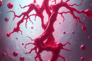
- Hereditary Hemorrhagic Telangiectasia (HHT), or Osler-Weber-Rendu disease, marks a type of blood vessel malformation.
- It involves genetic mutations and is an autosomal dominant trait.
- In HHT, there is a reduced amount of functional proteins available in the tissue lining the blood vessels.
- HHT results in skin and mucous membrane-associated epistaxis and other bleeding conditions.
- ENG, ACVRL1, and SMAD4 genes interact with growth factors that control blood vessel development.
- Type 1 HHT is associated with the ENG gene, while Type 2 is linked to the ACVRL1 gene.
- Type 3 HHT is currently unknown.
- Juvenile polyposis/HHT syndrome is caused by mutations in the gene SMAD4.
Other Vasculopathies
- These include conditions seen in patients with Multiple Myeloma or Systemic Amyloidosis.
- Moyamoya disease is also included in vasculopathies.
- Cerebral small vessel disease is another type.
Vasculitis
- Anti-neutrophil Cytoplasmic Antibody-Positive Vasculitis (ANCA) is a form of vasculitis.
- Other vasculitis forms include cryoglobulinemia.
- Hypergammaglobulinemic purpura is also a type of vasculitis.
- Henoch-Schönlein purpura (HSP) also falls under vasculitis.
Anti-neutrophil Cytoplasmic Antibody – Positive Vasculitis (ANCA)
- ANCA includes c-ANCA and p-ANCA types.
- ANCA is seen in SLE and Wegener's granulomatosis.
- It serves as serologic markers of primary necrotizing systemic vasculitis.
Cryoglobulinemia
- This condition involves cryoglobulin immune deposits that precipitate in dermal vessels.
- 90% of patients with Waldenström's primary macroglobulinemia (WM) have MYD88 L265P mutations.
- Symptoms involve cutaneous infarcts and petechiae.
- Platelet count remains normal, with chronic anemia and bleeding episodes.
- Bleeding is caused by abnormalities in platelet adhesiveness, while prothrombin time (PT) may be seen with low values of factor VIII.
- Intraluminal pressure is increased.
Hypergammaglobulinemic Purpura
- It is characterized by polyclonal hypergammaglobulinemia
- Secondary forms are associated with autoimmune disorders like Sjögren's syndrome and SLE, as well as chronic hepatitis C.
Henoch-Schönlein Purpura
- Henoch-Schönlein Purpura (HSP) occurs primarily in children aged 4 through 11 years old.
- Reddish-purple spots resembling bruises are a distinctive sign of HSP.
- HSP is an acute immunoglobulin A (IgA)-mediated disorder.
- Generalized small vessel vasculitis involves the skin, GI tract, kidneys, joints, and, rarely, the lungs and CNS.
Vascular Obstruction
- Thrombus, specifically blood clots, result in Disseminated Intravascular Coagulation.
- Emboli, by way of septic or thromboemboli, cause endocarditis or atrial fibrillation, respectively.
- Immunoglobulins, plasma proteins, fibrin, red blood cells, platelets, and fat cause multiple myeloma/Waldenström's primary macroglobulinemia, cryoglobulinemia, cryofibrinogenemia, polycythemia, thrombocytosis, and fat emboli syndrome, respectively.
DIC
- DIC is caused by a number of factors, including vascular damage leading to exposure of collagen, which activates XII.
- It is characterized by excess thrombin formation, conversion of fibrinogen to fibrin, and platelet deposition and consumption.
- Characterized by both thrombosis and hemolysis.
Cryofibrinogenemia
- It involves cryofibrinogen, which is a cold precipitable plasma protein.
- Lesions in associated cryofibrinogen disorders, such as Raynaud's phenomenon, result from fibrin thrombi obstructing small to medium-sized dermal blood vessels.
Septic Emboli
- Septic Emboli results from the development of tissue ischemia, necrosis of the skin and subcutaneous fate, and occasionally deeper tissues.
- They are associated with patients who have end-stage renal failure and undergoing hemodialysis.
Bleeding because of Perivascular Tissue
- Ehlers-Danlos syndrome (EDS) can cause bleeding from perivascular tissue.
- Marfan's syndrome is another syndrome that may lead to bleeding from perivascular tissue.
- Other syndromes that can cause bleeding from perivascular tissue include Loeys-Dietz syndrome
- Osteogenesis imperfecta
- Pseudoxanthoma elasticum
- Scurvy
- Steroid-induced purpura
- Solar purpura (senile purpura)
Ehlers-Danlos Syndrome (EDS)
- EDS involves abnormalities of collagen synthesis or processing.
- Defects in type 3 collagen, particularly abundant in the arterial wall and intestine.
- EDS results in excessive bleedings.
- Type IV EDS is prone to arterial aneurysms and dissections, and significant bleeding from spontaneous rupture of medium-sized abdominal arteries, and intestinal rupture.
- EDS IV involves structural defects in the proa1(III) chain of collagen type III encoded by COL3A1, inherited in an autosomal dominant pattern.
Pseudoxanthoma Elasticum (PXE)
- PXE is also known as Gröenblad - Strandberg syndrome, Grönblad-Strandberg syndrome.
- It involves an inherited connective tissue disorder resulting in calcification and mineralization of elastic fibers, especially in the internal elastic lamina of medium-sized arteries with subsequent rupture of the blood vessel.
- PXE is inherited as an autosomal recessive pattern – both copies of the genes in each cell have mutations.
- It is characterized by mutations in the ABC-C6 gene
- This gene results in nonfunctional MRP6 protein
Scurvy
- The absence of vitamin C in scurvy weakens collagen strands as a result of abnormal triple helical structures.
- This causes abnormal collagen due to defective perivascular supportive tissues.
- Patients are predisposed to capillary fragility, delayed wound healing, petechiae, and purpura.
Solar Purpura (Senile Purpura)
- In solar purpura, collagen and elastin are degraded.
Purpura Associated with Infections
- These infections include acute febrile illness with petechiae.
- Rickettsial diseases can cause purpura
- Brazilian purpura fever.
- Rat bite fever
- Hemorrhagic fever
- Vibrio vulnificus infection
- Strongyloides infection
- Bacterial toxin produce deendothelialization induced by an endotoxin
Purpura Related to Miscellaneous Causes
- Contact dermatitis can lead to purpura.
- Purpura can result from steroid use.
- Drug reactions can cause purpura.
- Vascular malignancy, specifically Kaposi sarcoma and vascular tumors.
- Purpura simplex
- Factitious or self-imposed purpura
- Religious stigmata.
Quantitative Platelet Disorders
- This category includes thrombocytopenia.
- Also, thrombocytosis is a quantitative platelet disorder.
Thrombocytopenia
- This may be due to artifactual causes like platelet clumping, giant platelets, and platelet satellitism.
- Decreased production of platelets, such as in hereditary thrombocytopenia, hypoplasia of megakaryocytes, or disorders of thrombopoiesis.
- Increased destruction of platelets (immunological or nonimmunological cause)
- Abnormal platelet distribution or pooling in splenic disorders, hypothermia, or dilution with massive transfusion.
Major Categories of Thrombocytopenic Conditions
- These conditions include disorders of production.
- Also, disorders of destruction or utilization.
- Disorders of platelet distribution and dilution.
Disorders of Production
- These are caused by hypoproliferation of the megakaryocytic cell line or ineffective thrombopoiesis.
- Acquired conditions or hereditary factors may cause hypoproliferation of the megakaryocytic cell line or ineffective thrombopoiesis.
- Acquired damage to hematopoietic cells of the bone arrow caused by factors such as irradiation, drugs (e.g., chloramphenicol and chemotherapeutic agents), chemicals (e.g., insecticides), and alcohol.
Disorders of Destruction or Utilization
- May be caused by immune mechanisms where there are antigens, antibodies, or complement.
- Posttransfusion purpura
- Can be drug induced.
- Bacterial sepsis.
- Immune thrombocytopenia
- Isoimmune Neonatal Thrombocytopenia
- Thrombocytopenia in pregnancy
Heparin-Induced Thrombocytopenia (HIT)
- There are two types - Nonimmune HIT (type I) and immune HIT (type II).
Nonimmune HIT
- It is a benign disorder affecting up to 10% of patients receiving heparin anticoagulant therapy.
- The mechanism of action is direct interaction between heparin and platelets.
- Platelet is >100,000/μL, with a rapid decline observed within the first 2 days of heparin administration.
- Platelet count returns to normal levels within 5 days despite continued heparin use or within 2 days if heparin therapy is discontinued.
Immune HIT
- The platelet count range falls between 20,000 to 150,000/μL.
- The lowest count is reached at about 5 days after the onset of the declining platelet count.
- The platelet count begins to rise approximately 2 days after heparin therapy is discontinued.
- Platelet count usually returns to normal within 4 to 10 days after discontinuing heparin.
Pathophysiology of HIT
- Immune HIT is caused by an antibody that recognizes heparin bound to PF4 on the platelet surface.
- The antibody binds to the heparin-PF4 complex, which then allows the antibody to bind the Fc receptor on the platelet.
- Interaction with the Fc receptor activates the platelet.
- Can results in loss of platelets, thrombocytopenia, and platelet aggregation (thrombosis).
Laboratory Data of HIT
- An enzyme-linked immunosorbent assay (ELISA) can provide labatory data.
- Other sources are platelet aggregation and serotonin release
Increased Utilization of Platelets
-
Immune Thrombocytopenic Purpura (ITP) is a cause
-
Can also arise due to Idiopathic Thrombocytopenic Purpura
-
It is acquired immune-mediated disorder characterized by isolated thrombocytopenia along with a platelet count less than 100 × 10º /L and the absence of potential initiating and/or underlying causes of the thrombocytopenia.
-
ITP is characterized by a low platelet count, normal bone marrow, and the absence of other causes of thrombocytopenia in both children and adults.
-
ITP is classified by duration into newly diagnosed, persistent (3 to 12 months duration), and chronic (≥12 months).
-
ITP is more common in females aged 10-40.
-
Immune Thrombocytopenic Purpura occurs more often and is longer in adults than in children.
Clinical Signs and Symptoms of ITP
- Signs include purpura, Epistaxis, gingival bleeding, hematuria, GI bleeding, and intracerebral hemorrhage
Pathophysiology of ITP
- Platelet destruction occurs due to antibodies produced
- ITP occurs in children and adults. Adults have a chronic ITP that is a destructive thrombocytopenia cause by antibodies produced.
Types of ITP
- Acute ITP – 2 – 6 y/o, just recovered from a viral illness.
- Chronic ITP – 20 – 50 y/o; produce an IgG antibody that coats the platelets, causing them to be sequestered and subsequently destroyed in spleen.
Laboratory Data of ITP
- Presence of thrombocytopenia, lack of anemia unless blood loss has occurred, and lack of white cell abnormalities
- Absence of other causes of thrombocytopenias (e.g.) collagen vascular diseases or lymphoproliferative disorders)
- Absence of infections, particularly human immunodefficiency virus (HIV)
Treatment for ITP
- May involve intravenous immunoglobulin (IVIg or WinRho, anti-D immune globulin)
- Splenectomy
- Platelet transfusion
Utilizations of Platelets
- Intravascular coagulation
- Vascular injury or occlusion
- Tissue injury
- Trauma
- Obstetrical complications
- Microbial sepsis
Thrombotic Thrombocytopenic Purpura
- It is characterized by formation of microthrombi in the microvasculature, and a high mortality rate
- Common signs are severe thrombocytopenia, microangiopathic hemolytic anemia, and fever.
- Neurologic symptoms, such as headache, stroke, and disease are common.
Types of TTP
- One type involves an Idiopathic cause where it is linked to enzyme ADAMTS13 known as a disintegrin-like and metalloprotease domain with thrombospondin-type motifs).
- It is responsible for the breakdown of large von Willebrand factor (vWF) multimers.
- A Secondary may be due to diagnosis in patients with a history of medications; such as with patients that have HIV, autoimmune disorders, allogeneic bone marrow transplant
- There is also a Hereditary cause in patients with (Upshaw-Schulman), and caused in result of inheritance of a deficiency of ADAMTS13.
Disorders of Platelet Distribution and Dilution
- Occur from the pooling of platelets in the spleen, which is frequent if splenomegaly is present.
- Develops when more than a double or triple increase in platelet production is required to maintain the normal quantity of circulating platelets.
- Disorders that produce splenomegaly with resultant splenic pooling or delayed intrasplenic transit include alcoholic or posthepatic cirrhosis with portal hypertension, lymphomas and leukemias, and lipid disorders such as Gaucher's disease.
Posttransfusion Thrombocytopenia
- Arises due to massive blood transfusion of units of packed RBC and plasma expanders.
- Results in reduced platelet count, or dilutional Thrombocytopenia.
Hereditary Defects of Platelet Function with Thrombocytopenia
- Include Fanconi's syndrome, constitutional aplastic anemia and its variants, ameiosis thrombocytopenia (TAR syndrome), X-linked amegakaryocytic thrombocytopenia, WAS, May-Hegglin anomaly, and Hereditary macrothrombocytopenia (e.g., Alport syndrome)
- Platelet morphology needs to be observed while examining a peripheral blood smear; abnormal variation in size should be noted.
Disorders of platelet size
- WAS - which demonstrate smallest platelets seen
- characterized large platelets and teh presence of Dohle-like bodies
- giant platelets and thrombocytopenia- Alport
- Bernard Soulier is largest. giant platelet.
Wiskott-Aldrich Syndrome (WAS)
-
WAS involves susceptibility to infections and abnormal bleeding.
-
There is a reduced ability to form blood clots as the result of unusually small, dysfunctional platelets.
-
Defective gene is on short arm of chromosome where X-Linked recessive mutations occur
-
WAS gene has protein call WASp, that relay signals from surface of the blood cells to the actin cytoskeleton
-
A lack of the WASp causes impairment of platelet development.
-
It can lead to a reduced size, microthrombocytonia, and dysfunctional platelets that have an intrinsic defect.
-
Signs include purpura, increased infections-fungal, viral and bacterial, and atopic dermatitis.
-
Hemolytic animea or idopathic thrombocytopenic.
-
More risks for malignancy
Labratory data include sequencing
- Lab data determines presence of protein cells
MYH9-Related Thrombocytopenia Syndromes (May-Hegglin Anomaly)
- Autsomoal Dominant, SEbastion, or Fechctner systems.
- Platelet cytoskeletal protein can common mutations
- Some inheritance.
- Premature release of platelets Platelets small
- 22 chrom, - 40 econs.
- 31 mutations in 11 excons.
Clinical Signs and Symptoms of MYH9
- Mild symptoms. bleeding. Hemostatic deficit.
Lab Findings in MYH9
-
Presence of Macrothrombocytopenia
-
large and poorly granulated platelets coexist.
-
Also presence of Dohle.
-
ITP most important. platelet size.
-
Immunostaininh good standards -platelets do not sowmajor deficits. .
-
Change of platelets. curve is absent but can be found.
-
A bone marrow may no be needeed
-
Marrow NMM
-
A work up for MYH9 should exclude anemia.
-
Assess cataracts
-
Test kidneys
Studying That Suits You
Use AI to generate personalized quizzes and flashcards to suit your learning preferences.