Podcast
Questions and Answers
Which of the following sequences correctly orders the four main functions of the digestive system?
Which of the following sequences correctly orders the four main functions of the digestive system?
- Ingestion, digestion & absorption, egestion, propulsion.
- Ingestion, digestion & absorption, propulsion, egestion.
- Ingestion, propulsion, digestion & absorption, egestion. (correct)
- Propulsion, ingestion, digestion & absorption, egestion.
Match the layer of the alimentary canal with its description:
Match the layer of the alimentary canal with its description:
Serosa = Reduces friction as digestive organs contract and move Muscularis = Layer of muscle extending in two directions: circular and longitudinal Submucosa = Contains glands, nerve fibers, and blood vessels Mucosa = Innermost layer; secretes mucus, digestive enzymes, and hormones; protects from pathogens
Teeth primarily contribute to chemical digestion through the secretion of enzymes.
Teeth primarily contribute to chemical digestion through the secretion of enzymes.
False (B)
Which type of teeth is primarily responsible for tearing and piercing food?
Which type of teeth is primarily responsible for tearing and piercing food?
The hard, protective outer layer of a tooth is called ______.
The hard, protective outer layer of a tooth is called ______.
How many teeth do toddlers typically have?
How many teeth do toddlers typically have?
What is the primary function of the uvula during swallowing?
What is the primary function of the uvula during swallowing?
Deglutition solely relies on voluntary muscle actions to move food from the mouth to the stomach.
Deglutition solely relies on voluntary muscle actions to move food from the mouth to the stomach.
What is the name given to the lump of food after chewing and mixing with saliva?
What is the name given to the lump of food after chewing and mixing with saliva?
The process by which the bolus is propelled down the esophagus using waves of muscle contractions is called ______.
The process by which the bolus is propelled down the esophagus using waves of muscle contractions is called ______.
What is the function of sphincters in the digestive system?
What is the function of sphincters in the digestive system?
Which of the following is NOT a layer of muscle in the stomach?
Which of the following is NOT a layer of muscle in the stomach?
Maceration is the process in the stomach that primarily involves chemical digestion through enzyme secretion.
Maceration is the process in the stomach that primarily involves chemical digestion through enzyme secretion.
Which cells in the stomach secrete hydrochloric acid (HCl)?
Which cells in the stomach secrete hydrochloric acid (HCl)?
The soupy mixture formed from the squeezing of the stomach and addition of gastric juices is known as ______.
The soupy mixture formed from the squeezing of the stomach and addition of gastric juices is known as ______.
What is the primary function of villi in the small intestine?
What is the primary function of villi in the small intestine?
Segmentation in the small intestine involves longitudinal muscles and results in peristaltic contractions.
Segmentation in the small intestine involves longitudinal muscles and results in peristaltic contractions.
What is the name of the sphincter that regulates the movement of chyme from the small intestine to the large intestine?
What is the name of the sphincter that regulates the movement of chyme from the small intestine to the large intestine?
What is the main function of the large intestine?
What is the main function of the large intestine?
The pouches that give the large intestine its lumpy appearance are called ______.
The pouches that give the large intestine its lumpy appearance are called ______.
Flashcards
Ingestion
Ingestion
Taking food into the body.
Propulsion
Propulsion
Movement of food through the digestive system.
Digestion & Absorption
Digestion & Absorption
Breaking down food and absorbing nutrients.
Egestion
Egestion
Signup and view all the flashcards
Serosa (Visceral Peritoneum)
Serosa (Visceral Peritoneum)
Signup and view all the flashcards
Muscularis
Muscularis
Signup and view all the flashcards
Submucosa
Submucosa
Signup and view all the flashcards
Mucosa
Mucosa
Signup and view all the flashcards
Teeth
Teeth
Signup and view all the flashcards
Incisors
Incisors
Signup and view all the flashcards
Canines
Canines
Signup and view all the flashcards
Premolars (Bicuspids)
Premolars (Bicuspids)
Signup and view all the flashcards
Molars (Tricuspids)
Molars (Tricuspids)
Signup and view all the flashcards
Enamel
Enamel
Signup and view all the flashcards
Dentin
Dentin
Signup and view all the flashcards
Digestion beginning in the mouth
Digestion beginning in the mouth
Signup and view all the flashcards
Lysozyme
Lysozyme
Signup and view all the flashcards
Amylase
Amylase
Signup and view all the flashcards
Study Notes
Intro & Teeth
- The digestive system functions include ingestion(taking in food), propulsion(movement of food), digestion and absorption(breaking down food), and egestion(elimination of waste).
- The alimentary canal layers, from superficial to deep, are: serosa, muscularis, submucosa, and mucosa.
- Serosa (peritoneum) reduces friction as digestive organs contract.
- Muscularis has two layers of muscle: circular and longitudinal.
- The submucosa contains glands, nerve fibers, and blood vessels.
- The mucosa is the innermost layer and protects from pathogens and secretes mucus, digestive enzymes, and hormones for nutrient absorption.
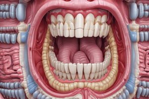
- Teeth are accessory digestive organs and start the mechanical digestion process.
- Incisors cut food.
- Canines tear and pierce food.
- Premolars (bicuspids) crush food.
- Molars (tricuspids) grind food.
- Tooth anatomy includes the enamel (hard, protects teeth), dentin (calcified connective tissue), pulp cavity (contains blood vessels and nerves), cementum (covers the dentin on the root), and the gum.
- The digestive system has multiple functions and is generally divided into two portions: the alimentary canal and the accessory organs.
- The teeth begin the process of digestion by tearing and grinding food.
Mouth & Esophagus
- Toddlers have 20 teeth, while adults have 32.
- The incisors come in first, followed by canines and molars and eventually replaced. Molars erupt during the late teens and early twenties.
- Chemical digestion starts in the mouth.
- Salivary glands produce saliva made of water, mucus which lubricates food, lysozyme which kills bacteria, and amylase which breaks down starch.
- The hard palate forms the bony roof of the mouth, while the soft palate forms the rest of the mouth.
- The uvula prevents swallowed food from entering the nasal cavity. The tongue helps push food down towards the esophagus.
- Deglutition (swallowing) has two phases: buccal and pharyngeal-esophageal.
- After chewing (mastication) and mixing with saliva, a food lump called a bolus forms. It is forced into the pharynx by the tongue.
- A thin flap of skin called the epiglottis blocks the larynx, and the uvula blocks the nasal cavity; this causes food to travel to the esophagus instead of the respiratory system.
- The bolus is propelled down the esophagus via waves of muscle contractions called peristalsis.
- Mastication forms a bolus inside the mouth, which is then swallowed.
- After deglutition, the bolus moves past the epiglottis and down the esophagus via involuntary contractions called peristalsis.
Stomach
- The stomach is a muscular sac with thick walls and continues mechanical and chemical digestion.
- Sphincters serve as gatekeepers and are thick muscle rings that allow food to enter or leave at the appropriate time for digestion.
- The two sphincters located in the stomach are the cardiac and pyloric sphincters.
- The stomach has a slippery outer serosa layer followed by three muscle layers: longitudinal, circular, and oblique.
- These muscles help churn food and propel it towards the small intestine via a process known as maceration.
- Rugae are wrinkles that stretch when the stomach fills.
- The mucosa layer of the stomach contains specialized gastric gland cells.
- Mucous cells secrete mucus to protect the stomach lining.
- Chief cells secrete pepsinogen (inactive enzyme).
- Parietal cells secrete HCl to kill microbes in food and convert pepsinogen into the digestive enzyme pepsin, which breaks down proteins.
- The soupy mixture formed from the squeezing of the stomach and the addition of these gastric juices is known as chyme.
- The stomach is made of three muscular layers and an outer serosa layer.
- During maceration, the stomach churns the food, and gastric gland cells add chemicals and enzymes, leading to the formation of chyme.
Small Intestine
- The small intestine is the location of most digestion and nutrient absorption.
- The small intestine is about 10 ft long in an adult and has 3 sections: duodenum, jejunum, and ileum.
- After the ileum, it joins the large intestine at the ileocecal sphincter.
- Villi are tiny, finger-like projections in the lining of the intestine filled with blood vessels.
- Many glands in the small intestine secrete digestive hormones.
- Mucus is alkaline and secreted from mucous gland and duodenal glands for protection.
- Secretin inhibits release of gastric juices when chyme is very acidic.
- Cholecystokinin (CCK) causes the gallbladder to release bile and causes fat breakdown
- Maltase, sucrase, and lactase break down sugars.
- Peptidase and enterokinase break down proteins.
- The chyme must be slowed down so the small intestine has time to absorb nutrients.
- Circular muscles in the intestinal wall cause segmentation of the chyme, which is in contrast to peristaltic contractions, which involve longitudinal muscles.
- The small intestine is very long and divided into three sections and contains villi which absorb nutrients, and glands that secrete hormones. Segmentation slows down chyme movement for proper absorption.
Large Intestine & Anus
- After food leaves the small intestine, it enters the large intestine, which has 3 sections: cecum, colon, and rectum.
- Colon is broken into ascending colon, transverse colon and descending colon
- Two major functions of the large intestine are water reabsorption and waste removal.
- Large amounts of bacteria are located in the large intestine to produce vitamins B and K.
- Haustra are the small pouches that give the large intestine its lumpy appearance.
- Haustral contractions are slow, segmentation movements that last thirty minutes after food moves into the large intestine.
- Mass movements push waste towards the rectum three to four times a day.
- The rectum collects and stores undigested waste and bacteria.
- As the rectum fills, pressure is placed on the internal anal sphincter, causing it to relax.
- The external anal sphincter must be voluntarily relaxed to release the feces through the anus.
- The large intestine is divided into the cecum, colon, and rectum.
- It is responsible for reabsorbing water and removing waste through haustral contractions and mass movements toward the anus.
Accessory Organs
- The liver is the largest internal organ; it has left and right lobes and contains hepatic sinusoids full of blood
- Liver cells remove bacteria and old blood cells, detoxify blood from drugs and poisons, store glycogen, produce fats, manufacture proteins, and store iron and copper; the liver also secretes bile to break down fats.
- The gallbladder is a small, green sac attached to the liver that stores bile from the liver before sending it to the small intestine.
- The gallbladder concentrates bile by removing water; gallstones (made of cholesterol) may form if too much water is removed.
- The pancreas is a long, thin gland behind the stomach with two functions: to secrete digestive enzymes and sodium bicarbonate (which neutralizes stomach acid), and to secrete hormones that regulate blood sugar levels.
- The major digestive system accessory organs include the liver, gallbladder, and pancreas.
- The liver secretes bile that is then stored by the gallbladder to be released into the small intestine
- The pancreas secretes digestive enzymes and hormones.
Nutrition
- Metabolism refers to the life-sustaining chemical reactions of the body and is categorized into catabolism (breakdown releasing energy) and anabolism (formation using energy).
- Nutrients are substances that provide the body with energy and can be used for growth.
- The four types of macronutrients are carbs, lipids, proteins, and water.
- The two types of micronutrients are vitamins and minerals.
- Carbohydrates are sugars (glucose) and are the body’s main energy source. Carbohydrates are broken down by salivary amylase in the mouth and pancreatic amylase in the small intestine.
- Lipids/fats are a secondary energy source used for cell membranes, nerves, insulation, and hormone production and are broken down by bile salts (liver) and pancreatic lipase (small intestine).
- Proteins are used for cellular structures and enzymes; they’re broken down into polypeptide chains or amino acids.
- Water is used for chemical reactions, dissolving foods for digestion, maintaining blood pH levels and regulating body temperature.
- Vitamins are organic molecules needed in small quantities.
- Minerals are inorganic molecules needed in small quantities: calcium, chloride, fluorine, iodine, iron, magnesium, and phosphorus.
- Metabolic processes are required for life. The macronutrients metabolized in the digestive system are carbohydrates, lipids, and proteins. Water, vitamins, and minerals are also required for maintaining homeostasis.
Studying That Suits You
Use AI to generate personalized quizzes and flashcards to suit your learning preferences.