Podcast
Questions and Answers
What type of radiation is used in X-ray (radiography) to produce images of the inside of the body?
What type of radiation is used in X-ray (radiography) to produce images of the inside of the body?
- Magnetic fields
- Gamma rays
- Ionizing radiation (correct)
- Radio waves
What is Magnetic Resonance Imaging (MRI) especially useful for diagnosing?
What is Magnetic Resonance Imaging (MRI) especially useful for diagnosing?
- Pneumonia and lung cancer
- Tumors and infections
- Bone fractures and lung diseases
- Brain and spinal cord abnormalities, joint and musculoskeletal disorders, and heart and blood vessel issues (correct)
What is the primary purpose of a lead apron in X-ray (radiography)?
What is the primary purpose of a lead apron in X-ray (radiography)?
- To shield the patient from radiation
- To detect bone fractures
- To shield other parts of the body from radiation (correct)
- To position the patient correctly
Why must the patient be still during the MRI imaging process?
Why must the patient be still during the MRI imaging process?
What is prohibited in the MRI room?
What is prohibited in the MRI room?
Why may patients with claustrophobia require sedation during an MRI exam?
Why may patients with claustrophobia require sedation during an MRI exam?
What is the primary purpose of applying a water-based gel to the skin during an ultrasound procedure?
What is the primary purpose of applying a water-based gel to the skin during an ultrasound procedure?
What should be checked for before an MRI exam?
What should be checked for before an MRI exam?
What is the purpose of ear protection during an MRI exam?
What is the purpose of ear protection during an MRI exam?
Why is a full bladder required for some ultrasound exams?
Why is a full bladder required for some ultrasound exams?
What is the primary difference between a CT scan and an ultrasound?
What is the primary difference between a CT scan and an ultrasound?
What is a common application of nuclear medicine?
What is a common application of nuclear medicine?
What is the primary advantage of ultrasound over CT scans?
What is the primary advantage of ultrasound over CT scans?
What is the role of the transducer in an ultrasound procedure?
What is the role of the transducer in an ultrasound procedure?
What is a critical consideration when performing a CT scan on a pregnant patient?
What is a critical consideration when performing a CT scan on a pregnant patient?
What is a common use of ultrasound in pregnancy?
What is a common use of ultrasound in pregnancy?
What is the primary purpose of SPECT Tomography?
What is the primary purpose of SPECT Tomography?
What is a common application of SPECT Tomography?
What is a common application of SPECT Tomography?
What is Elastography used for?
What is Elastography used for?
What is a precaution that should be taken during SPECT Tomography?
What is a precaution that should be taken during SPECT Tomography?
What is Magnetic Resonance Angiography (MRA) used for?
What is Magnetic Resonance Angiography (MRA) used for?
What is the purpose of a gamma camera in SPECT Tomography?
What is the purpose of a gamma camera in SPECT Tomography?
What is a potential complication of SPECT Tomography?
What is a potential complication of SPECT Tomography?
What is a preparation required for SPECT Tomography?
What is a preparation required for SPECT Tomography?
What is the primary purpose of functional MRI (fMRI)?
What is the primary purpose of functional MRI (fMRI)?
What is a common application of tomosynthesis?
What is a common application of tomosynthesis?
What is the primary purpose of diagnostic imaging?
What is the primary purpose of diagnostic imaging?
What type of imaging modality is used to detect bone fractures and lung conditions?
What type of imaging modality is used to detect bone fractures and lung conditions?
Why do patients with metal implants need to be cautious during an MRI?
Why do patients with metal implants need to be cautious during an MRI?
What is injected into the patient during a CT scan to highlight specific areas?
What is injected into the patient during a CT scan to highlight specific areas?
What is a common precaution taken during an MRI procedure?
What is a common precaution taken during an MRI procedure?
Why is it important for patients to remove metal objects during a CT scan?
Why is it important for patients to remove metal objects during a CT scan?
What type of images does tomosynthesis generate?
What type of images does tomosynthesis generate?
What is a common use of functional MRI (fMRI) in medical practice?
What is a common use of functional MRI (fMRI) in medical practice?
What is a critical consideration for patients undergoing a CT scan with contrast dye?
What is a critical consideration for patients undergoing a CT scan with contrast dye?
What is the primary difference between an MRI and a CT scan?
What is the primary difference between an MRI and a CT scan?
What is the purpose of CT scans in medical procedures?
What is the purpose of CT scans in medical procedures?
Why do patients need to remain still during an MRI procedure?
Why do patients need to remain still during an MRI procedure?
What is a common application of MRI and CT scans?
What is a common application of MRI and CT scans?
What sensation may patients experience during a CT scan with contrast dye?
What sensation may patients experience during a CT scan with contrast dye?
X-rays are used to diagnose complex bone fractures and detect cancer.
X-rays are used to diagnose complex bone fractures and detect cancer.
MRI is used to guide specific medical procedures.
MRI is used to guide specific medical procedures.
Tomosynthesis is used to detect soft tissue injuries.
Tomosynthesis is used to detect soft tissue injuries.
CT scans are used to diagnose lung conditions.
CT scans are used to diagnose lung conditions.
Patients need to fast before undergoing an MRI exam.
Patients need to fast before undergoing an MRI exam.
A gamma camera is used in CT scans to produce images.
A gamma camera is used in CT scans to produce images.
Ultrasound is used to monitor internal injuries.
Ultrasound is used to monitor internal injuries.
Metal objects are allowed in the MRI room.
Metal objects are allowed in the MRI room.
Ultrasound is a method that uses ionizing radiation to produce images of the inside of the body.
Ultrasound is a method that uses ionizing radiation to produce images of the inside of the body.
Nuclear medicine is used to diagnose and treat diseases using small amounts of radioactive materials.
Nuclear medicine is used to diagnose and treat diseases using small amounts of radioactive materials.
Computed Tomography (CT) Scans are critical for diagnosing injuries to bones and soft tissues.
Computed Tomography (CT) Scans are critical for diagnosing injuries to bones and soft tissues.
Ultrasound is frequently used to examine the lungs during pregnancy.
Ultrasound is frequently used to examine the lungs during pregnancy.
A radiopharmaceutical is administered to the patient orally or via injection during a CT Scan.
A radiopharmaceutical is administered to the patient orally or via injection during a CT Scan.
Ultrasound is a safe and non-invasive method of diagnostic imaging.
Ultrasound is a safe and non-invasive method of diagnostic imaging.
Nuclear Medicine is used to assess liver function.
Nuclear Medicine is used to assess liver function.
CT Scans are used to guide biopsies.
CT Scans are used to guide biopsies.
Gamma cameras are used in Mammography to capture images.
Gamma cameras are used in Mammography to capture images.
Fluoroscopy is used to detect and diagnose breast cancer.
Fluoroscopy is used to detect and diagnose breast cancer.
Compression of the breast is necessary for clear images in Mammography.
Compression of the breast is necessary for clear images in Mammography.
The patient should use deodorants and perfumes on the day of the Mammography exam.
The patient should use deodorants and perfumes on the day of the Mammography exam.
Fluoroscopy is used to guide catheter placement and assess joint movement.
Fluoroscopy is used to guide catheter placement and assess joint movement.
Mammography produces detailed images of the internal organs.
Mammography produces detailed images of the internal organs.
Patient preparation for Fluoroscopy involves following specific instructions for the type of scan.
Patient preparation for Fluoroscopy involves following specific instructions for the type of scan.
Mammography is a type of Nuclear Medicine.
Mammography is a type of Nuclear Medicine.
Magnetic Resonance Imaging (MRI) is used to assess coronary artery disease and cardiovascular risk.
Magnetic Resonance Imaging (MRI) is used to assess coronary artery disease and cardiovascular risk.
Tomosynthesis is a type of advanced MRI that generates 3D images of the breast.
Tomosynthesis is a type of advanced MRI that generates 3D images of the breast.
Functional MRI (fMRI) uses X-ray images to measure and map brain activity.
Functional MRI (fMRI) uses X-ray images to measure and map brain activity.
Elastography is used to assess liver fibrosis, tumors, and other soft tissue abnormalities.
Elastography is used to assess liver fibrosis, tumors, and other soft tissue abnormalities.
Metal objects are permitted in the MRI room.
Metal objects are permitted in the MRI room.
Tomosynthesis is used to detect bone fractures and lung conditions.
Tomosynthesis is used to detect bone fractures and lung conditions.
Ultrasound is used to guide specific medical procedures.
Ultrasound is used to guide specific medical procedures.
Functional MRI (fMRI) is used to investigate brain function and study neurological and psychiatric disorders.
Functional MRI (fMRI) is used to investigate brain function and study neurological and psychiatric disorders.
Cardiac CT (Coronary CT Angiography) combines PET and CT scans with FDG, a radioactive glucose analog.
Cardiac CT (Coronary CT Angiography) combines PET and CT scans with FDG, a radioactive glucose analog.
Fluorodeoxyglucose (FDG) PET/CT is used to assess coronary artery disease.
Fluorodeoxyglucose (FDG) PET/CT is used to assess coronary artery disease.
Metal objects are not allowed in the area during a Cardiac CT (Coronary CT Angiography) procedure.
Metal objects are not allowed in the area during a Cardiac CT (Coronary CT Angiography) procedure.
Patients with claustrophobia may require sedation during a Fluorodeoxyglucose (FDG) PET/CT procedure.
Patients with claustrophobia may require sedation during a Fluorodeoxyglucose (FDG) PET/CT procedure.
The patient is given a contrast dye injection during a Fluorodeoxyglucose (FDG) PET/CT procedure.
The patient is given a contrast dye injection during a Fluorodeoxyglucose (FDG) PET/CT procedure.
The procedure for Cardiac CT (Coronary CT Angiography) is generally safe, with only minor risks from the contrast dye.
The procedure for Cardiac CT (Coronary CT Angiography) is generally safe, with only minor risks from the contrast dye.
Fluorodeoxyglucose (FDG) PET/CT is used to assess cardiac function and congenital heart defects.
Fluorodeoxyglucose (FDG) PET/CT is used to assess cardiac function and congenital heart defects.
The patient needs to fast before undergoing a Fluorodeoxyglucose (FDG) PET/CT procedure.
The patient needs to fast before undergoing a Fluorodeoxyglucose (FDG) PET/CT procedure.
What type of imaging modality employs low-dose X-rays to examine breast tissue?
What type of imaging modality employs low-dose X-rays to examine breast tissue?
What is the purpose of compressing the breast tissue during a mammography procedure?
What is the purpose of compressing the breast tissue during a mammography procedure?
What is a common application of fluoroscopy?
What is a common application of fluoroscopy?
What instruction should be given to patients before a mammography exam?
What instruction should be given to patients before a mammography exam?
What type of imaging modality uses a continuous X-ray beam to produce real-time images of the internal organs?
What type of imaging modality uses a continuous X-ray beam to produce real-time images of the internal organs?
What is a warning that should be given to patients before a mammography exam?
What is a warning that should be given to patients before a mammography exam?
What is a potential complication of catheter insertion during a thermography procedure?
What is a potential complication of catheter insertion during a thermography procedure?
What is an essential aspect of patient preparation for a fluoroscopy exam?
What is an essential aspect of patient preparation for a fluoroscopy exam?
What is a common consideration for patients undergoing a mammography exam?
What is a common consideration for patients undergoing a mammography exam?
What is the purpose of Dual-Energy X-ray Absorptiometry (DEXA)?
What is the purpose of Dual-Energy X-ray Absorptiometry (DEXA)?
Why does the patient need to be positioned and compressed during a DEXA procedure?
Why does the patient need to be positioned and compressed during a DEXA procedure?
What is a precaution that should be taken by patients undergoing a DEXA procedure?
What is a precaution that should be taken by patients undergoing a DEXA procedure?
What is the primary purpose of Electrocardiography (ECG/EKG)?
What is the primary purpose of Electrocardiography (ECG/EKG)?
What is the primary benefit of using MRI over X-ray in diagnosing joint and musculoskeletal disorders?
What is the primary benefit of using MRI over X-ray in diagnosing joint and musculoskeletal disorders?
Why is it essential to check patients for metal objects before an MRI exam?
Why is it essential to check patients for metal objects before an MRI exam?
What is a characteristic of Interventional Radiology?
What is a characteristic of Interventional Radiology?
What is a critical consideration for patients undergoing an MRI exam?
What is a critical consideration for patients undergoing an MRI exam?
What is a common application of SPECT (Single Photon Emission Computed Tomography)?
What is a common application of SPECT (Single Photon Emission Computed Tomography)?
What is a characteristic of DEXA and SPECT procedures?
What is a characteristic of DEXA and SPECT procedures?
What is the primary purpose of using a lead apron in X-ray imaging?
What is the primary purpose of using a lead apron in X-ray imaging?
Which of the following is a common application of MRI in medical practice?
Which of the following is a common application of MRI in medical practice?
What is a primary difference between MRI and X-ray imaging?
What is a primary difference between MRI and X-ray imaging?
Why do patients need to remain still during an MRI exam?
Why do patients need to remain still during an MRI exam?
What is a common application of diagnostic imaging in medical practice?
What is a common application of diagnostic imaging in medical practice?
What is the primary purpose of Positron Emission Tomography (PET) scans?
What is the primary purpose of Positron Emission Tomography (PET) scans?
What precaution should be taken during fluoroscopy?
What precaution should be taken during fluoroscopy?
What is the primary purpose of Bone Densitometry (DXA)?
What is the primary purpose of Bone Densitometry (DXA)?
What is Angiography used for?
What is Angiography used for?
What is a warning/consideration for PET scans?
What is a warning/consideration for PET scans?
What is a preparation required for Bone Densitometry (DXA)?
What is a preparation required for Bone Densitometry (DXA)?
What is a critical component of Positron Emission Tomography (PET) scans?
What is a critical component of Positron Emission Tomography (PET) scans?
What is a common application of Bone Densitometry (DXA)?
What is a common application of Bone Densitometry (DXA)?
What is a precaution that should be taken during Cardiac CT (Coronary CT Angiography)?
What is a precaution that should be taken during Cardiac CT (Coronary CT Angiography)?
What is a common application of Fluorodeoxyglucose (FDG) PET/CT?
What is a common application of Fluorodeoxyglucose (FDG) PET/CT?
What is a benefit of Fluorodeoxyglucose (FDG) PET/CT?
What is a benefit of Fluorodeoxyglucose (FDG) PET/CT?
Why may patients with claustrophobia require sedation during Cardiac CT (Coronary CT Angiography)?
Why may patients with claustrophobia require sedation during Cardiac CT (Coronary CT Angiography)?
What is a common feature of both Cardiac CT (Coronary CT Angiography) and Fluorodeoxyglucose (FDG) PET/CT?
What is a common feature of both Cardiac CT (Coronary CT Angiography) and Fluorodeoxyglucose (FDG) PET/CT?
What is the purpose of injecting contrast dye in Cardiac CT (Coronary CT Angiography)?
What is the purpose of injecting contrast dye in Cardiac CT (Coronary CT Angiography)?
What is a benefit of Cardiac CT (Coronary CT Angiography) over Fluorodeoxyglucose (FDG) PET/CT?
What is a benefit of Cardiac CT (Coronary CT Angiography) over Fluorodeoxyglucose (FDG) PET/CT?
What is a common application of Cardiac CT (Coronary CT Angiography)?
What is a common application of Cardiac CT (Coronary CT Angiography)?
Diagnostic imaging refers to a variety of techniques that allow healthcare professionals to visualize what part of the body?
Diagnostic imaging refers to a variety of techniques that allow healthcare professionals to visualize what part of the body?
What is the primary purpose of CT scans in medical procedures?
What is the primary purpose of CT scans in medical procedures?
What is injected into the patient during a CT scan?
What is injected into the patient during a CT scan?
Why must the patient be still during a CT scan?
Why must the patient be still during a CT scan?
What is a critical consideration when performing a CT scan on a patient?
What is a critical consideration when performing a CT scan on a patient?
What is the purpose of diagnostic imaging?
What is the purpose of diagnostic imaging?
Why do metal objects interfere with CT scans?
Why do metal objects interfere with CT scans?
What should patients do before undergoing a CT scan?
What should patients do before undergoing a CT scan?
What is the primary use of mammography?
What is the primary use of mammography?
What should patients avoid using on the day of a mammography exam?
What should patients avoid using on the day of a mammography exam?
What is fluoroscopy used to guide?
What is fluoroscopy used to guide?
Why is compression of the breast necessary during mammography?
Why is compression of the breast necessary during mammography?
What is the purpose of explaining the procedure to the patient before fluoroscopy?
What is the purpose of explaining the procedure to the patient before fluoroscopy?
What type of images does fluoroscopy produce?
What type of images does fluoroscopy produce?
Why is it important for patients to understand the low level of radiation exposure during mammography?
Why is it important for patients to understand the low level of radiation exposure during mammography?
What is the primary application of mammography?
What is the primary application of mammography?
What is the primary purpose of Electroencephalography (EEG)?
What is the primary purpose of Electroencephalography (EEG)?
What is Cholangiopancreatography used to diagnose?
What is Cholangiopancreatography used to diagnose?
What is the purpose of electrodes in Electroencephalography (EEG)?
What is the purpose of electrodes in Electroencephalography (EEG)?
What is a risk associated with Cholangiopancreatography?
What is a risk associated with Cholangiopancreatography?
What is the purpose of patient preparation for Cholangiopancreatography?
What is the purpose of patient preparation for Cholangiopancreatography?
What is the purpose of a catheter in diagnosing vascular conditions?
What is the purpose of a catheter in diagnosing vascular conditions?
What is the purpose of Electroencephalography (EEG) in medical practice?
What is the purpose of Electroencephalography (EEG) in medical practice?
What is the difference between ERCP and MRCP?
What is the difference between ERCP and MRCP?
What is SPECT Tomography used for, besides detecting brain disorders?
What is SPECT Tomography used for, besides detecting brain disorders?
What is Magnetic Resonance Angiography (MRA) primarily used for?
What is Magnetic Resonance Angiography (MRA) primarily used for?
What is Elastography used for, besides measuring liver fibrosis?
What is Elastography used for, besides measuring liver fibrosis?
What is a precaution taken during SPECT Tomography?
What is a precaution taken during SPECT Tomography?
What guides the intervention during SPECT Tomography?
What guides the intervention during SPECT Tomography?
What is prepared during SPECT Tomography?
What is prepared during SPECT Tomography?
What is detected by a gamma camera in SPECT Tomography?
What is detected by a gamma camera in SPECT Tomography?
What is performed after the patient receives a radiotracer injection in SPECT Tomography?
What is performed after the patient receives a radiotracer injection in SPECT Tomography?
What is cardiac CT used to assess?
What is cardiac CT used to assess?
What is fluorodeoxyglucose (FDG) used for?
What is fluorodeoxyglucose (FDG) used for?
What is a consideration for patients with claustrophobia undergoing a cardiac CT scan?
What is a consideration for patients with claustrophobia undergoing a cardiac CT scan?
What is injected into the patient during a cardiac CT scan?
What is injected into the patient during a cardiac CT scan?
What is a warning for patients undergoing a cardiac CT scan?
What is a warning for patients undergoing a cardiac CT scan?
Why is fasting required before a cardiac CT scan?
Why is fasting required before a cardiac CT scan?
What is a consideration for patients undergoing an FDG PET/CT scan?
What is a consideration for patients undergoing an FDG PET/CT scan?
What is the primary application of an FDG PET/CT scan?
What is the primary application of an FDG PET/CT scan?
Flashcards are hidden until you start studying
Study Notes
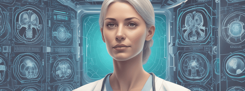
Diagnostic Imaging
- Diagnostic imaging refers to various techniques that allow healthcare professionals to visualize the internal structures of the body, resulting in more accurate diagnoses and effective treatment planning.
X-ray (Radiography)
- Uses ionizing radiation to produce images of the inside of the body.
- Primarily used to detect bone fractures, infections, tumors, and lung diseases like pneumonia and lung cancer.
- Procedure: The patient is positioned on a table that slides into a tunnel-shaped scanner.
- Warnings and considerations: Limit ionizing radiation exposure, particularly for pregnant patients.
Computed Tomography (CT) Scan
- Combines multiple X-ray images taken from various angles and employs computer processing to produce cross-sectional images of bones, blood vessels, and soft tissues.
- Critical for diagnosing complex bone fractures, detecting cancer, monitoring internal injuries, and guiding specific medical procedures.
- Procedure: The patient is placed on a motorized table that slides into a tunnel-shaped scanner.
- Patient Preparation: Ensure that the patient is not allergic to any contrast dyes that may be used and that any fasting instructions have been followed.
- Warnings and Considerations: Inform the patient that contrast dye may cause a brief sensation of warmth, and metal objects can interfere with imaging.
Magnetic Resonance Imaging (MRI)
- Uses strong magnetic fields and radio waves to create detailed images of organs and tissues in the body.
- Especially useful for diagnosing brain and spinal cord abnormalities, joint and musculoskeletal disorders, and heart and blood vessel issues.
- Procedure: The patient is positioned on a table that slides into a large tube-shaped magnet.
- Patient Preparation: Check the patient for any metal implants, pacemakers, or other metal objects that may interfere with the magnetic field.
- Warnings and Considerations: Metal objects are strictly prohibited in the MRI room, and patients with claustrophobia may require sedation.
Ultrasound (Sonography)
- Uses high-frequency sound waves to produce images of the inside of the body.
- Frequently used to examine fetuses during pregnancy, assess organs like the liver, kidneys, and heart, and guide biopsies.
- Procedure: A water-based gel is applied to the skin to aid the transducer in making secure contact.
- Warnings and Considerations: Ultrasound is a safe, non-invasive procedure.
Nuclear Medicine
- Uses small amounts of radioactive materials (radiopharmaceuticals) to diagnose and treat diseases.
- Effective for assessing thyroid function, detecting bone abnormalities, and diagnosing heart disease.
- Procedure: A radiopharmaceutical is administered to the patient orally or via injection.
- Applications: Angioplasty, stent placements, biopsies, and tumor ablation procedures.
SPECT (Single Photon Emission Computed Tomography)
- Creates 3D images of the body by detecting gamma rays emitted by a radiotracer.
- Used to detect and monitor brain and heart disorders, as well as bone diseases.
- Procedure: The patient lies on a table while a gamma camera rotates around them.
Magnetic Resonance Angiography (MRA)
- An MRI technique used to visualize blood vessels.
- Assesses aneurysms, stenosis, and other vascular abnormalities.
- Procedure: The patient lies on a table that slides into the MRI machine and remains still throughout the imaging.
- Patient Preparation: Check for metal implants and make sure the patient has followed all dietary restrictions.
Elastography
- An ultrasound or MRI-based imaging technique for measuring tissue stiffness.
- Useful for determining liver fibrosis, detecting tumors, and assessing other soft tissue abnormalities.
- Procedure: The patient remains still while the ultrasound or MRI device measures tissue stiffness.
Functional MRI (fMRI)
- Uses blood flow changes to measure and map brain activity.
- Investigates brain function, plans brain surgery, and studies neurological and psychiatric disorders.
- Procedure: The patient engages in tasks or responds to stimuli while in the MRI machine, which measures brain activity.
Tomosynthesis
- A type of advanced mammography that generates 3D images of the breast by combining multiple X-ray images from various angles.
- Enhances breast cancer detection and diagnosis, particularly in dense breast tissue.
Cardiac Imaging
- Uses imaging modalities such as CT and MRI to visualize the heart and its vessels.
- Applications include the early detection of coronary artery disease and the assessment of cardiovascular risk.
Diagnostic Imaging
- Diagnostic imaging techniques allow healthcare professionals to visualize internal body structures, enabling accurate diagnoses and effective treatment planning.
Computed Tomography (CT) Scan
- Combines multiple X-ray images taken from various angles and uses computer processing to produce cross-sectional images of bones, blood vessels, and soft tissues.
- Critical for diagnosing complex bone fractures, detecting cancer, monitoring internal injuries, and guiding specific medical procedures.
- Procedure: Patient is placed on a motorized table that slides into a tunnel-shaped scanner and must remain still during the scan.
- Patient preparation: Ensure patient is not allergic to contrast dyes and has followed fasting instructions.
- Warnings/considerations: Inform patient that contrast dye may cause a brief sensation of warmth, and metal objects can interfere with imaging.
Ultrasound (Sonography)
- Uses high-frequency sound waves to produce images of the inside of the body.
- Frequently used to examine fetuses during pregnancy, assess organs like the liver, kidneys, and heart, and guide biopsies.
- Procedure: Transducer is moved over the skin, sending sound waves into the body, which bounce back to form images.
- Patient preparation: No special preparation is usually required, but some exams may necessitate a full bladder.
- Warnings/considerations: Ultrasound is a safe, non-invasive procedure with no known risks from sound waves.
Nuclear Medicine
- Uses small amounts of radioactive materials to diagnose and treat diseases.
- Effective for assessing thyroid function, detecting bone abnormalities, and diagnosing heart disease.
- Procedure: Radiopharmaceutical is administered to the patient orally or via injection, and images are captured using a gamma camera.
- Patient preparation: Follow specific preparation instructions for the type of scan.
- Warnings/considerations: Make sure patient understands the low level of radiation exposure and follows all post-procedure safety instructions.
Mammography
- Uses low-dose X-rays to examine breast tissue, producing detailed images that can reveal abnormalities in breast tissue.
- Essential for screening and diagnosing breast cancer.
- Procedure: Patient's breast is placed on a flat surface and compressed with a paddle to spread the tissue, and X-rays are taken from various angles.
- Patient preparation: Inform patient that they should not use deodorants, perfumes, or powders on the day of the exam.
- Warnings/considerations: Although compression of the breast can be uncomfortable, it is necessary for clear images.
Fluoroscopy
- Uses a continuous X-ray beam to produce real-time images of the internal organs.
- Frequently used to guide catheter placement, examine the gastrointestinal tract, and assess joint movement.
- Procedure: Patient is positioned on an examination table, and a continuous X-ray beam is focused on the area of interest.
- Patient preparation: Explain the procedure and make sure the patient follows any pre-procedure instructions, such as fasting.
- Warnings/considerations: None mentioned.
Magnetic Resonance Imaging (MRI)
- Uses magnetic fields and radio waves to produce images of the body's internal structures.
- Used to assess liver fibrosis, tumors, and other soft tissue abnormalities.
- Procedure: Patient lies on a table that slides into the MRI machine and remains still throughout the imaging process.
- Patient preparation: Check for metal implants and make sure the patient has followed all dietary restrictions.
- Warnings/considerations: Metal objects are not permitted in the MRI room, and patients with claustrophobia may require sedation.
Functional MRI (fMRI)
- Uses blood flow changes to measure and map brain activity.
- Used to investigate brain function, plan brain surgery, and study neurological and psychiatric disorders.
- Procedure: Patient engages in tasks or responds to stimuli while in the MRI machine, which measures brain activity.
- Patient preparation: Follow the standard MRI preparation guidelines.
- Warnings/considerations: The procedure is safe, but metal objects are not allowed, and patients with claustrophobia may require sedation.
Tomosynthesis
- A type of advanced mammography that generates 3D images of the breast by combining multiple X-ray images from various angles.
- Enhances breast cancer detection and diagnosis, particularly in dense breast tissue.
- Procedure: Patient's breast is placed on a flat surface, and X-rays are taken from various angles.
- Patient preparation: Follow the standard mammography preparation guidelines.
- Warnings/considerations: None mentioned.
Cardiac CT (Coronary CT Angiography)
- Examines the heart and its blood vessels, which are often enhanced with contrast material.
- Used to assess coronary artery disease, cardiac function, and congenital heart defects.
- Procedure: Patient lies on a table that slides into the CT scanner, and contrast dye is injected to highlight the heart's structures.
- Patient preparation: Ensure that the patient follows fasting instructions and is not allergic to contrast dye.
- Warnings/considerations: Inform the patient that the contrast dye may cause a brief sensation of warmth.
Fluorodeoxyglucose (FDG) PET/CT
- Combines PET and CT scans with FDG, a radioactive glucose analog, to evaluate metabolic activity and anatomical structure.
- Used to diagnose and stage cancer, track treatment outcomes, and assess neurological conditions.
- Procedure: Patient is given an FDG injection, waits for it to be distributed, and then lies on a table for the PET/CT scan.
- Patient preparation: Make sure the patient follows fasting instructions and understands the low risk of the radioactive material.
- Warnings/considerations: The procedure is generally safe, with only minor risk from the FDG.
Medical Imaging Techniques
- Ultrasound (Sonography) uses high-frequency sound waves to produce images of the body, commonly used to examine fetuses during pregnancy, assess organs, and guide biopsies.
- A water-based gel is applied to the skin to aid the transducer in making secure contact.
- Ultrasound is a safe, non-invasive technique with no significant risks.
Computed Tomography (CT) Scan
- Combines multiple X-ray images taken from various angles and employs computer processing to produce cross-sectional images of bones, blood vessels, and soft tissues.
- Critical for diagnosing coronary artery disease and assessing cardiovascular risk.
Nuclear Medicine
- Uses small amounts of radioactive materials (radiopharmaceuticals) to diagnose and treat diseases, effective for assessing thyroid function, detecting bone abnormalities, and diagnosing heart disease.
- A radiopharmaceutical is administered to the patient orally or via injection.
Electroencephalography (EEG)
- Measures the electrical activity of the brain to diagnose epilepsy, sleep disorders, and other neurological conditions.
- Electrodes are placed on the patient's scalp to measure brain wave patterns.
Cholangiopancreatography
- Includes both ERCP and MRCP, used to detect gallstones, tumors, and pancreatitis.
- ERCP diagnoses and treats bile and pancreatic duct conditions using endoscopy and fluoroscopy, while MRCP visualizes these ducts with MRI.
Electrocardiography (ECG/EKG)
- Measures the electrical activity of the heart over time, critical for diagnosing heart arrhythmias, myocardial infarctions, and other cardiac conditions.
Dual-Energy X-ray Absorptiometry (DEXA)
- An advanced bone densitometry technique that estimates bone density using two different X-ray beams.
- Measures bone mineral density (BMD) to diagnose osteoporosis and assess fracture risk.
Mammography and Tomosynthesis
- Tomosynthesis generates 3D images of the breast by combining multiple X-ray images from various angles, enhancing breast cancer detection and diagnosis, particularly in dense breast tissue.
- It improves breast cancer detection and diagnosis, particularly in dense tissue.
SPECT (Single Photon Emission Computed Tomography)
- Used to assess liver fibrosis, tumors, and other soft tissue abnormalities.
- Measures tissue stiffness using ultrasound or MRI.
Functional MRI (fMRI)
- Uses blood flow changes to measure and map brain activity, investigating brain function, planning brain surgery, and studying neurological and psychiatric disorders.
Interventional Radiology
- A subspecialty of radiology that employs imaging techniques to guide minimally invasive surgeries.
- Uses imaging to diagnose and treat various conditions, such as gallstones, tumors, and pancreatitis.
Molecular Imaging
- Visualizes biological processes at the molecular and cellular levels, used to detect and monitor cancer, measure metabolic activity, and assess the effectiveness of targeted therapies.
- Applications include cancer detection and monitoring, metabolic activity assessment, and the efficacy of targeted therapies.
Contrast-Enhanced Ultrasound (CEUS)
- Improves ultrasound images by utilizing microbubble contrast agents, enhancing the visualization of blood flow and organ perfusion, as well as the detection of liver tumors.
- Procedure involves administering a microbubble contrast injection during an ultrasound scan.
Dynamic Contrast-Enhanced MRI (DCE-MRI)
- An MRI technique that uses rapid imaging after injecting contrast material to assess tissue vascularity and permeability.
Ultrafast CT (Electron Beam CT)
- Scans the heart for coronary artery calcification, used to detect coronary artery disease and assess cardiovascular risk.
Diagnostic Imaging Techniques
- Diagnostic imaging and radiology are used to visualize the inside of the body for clinical analysis and medical intervention.
X-ray (Radiography)
- Uses ionizing radiation to produce images of the inside of the body.
- Primarily used to detect bone fractures, infections, tumors, and lung diseases like pneumonia and lung cancer.
- Procedure: Patient is positioned on a table, and X-rays are taken from various angles.
- Patient Preparation: Remove jewelry or metal objects, position patient correctly, and use a lead apron to shield other parts of the body from radiation.
Magnetic Resonance Imaging (MRI)
- Uses strong magnetic fields and radio waves to create detailed images of organs and tissues in the body.
- Especially useful for diagnosing brain and spinal cord abnormalities, joint and musculoskeletal disorders, and heart and blood vessel issues.
- Procedure: Patient is positioned on a table that slides into a large tube-shaped magnet, and must remain still during the imaging process.
- Patient Preparation: Check patient for metal implants, pacemakers, or other metal objects, and provide ear protection due to loud noises.
Mammography
- Uses low-dose X-rays to examine breast tissue and is essential for screening and diagnosing breast cancer.
- Procedure: Patient's breast is placed on a flat surface and compressed with a paddle to spread tissue, and X-rays are taken from various angles.
- Patient Preparation: Inform patient not to use deodorants, perfumes, or powders on the day of the exam, and explain the procedure.
Fluoroscopy
- Uses a continuous X-ray beam to produce real-time images of internal organs.
- Frequently used to guide catheter placement, examine the gastrointestinal tract, and assess joint movement.
- Procedure: Patient is positioned on an examination table, and a continuous X-ray beam is focused on the area of interest.
- Patient Preparation: Explain the procedure, ensure patient follows pre-procedure instructions, and keep procedures brief to minimize radiation risk.
Positron Emission Tomography (PET)
- Uses a small amount of radioactive material to visualize and measure metabolic processes in the body.
- Critical for detecting cancer, assessing brain disorders, and determining heart function.
- Procedure: Radioactive tracer is injected into the patient, allowed to circulate, and then the patient lies on a table that fits into the PET scanner.
- Patient Preparation: Inform patient to follow dietary restrictions and fasting instructions, and ensure they understand the low risk of the radioactive material.
Bone Densitometry (DXA)
- Measures bone density with a low dose of ionizing radiation.
- Primarily used to diagnose osteoporosis and determine fracture risk.
- Procedure: Patient lies on a table while a scanner scans the spine and hips.
- Patient Preparation: Inform patient to avoid calcium supplements for 24 hours prior to the examination.
Angiography
- Uses X-rays to visualize blood vessels following the injection of a contrast material.
- Used to diagnose heart arrhythmias, myocardial infarctions, and other heart problems.
- Warnings and Considerations: May cause allergic reactions and complications during catheter insertion.
Electrocardiography (ECG/EKG)
- Measures the electrical activity of the heart over time.
- Critical for detecting heart arrhythmias, myocardial infarctions, and other heart problems.
Dual-Energy X-ray Absorptiometry (DEXA)
- Advanced bone densitometry technique that estimates bone density using two different X-ray beams.
- Measures bone mineral density to diagnose osteoporosis and assess fracture risk.
- Procedure: Patient's breast is positioned and compressed while several X-ray images are taken.
- Patient Preparation: Inform patient not to use deodorants, perfumes, or powders on the day of the exam.
Interventional Radiology
- Subspecialty of radiology that employs imaging techniques to guide minimally invasive surgeries.
- Warnings and Considerations: Procedure is safe, but metal objects are not allowed, and patients with claustrophobia may require sedation.
Fluorodeoxyglucose (FDG) PET/CT
- Combines PET and CT scans with FDG, a radioactive glucose analog, to evaluate metabolic activity and anatomical structure.
- Used to diagnose and stage cancer, track treatment outcomes, and assess neurological conditions.
- Procedure: Patient is given an FDG injection, waits for it to be distributed, and then lies on a table for the PET/CT scan.
- Patient Preparation: Make sure patient follows fasting instructions and understands the low risk of the radioactive material.
Cardiac CT (Coronary CT Angiography)
- Examines the heart and its blood vessels, often enhanced with contrast material.
- Used to assess coronary artery disease, cardiac function, and congenital heart defects.
- Procedure: Patient lies on a table that slides into the CT scanner, and contrast dye is injected to highlight the heart's structures.
- Patient Preparation: Ensure patient follows fasting instructions and is not allergic to contrast dye.
SPECT (Single Photon Emission Computed Tomography)
- Uses a small amount of radioactive material to visualize and measure metabolic processes in the body.
- Quick and safe procedure with minimal radiation exposure.
- Warnings and Considerations: Metal objects are not allowed, and patients with claustrophobia may require sedation.
Diagnostic Imaging
- Diagnostic imaging allows healthcare professionals to visualize internal body structures, leading to more accurate diagnoses and effective treatment planning.
- Various imaging modalities are used to detect and diagnose complex bone fractures, cancer, internal injuries, and guide specific medical procedures.
X-ray
- Aids in the detection of bone fractures and lung conditions
- Procedure: Patient is placed on a motorized table that slides into a tunnel-shaped scanner
- Patient must be still during the scan
- Sometimes a contrast dye is injected to highlight specific areas
- Patient Preparation: Ensure patient is not allergic to contrast dyes and follows fasting instructions
- Warnings/Considerations: Inform patient that contrast dye may cause a brief sensation of warmth, and metal objects can interfere with imaging
CT Scan
- Used to diagnose complex bone fractures, detect cancer, monitor internal injuries, and guide specific medical procedures
- Procedure: Patient is placed on a motorized table that slides into a tunnel-shaped scanner
- Patient must be still during the scan
- Sometimes a contrast dye is injected to highlight specific areas
- Patient Preparation: Ensure patient is not allergic to contrast dyes and follows fasting instructions
- Warnings/Considerations: Inform patient that contrast dye may cause a brief sensation of warmth, and metal objects can interfere with imaging
Mammography
- Uses low-dose X-rays to examine breast tissue
- Essential for screening and diagnosing breast cancer
- Procedure: Patient's breast is placed on a flat surface and compressed with a paddle to spread the tissue
- X-rays are taken from various angles
- Patient Preparation: Inform patient not to use deodorants, perfumes, or powders on the day of the exam
- Warnings/Considerations: Although compression of the breast can be uncomfortable, it is necessary for clear images
Fluoroscopy
- Uses a continuous X-ray beam to produce real-time images of internal organs
- Frequently used to guide catheter placement, examine the gastrointestinal tract, and assess joint movement
- Procedure: Patient is positioned on an examination table, and a continuous X-ray beam is focused on the area of interest
- Patient Preparation: Explain the procedure and ensure patient follows pre-procedure instructions
- Warnings/Considerations: Make sure patient understands the low level of radiation exposure and follows post-procedure safety instructions
Angiography
- Used to diagnose heart arrhythmias, myocardial infarctions, and other cardiac conditions
- Detects blockages in blood vessels, aneurysms, and other vascular conditions
- Electrodes are placed on the patient's chest, arms, and legs to record electrical signals from the heart
- Catheter is inserted into a blood vessel, and contrast dye is administered while X-rays are taken
- Patient Preparation: Ensure patient is relaxed and still throughout the procedure
- Warnings/Considerations: Risk of bleeding, infection, and allergic reactions to the dye
Electroencephalography (EEG)
- Measures the electrical activity of the brain
- Used to diagnose epilepsy, sleep disorders, and other neurological conditions
- Electrodes are placed on the patient's scalp to measure brain wave patterns
- Patient Preparation: Make sure patient has clean hair and follows any specific instructions
- Warnings/Considerations: No significant risks, and the procedure is quick and non-invasive
Cholangiopancreatography
- Includes ERCP and MRCP
- ERCP diagnoses and treats bile and pancreatic duct conditions using endoscopy and fluoroscopy
- MRCP visualizes these ducts with MRI
- Used to detect gallstones, tumors, and pancreatitis
- Procedure: Patient is positioned as directed, usually under local anesthesia, while real-time imaging guides the intervention
- Patient Preparation: Ensure patient follows any fasting or medication guidelines
- Warnings/Considerations: Keep an eye out for complications after the procedure, such as bleeding or infection
SPECT (Single Photon Emission Computed Tomography)
- Creates 3D images of the body by detecting gamma rays emitted by a radiotracer
- Used to detect and monitor brain disorders, heart conditions, and bone diseases
- Procedure: Patient lies on a table while a gamma camera rotates around them
- Patient Preparation: Confirm patient has followed any specific instructions, such as fasting
- Warnings/Considerations: Inform patient that the radioactive material poses a low risk, even though it is generally considered safe
Magnetic Resonance Angiography (MRA)
- Visualizes blood vessels
- Used to assess aneurysms, stenosis, and other vascular abnormalities
- Procedure: Patient lies on a table that slides into the MRI scanner
- Patient Preparation: Ensure patient has followed any specific instructions, such as fasting
- Warnings/Considerations: Inform patient that metal objects are not allowed, and patients with claustrophobia may require sedation
Elastography
- Measures tissue stiffness
- Used for determining liver fibrosis, detecting tumors, and assessing other soft tissue abnormalities
- Procedure: Patient undergoes an ultrasound or MRI-based imaging technique
- Patient Preparation: Ensure patient has followed any specific instructions
- Warnings/Considerations: The procedure is quick and safe, with minimal radiation exposure
Cardiac CT (Coronary CT Angiography)
- Examines the heart and its blood vessels
- Used to assess coronary artery disease, cardiac function, and congenital heart defects
- Procedure: Patient lies on a table that slides into the CT scanner, and contrast dye is injected to highlight the heart's structures
- Patient Preparation: Ensure patient follows fasting instructions and is not allergic to contrast dye
- Warnings/Considerations: Inform patient that contrast dye may cause a brief sensation of warmth
Fluorodeoxyglucose (FDG) PET/CT
- Combines PET and CT scans with FDG, a radioactive glucose analog
- Used to evaluate metabolic activity and anatomical structure
- Procedure: Patient is given an FDG injection, waits for it to be distributed, and then lies on a table for the PET/CT scan
- Patient Preparation: Ensure patient follows any fasting instructions and understands the low risk of the radioactive material
- Warnings/Considerations: The procedure is generally safe, with only minor risk from the FDG
Studying That Suits You
Use AI to generate personalized quizzes and flashcards to suit your learning preferences.


