Podcast
Questions and Answers
Which germ layer develops into the skin and nervous system?
Which germ layer develops into the skin and nervous system?
- Mesoderm
- Notochord
- Ectoderm (correct)
- Endoderm
What structure defines the midline of the embryo?
What structure defines the midline of the embryo?
- Amniotic cavity
- Epiblast
- Primitive streak and notochord (correct)
- Neural tube
The neural plate is induced by signals from what structure?
The neural plate is induced by signals from what structure?
- Notochord (correct)
- Neural crest
- Surface ectoderm
- Amniotic cavity
The neural tube will eventually develop into what?
The neural tube will eventually develop into what?
What is the term for the formation of the neural tube?
What is the term for the formation of the neural tube?
Neural crest cells separate from the neural folds and form a layer draped over the neural tube, between it and the:
Neural crest cells separate from the neural folds and form a layer draped over the neural tube, between it and the:
Lack of closure of the rostral end of the neural tube results in what condition?
Lack of closure of the rostral end of the neural tube results in what condition?
Which vitamin is essential for preventing neural tube defects?
Which vitamin is essential for preventing neural tube defects?
What are the three primary brain vesicles?
What are the three primary brain vesicles?
The prosencephalon develops into which of the following?
The prosencephalon develops into which of the following?
Which primary vesicle forms the pons and cerebellum?
Which primary vesicle forms the pons and cerebellum?
The cavity of the neural tube becomes what?
The cavity of the neural tube becomes what?
What is the cephalic flexure?
What is the cephalic flexure?
Which structure divides into the thalamus and hypothalamus?
Which structure divides into the thalamus and hypothalamus?
Massive growth of which structure dominates CNS development after the embryonic vesicles form?
Massive growth of which structure dominates CNS development after the embryonic vesicles form?
What are the final cells left near the lumen after neurogenesis?
What are the final cells left near the lumen after neurogenesis?
What induces the neural tube to form the roof and floor plates?
What induces the neural tube to form the roof and floor plates?
Major derivatives of the alar plate form:
Major derivatives of the alar plate form:
The cavity of the neural tube shrinks down into a tiny:
The cavity of the neural tube shrinks down into a tiny:
The dura mater attaches to:
The dura mater attaches to:
What are the relatively thin collagenous sheaths covering entire peripheral nerves, here too providing most of the mechanical strength?
What are the relatively thin collagenous sheaths covering entire peripheral nerves, here too providing most of the mechanical strength?
What is the most common benign intracranial tumor?
What is the most common benign intracranial tumor?
The brain is suspended in CSF. What about the density of the brain and CSF makes it possible for the arachnoid trabeculae to stabilize the position of the brain?
The brain is suspended in CSF. What about the density of the brain and CSF makes it possible for the arachnoid trabeculae to stabilize the position of the brain?
What are the major venous drainage paths for the brain called?
What are the major venous drainage paths for the brain called?
What do dural septa do?
What do dural septa do?
What is the MOST favored cite for sampling CSF?
What is the MOST favored cite for sampling CSF?
What structure is continuous through an interventricular foramen with the third ventricle?
What structure is continuous through an interventricular foramen with the third ventricle?
Which best describes CSF?
Which best describes CSF?
What creates CSF?
What creates CSF?
The arteries supply what percentage of the body weight with blood?
The arteries supply what percentage of the body weight with blood?
The middle cerebral artery heads off laterally, reaches the ? and divides into several branches
The middle cerebral artery heads off laterally, reaches the ? and divides into several branches
If both internal carotid arteries became occluded, the vertebral and basilar could likely supply the territory of both internal carotids via:
If both internal carotid arteries became occluded, the vertebral and basilar could likely supply the territory of both internal carotids via:
What is the MOST likely location of cerebral aneurysms?
What is the MOST likely location of cerebral aneurysms?
What supplies anterior 2/3 of the spinal cord?
What supplies anterior 2/3 of the spinal cord?
Veins of the brain fall into two categories, including superficial veins, and ?
Veins of the brain fall into two categories, including superficial veins, and ?
Which of the meninges continue into peripheral nerves, as the epineurium?
Which of the meninges continue into peripheral nerves, as the epineurium?
Which of the following best defines an arachnoid barrier?
Which of the following best defines an arachnoid barrier?
The capillaries of the ___ have no blood-brain barrier.
The capillaries of the ___ have no blood-brain barrier.
What is something a pharmaceutical company may include to its medication if it needs to break the blood brain barrier?
What is something a pharmaceutical company may include to its medication if it needs to break the blood brain barrier?
Where does the blood-nerve barrier end?
Where does the blood-nerve barrier end?
Flashcards
Notochord
Notochord
A mesodermal rod formed by cells moving through the primitive node.
Gastrulation
Gastrulation
The process of gastrulation during the second week establishes the three tissue layers of the embryo (ectoderm, mesoderm, and endoderm), as well as the midline and the rostral-caudal axis of the future body.
Neural Plate
Neural Plate
Thickening of the ectoderm overlying the notochord. Induced by signaling molecules. Develops into the neural tube.
Neurulation
Neurulation
Signup and view all the flashcards
Neural Crest Cells
Neural Crest Cells
Signup and view all the flashcards
Neural Groove
Neural Groove
Signup and view all the flashcards
Primary Brain Vesicles
Primary Brain Vesicles
Signup and view all the flashcards
The 3 Primary vesicles
The 3 Primary vesicles
Signup and view all the flashcards
Secondary Vesicles from the Prosencephalon
Secondary Vesicles from the Prosencephalon
Signup and view all the flashcards
Secondary Vesicles from the Rhombencephalon
Secondary Vesicles from the Rhombencephalon
Signup and view all the flashcards
Peripheral Nervous System (PNS)
Peripheral Nervous System (PNS)
Signup and view all the flashcards
Neural Tube Flexures
Neural Tube Flexures
Signup and view all the flashcards
Sonic Hedgehog
Sonic Hedgehog
Signup and view all the flashcards
Ventricles (Brain)
Ventricles (Brain)
Signup and view all the flashcards
Cerebrospinal Fluid (CSF)
Cerebrospinal Fluid (CSF)
Signup and view all the flashcards
Dura Mater
Dura Mater
Signup and view all the flashcards
Arachnoid Mater
Arachnoid Mater
Signup and view all the flashcards
Pia Mater
Pia Mater
Signup and view all the flashcards
Arachnoid Trabeculae
Arachnoid Trabeculae
Signup and view all the flashcards
Superior Sagittal Sinus
Superior Sagittal Sinus
Signup and view all the flashcards
Dural Venous Sinuses
Dural Venous Sinuses
Signup and view all the flashcards
Collateral Circulation
Collateral Circulation
Signup and view all the flashcards
Defective Neurulation
Defective Neurulation
Signup and view all the flashcards
Hirschsprung's Disease
Hirschsprung's Disease
Signup and view all the flashcards
Alpha-fetoprotein (AFP)
Alpha-fetoprotein (AFP)
Signup and view all the flashcards
Choroid Plexus
Choroid Plexus
Signup and view all the flashcards
Holoprosencephaly
Holoprosencephaly
Signup and view all the flashcards
Lissencephaly
Lissencephaly
Signup and view all the flashcards
Study Notes
- Humans have around 10^11 neurons.
- These neurons are wired predictably and arranged similarly in brains.
- Chemical signals induce stem cells to differentiate into parts of the nervous system.
- The CNS develops from a simple tube, understanding this aids in comprehending the arrangement of the brain and malformations.
Notochord and Neural Plate
- At the end of the first week, cells for tissues and organs reside in an epithelial disk (epiblast).
- Gastrulation during the second week establishes the three tissue layers: ectoderm, mesoderm, and endoderm
- It also creates the midline and rostral-caudal axis.
- Around day 8, a band of rapidly dividing cells forms the primitive streak
- It's arranged like a radius across the epiblast, ending in the primitive node.
- Newborn cells dive through the epiblast at the primitive streak and node.
- They spread to form mesodermal and endodermal layers.
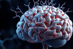
- Epiblast cells staying behind become the ectoderm, for skin and the nervous system.
- Cells moving through the primitive node form a mesodermal rod, the notochord, extending from the primitive streak.
- The primitive streak and notochord define the embryo's midline.
- Notochord remnants persist post-birth as the nucleus pulposus within intervertebral disks.
- The notochord marks the rostral-caudal axis of the CNS.
- Its origin near the primitive node underlies the lumbar spinal cord.
- The notochord ends near the future diencephalon.
- The notochord secretes signaling molecules like sonic hedgehog.
- This signals the overlying midline ectoderm to thicken into the neural plate.
- As the notochord induces the neural plate, the ectoderm and nearby tissues release their signaling molecules.
- Opposing gradients from these molecules change differentiation and growth.
- Cells near the midline become motor neurons.
- Cells in more lateral parts of the neural plate become sensory neurons.
- Cells at the junction of the neural plate and ectoderm become neural crest cells.
Neural Tube Formation
- During the third week, a longitudinal groove (neural groove) appears in the neural plate, flanked by a neural fold on either side.
- Neural crest cells grow at the apex of each neural fold.
- The neural folds enlarge and meet in the midline at the level of the future cervical spinal cord, forming the beginning of the neural tube.
- Additional closure sites appear and zip up in rostral and caudal directions.
- As closure progresses, neural crest cells separate from the neural folds and form a layer over the neural tube, between it and the surface ectoderm.
- The neural tube closes up and detaches from the surface ectoderm by the end of the fourth week.
- Neurulation, the formation of the neural tube, is a major developmental event
- The neural tube will become the entire CNS
- Its cavity becomes the ventricular system of the brain.
- The sacral spinal cord forms after neural tube closure through secondary neurulation
- The neural tube extends into a stem cell mass at the caudal end of the spinal cord.
- Neural crest cells migrate to form the neurons and glial cells of the peripheral nervous system
- They also form cutaneous melanocytes, the adrenal medulla, and nonneural tissues in the head.
- This includes primary sensory neurons of spinal and cranial nerve ganglia.
- It also includes autonomic neurons and Schwann and satellite cells of peripheral nerves and ganglia.
- Placodes of neural crest-like cells initially in the surface ectoderm detach later and form the remainder of the PNS in the head.
Defective Neurulation
- Defective neurulation is relatively common, affecting approximately 0.1% of infants.
- It effects even higher percentages of fetuses that do not survive to term.
- Neural tube defects lead to different abnormalities in the CNS.
- Neural tube defects also lead to abnormalities in the skull and vertebrae.
- Craniorachischisis is a fatal condition where the neural tube does not close, and neural tissue is continuous with the skin.
- Anencephaly results if only the rostral end of the neural tube fails to close.
- In this case, the skull is absent and most of the cerebrum is replaced by exposed neural tissue.
- The face and the rest of the skull develop because of the notochord and ventral neural tube.
- Spina bifida occurs if the caudal end of the neural tube fails to close
- This ranges from spinal cord exposure to vertebral arch deficiencies with herniation of spinal cord or meninges.
- Milder forms may be asymptomatic with a single defective vertebra, indicated by a tuft of hair or dimple.
- Hirschsprung's disease is due to a proliferation or migration defect of neural crest cells, with defective innervation of a colon segment.
- The affected segment constricts, restricting passage of intestinal contents, causing proximal dilation (congenital megacolon).
Clinical Note: Detecting Neural Tube Defects
- Alpha-fetoprotein (AFP) is synthesized in the fetal liver and is a major component of fetal serum.
- Some is normally excreted into amniotic fluid through fetal urine.
- In open neural tube defects, more than the normal amount of AFP leaks into cerebrospinal fluid, amniotic fluid, and maternal serum.
- Measuring AFP levels during gestation is a useful screening test for neural tube defects.
Biochemistry Note: Preventing Neural Tube Defects
- Adequate folic acid is essential for rapidly dividing cells because of its role in DNA synthesis.
- Folic acid supplementation may prevent more than 50% of neural tube defects.
- Because neurulation occurs early, supplementation must be provided near the time of conception.
- Folate is added to parts of the food supply in some countries for this reason.
Brain Vesicles
- The neural tube is not straight.
- Even before closing, the brain part of the tube develops curves and creases, laying the groundwork for the arrangement of its future parts.
- During the fourth week, three primary vesicles appear: the prosencephalon (forebrain), mesencephalon (midbrain), and rhombencephalon (hindbrain).
- The prosencephalon becomes the cerebrum.
- The mesencephalon forms the midbrain.
- The rhombencephalon forms the pons, medulla, and cerebellum.
- Less than a week later, bulges grow on each side of the prosencephalon, and differences appear in the rostral and caudal halves of the rhombencephalon.
- The prosencephalon gives rise to the telencephalon and diencephalon.
- The rhombencephalon gives rise to the metencephalon and myelencephalon.
- The mesencephalon remains undivided.
- Each telencephalic vesicle develops into a cerebral hemisphere.
- The diencephalon becomes the thalamus, hypothalamus, and other structures.
- The metencephalon becomes the pons and cerebellum.
- The myelencephalon becomes the medulla.
- The neural tube cavity persists as a system of ventricles.
Flexures in the Neural Tube
- There are three prominent bends that appear when embryonic vesicles are forming.
- The midbrain bends around the rostral end of the notochord, forming the cephalic flexure.
- The cephalic flexure persists as the bend in the CNS between the brainstem and cerebrum.
- The cervical flexure appears between the spinal cord and brainstem and straightens out later.
- The brainstem bends anteriorly in the pontine flexure.
- The pontine flexure does not cause a long-term bend but determines the shape of the fourth ventricle and the position of the cerebellum.
"Rotation” of the Cerebral Hemispheres
- After embryonic vesicles form, massive growth of the telencephalon dominates remaining CNS development.
- Each telencephalic vesicle expands in all directions from the diencephalon.
- The medial and inferior vesicle parts fold down and fuse with the diencephalon where the basal ganglia will form.
- This fusion develops a cortical covering and sets a fixed pivot point as the hemisphere expands.
- The insula is overgrown by parts of the frontal, parietal, and temporal lobes.
- Structures like the lateral ventricle and the caudate nucleus are dragged into a C shape.
Malformation of the Prosencephalon
- The prosencephalon subdivides early.
- Problems with this subdivision cause holoprosencephaly.
- In severe forms, olfactory bulbs, tracts, and the optic chiasm are missing.
- Cerebral hemispheres are undivided, and the corpus callosum is absent.
- There is a single ventricle that spans the midline.
- Effects on the skull and face are opposite to those in anencephaly
- The development on in holoprosencephaly dorsal and posterior skull is present.
Proliferation, Migration, and Differentiation of Neurons
- Neural tube closure and prosencephalon formation occur in the first month of development.
- This is followed by massive proliferation of neurons and glial cells during the third and fourth months and later.
- Cells than migrate to their final locations and differentiate into specific cell types.
Neurogenesis
- The simple neural tube has a pseudostratified columnar epithelium, a single cell thick.
- Early on, each cell spans the entire epithelium, its nucleus shuttling between the ventricular and external surfaces.
- As the nucleus moves outward, its DNA replicates.
- As it moves inward, the cell retracts its surface-directed process and divides.
- The two daughter cells either repeat the process, or one or both leave the mitotic cycle, commit to being a progenitor cell, and migrate to a final destination.
- The final round of mitosis happens before birth in almost all CNS areas
- The last cells left near the lumen of the neural tube transform into the ependymal cells.
- Neural progenitors accumulate outside the mitotic area, dividing the neural tube wall into three zones: ventricular, intermediate, and marginal.
- The ventricular zone contains stem cell nuclei that shuttle and divide.
- The intermediate zone contains committed precursor cells.
- The marginal zone is relatively cell-free with surface-directed processes of stem cells.
- In the spinal cord, the ventricular zone becomes the ependymal cells surrounding the central canal.
- The intermediate zone becomes the spinal gray matter.
- The marginal zone becomes the white matter, invaded by ascending and descending tracts.
- The sequence is complex in other CNS regions.
- In the telencephalon, for example, some neurons stay close to the lateral ventricles, forming the basal ganglia and amygdala.
- Other neurons migrate longer distances to form the cerebral cortex, with newborn neurons migrating past existing cortical neurons, forming the cortex from the inside out.
Abnormalities of Proliferation and Migration
- Neural stem cell divisions, timing of divisions, and migration of newborn neurons need control for proper CNS wiring.
- Micrencephaly ("small brain") results if too few cell divisions occur or too few stem cells are formed.
- If neuron migration to the cortex is abnormal, gyri do not form properly.
- Lissencephaly ("smooth brain") occurs when a large percentage of neurons stop before reaching the cortex.
- As a result, the surface area of the cortex is reduced, and gyri do not form.
- Heterotopic clumps of gray matter result if neurons get "stuck" in the white matter before reaching the cortex.
Longitudinal Zones in the Central Nervous System
- Signaling molecules induce the roof and floor plate from the nearest neural tube parts.
- The signaling molecules are the same and are released by the surface ectoderm and notochord
- The roof and floor plates thin out and the lateral walls develop into various parts of the CNS
- Seen most clearly in the spinal cord where alar and basal plate near roof and floor plate respectively.
- Alar and basal plate are separated by the sulcus limitans, running longitudinally
- Alar and basal plate form posterior and anterior horns containing sensory and motor neurons
- Preganglionic autonomic neurons are at intermediate gray matter near where the sulcus limitans used to be.
- The cavity becomes the central canal, where little tissue is left.
- The organization continues into the rhombencephalon which spreads the neural tube walls and stretches the roof plate out.
- The elongated roof plate becomes the top of the fourth ventricle.
- The alar and basal plates form the floor of 4th ventricle with alar plate lateral
- This extends development into sensory and motor functions.
Studying That Suits You
Use AI to generate personalized quizzes and flashcards to suit your learning preferences.