Podcast
Questions and Answers
Which condition primarily leads to cyanosis due to the shunting of deoxygenated blood into systemic circulation?
Which condition primarily leads to cyanosis due to the shunting of deoxygenated blood into systemic circulation?
- Atrial septal defect (ASD)
- Ventricular septal defect (VSD)
- Tetralogy of Fallot (correct)
- Patent ductus arteriosus (PDA)
What is a common symptom associated with acyanotic congenital heart defects?
What is a common symptom associated with acyanotic congenital heart defects?
- Failure to thrive (correct)
- Clubbing of the nails
- Cyanosis of the lips
- Pale gray or blue skin color
What is the primary mechanism of left-to-right shunts in acyanotic congenital heart defects?
What is the primary mechanism of left-to-right shunts in acyanotic congenital heart defects?
- Obstruction of blood flow from the heart
- Excessive blood flow to the lungs (correct)
- Decreased pressure in the right atrium
- Blocking the outflow of oxygenated blood
Which congenital defect is NOT classified as acyanotic?
Which congenital defect is NOT classified as acyanotic?
Which condition can result from right ventricular pressure overload from a left-to-right shunt?
Which condition can result from right ventricular pressure overload from a left-to-right shunt?
From which part of the embryo does the heart develop?
From which part of the embryo does the heart develop?
What significant event occurs on the 21-22nd day during heart development?
What significant event occurs on the 21-22nd day during heart development?
Which structure is the arterial end of the heart tube?
Which structure is the arterial end of the heart tube?
What type of cells induce vasculogenesis in the primary heart field?
What type of cells induce vasculogenesis in the primary heart field?
What happens to the two endothelial heart tubes during the 3rd week of embryonic development?
What happens to the two endothelial heart tubes during the 3rd week of embryonic development?
Which of the following correctly lists the five dilatations of the heart tube from cranial to caudal end?
Which of the following correctly lists the five dilatations of the heart tube from cranial to caudal end?
What does the unfused part of the sinus venosus form in the heart tube?
What does the unfused part of the sinus venosus form in the heart tube?
Which type of mesoderm is responsible for forming the myocardium and epicardium surrounding the primitive heart tube?
Which type of mesoderm is responsible for forming the myocardium and epicardium surrounding the primitive heart tube?
What is the name of the gap between the lower edge of septum primum and septum intermedium during the formation of the interatrial septum?
What is the name of the gap between the lower edge of septum primum and septum intermedium during the formation of the interatrial septum?
Which structure forms the upper part of the interatrial septum?
Which structure forms the upper part of the interatrial septum?
What physiological change occurs that leads to the closure of the foramen ovale after birth?
What physiological change occurs that leads to the closure of the foramen ovale after birth?
What structure is formed as the upper part of the septum primum breaks down during interatrial septum formation?
What structure is formed as the upper part of the septum primum breaks down during interatrial septum formation?
How does the foramen ovale function in the heart before birth?
How does the foramen ovale function in the heart before birth?
At what stage of fetal development does the septum primum begin to develop?
At what stage of fetal development does the septum primum begin to develop?
What happens to the foramen ovale after birth as the left atrium receives blood from the lungs?
What happens to the foramen ovale after birth as the left atrium receives blood from the lungs?
What anatomical feature represents the lower edge of the septum secundum in adults?
What anatomical feature represents the lower edge of the septum secundum in adults?
Which of the following statements is true regarding the formation of the interatrial septum?
Which of the following statements is true regarding the formation of the interatrial septum?
What is the role of septum primum during fetal development?
What is the role of septum primum during fetal development?
What structure is responsible for the formation of the atrioventricular septum?
What structure is responsible for the formation of the atrioventricular septum?
Which condition results from a defect in cardiac looping?
Which condition results from a defect in cardiac looping?
What happens to the bulboventricular sulcus during heart development?
What happens to the bulboventricular sulcus during heart development?
Which of the following is NOT a septum that partitions the primitive heart tube?
Which of the following is NOT a septum that partitions the primitive heart tube?
Where does the primitive atrium lie in relation to the truncus arteriosus?
Where does the primitive atrium lie in relation to the truncus arteriosus?
Which rare condition is characterized by the heart lying exposed on the surface of the thorax?
Which rare condition is characterized by the heart lying exposed on the surface of the thorax?
What is the primary function of the interatrial septum?
What is the primary function of the interatrial septum?
What happens to the primitive heart tube as it develops into the adult heart?
What happens to the primitive heart tube as it develops into the adult heart?
At what stage of development do the endocardial cushions appear to form the atrioventricular septum?
At what stage of development do the endocardial cushions appear to form the atrioventricular septum?
Which of the following describes dextrocardia?
Which of the following describes dextrocardia?
At what week does the formation of the interventricular septum typically begin?
At what week does the formation of the interventricular septum typically begin?
How many parts compose the interventricular septum?
How many parts compose the interventricular septum?
What is the correct order of development for the aorticopulmonary septum?
What is the correct order of development for the aorticopulmonary septum?
What effect does the fusion of truncal and bulbar ridges have on the ventricular outflow tract?
What effect does the fusion of truncal and bulbar ridges have on the ventricular outflow tract?
Which of the following is NOT a type of valvular anomaly?
Which of the following is NOT a type of valvular anomaly?
What can congenital heart defects (CHDs) lead to?
What can congenital heart defects (CHDs) lead to?
What is the formation of the interventricular septum characterized by?
What is the formation of the interventricular septum characterized by?
What is a potential outcome of the disruption of normal cardiac morphogenesis?
What is a potential outcome of the disruption of normal cardiac morphogenesis?
Which part of the interventricular septum is NOT one of its components?
Which part of the interventricular septum is NOT one of its components?
Which of the following not correctly describe a potential state of valvular anomalies?
Which of the following not correctly describe a potential state of valvular anomalies?
What parts make up the interventricular septum?
What parts make up the interventricular septum?
During which week does the formation of the aorticopulmonary septum typically begin?
During which week does the formation of the aorticopulmonary septum typically begin?
What outcome may occur due to Congenital Heart Defects (CHDs)?
What outcome may occur due to Congenital Heart Defects (CHDs)?
Which cells are involved in the formation of the aorticopulmonary septum?
Which cells are involved in the formation of the aorticopulmonary septum?
What is a characteristic of the interventricular septum's development compared to the atrial septation process?
What is a characteristic of the interventricular septum's development compared to the atrial septation process?
What establishes the initial attachment of the heart tube within the pericardial cavity?
What establishes the initial attachment of the heart tube within the pericardial cavity?
How does the heart tube relate to the pericardial cavity after the formation of the head fold?
How does the heart tube relate to the pericardial cavity after the formation of the head fold?
What happens to the cardiac jelly as development progresses?
What happens to the cardiac jelly as development progresses?
Which embryonic layer contributes to the formation of the endothelial heart tube?
Which embryonic layer contributes to the formation of the endothelial heart tube?
What structure forms after the mesocardium connecting the bulboventricular loop disappears?
What structure forms after the mesocardium connecting the bulboventricular loop disappears?
During heart tube formation, what is the role of the myoepicardial mantle?
During heart tube formation, what is the role of the myoepicardial mantle?
What embryological structure gives rise to the visceral layer of the pericardium?
What embryological structure gives rise to the visceral layer of the pericardium?
Which structure is formed from the proliferation of the splanchnopleuric mesoderm on the dorsal aspect of the pericardial cavity?
Which structure is formed from the proliferation of the splanchnopleuric mesoderm on the dorsal aspect of the pericardial cavity?
Which of the following describes the heart tube's shape during the formation of the bulboventricular loop?
Which of the following describes the heart tube's shape during the formation of the bulboventricular loop?
What was the initial origin of all components of the cardiovascular system (CVS)?
What was the initial origin of all components of the cardiovascular system (CVS)?
At what time does the development of the separate blood vascular system begin?
At what time does the development of the separate blood vascular system begin?
What is a key modification that occurs in lateral folding during heart development?
What is a key modification that occurs in lateral folding during heart development?
What does the right-left blood shunt primarily indicate in fetal circulation?
What does the right-left blood shunt primarily indicate in fetal circulation?
Which of the following components is involved in the development of the heart tube?
Which of the following components is involved in the development of the heart tube?
What aspect of the primitive ventricles is important to understand in their clinical correlations?
What aspect of the primitive ventricles is important to understand in their clinical correlations?
What is the primary function of the endocardial cushions during heart development?
What is the primary function of the endocardial cushions during heart development?
What structural feature is formed by the fusion of the truncus arteriosus and bulbar ridges?
What structural feature is formed by the fusion of the truncus arteriosus and bulbar ridges?
In which week does the separation of the atrioventricular septum occur in embryonic development?
In which week does the separation of the atrioventricular septum occur in embryonic development?
What condition is a result of a defect in cardiac looping?
What condition is a result of a defect in cardiac looping?
Which of the following is NOT a normal process during the development of the cardiovascular system?
Which of the following is NOT a normal process during the development of the cardiovascular system?
What anatomical position does the primitive atrium occupy in relation to the truncus arteriosus?
What anatomical position does the primitive atrium occupy in relation to the truncus arteriosus?
What leads to the formation of the septum intermedium in the heart?
What leads to the formation of the septum intermedium in the heart?
What happens to the bulboventricular sulcus during heart development?
What happens to the bulboventricular sulcus during heart development?
What is a characteristic of ectopia cordis?
What is a characteristic of ectopia cordis?
What is the role of microtubules and dynein arms in cardiac development?
What is the role of microtubules and dynein arms in cardiac development?
What major event occurs with the formation of the interatrial septum?
What major event occurs with the formation of the interatrial septum?
What is the fate of the primitive heart tube as it develops?
What is the fate of the primitive heart tube as it develops?
What is the foramen that is formed by the breakdown of the upper part of the septum primum as it fuses with the AV septum?
What is the foramen that is formed by the breakdown of the upper part of the septum primum as it fuses with the AV septum?
Which septum primarily forms the lower part of the interatrial septum?
Which septum primarily forms the lower part of the interatrial septum?
How does the foramen ovale function in the fetal heart?
How does the foramen ovale function in the fetal heart?
What occurs physiologically to close the foramen ovale after birth?
What occurs physiologically to close the foramen ovale after birth?
Which embryonic structure grows downward toward the septum intermedium and overlaps the foramen secundum?
Which embryonic structure grows downward toward the septum intermedium and overlaps the foramen secundum?
What is the gap between the lower edge of septum primum and septum intermedium called during fetal development?
What is the gap between the lower edge of septum primum and septum intermedium called during fetal development?
What anatomical feature represents the remnants of septum primum and septum secundum in adults?
What anatomical feature represents the remnants of septum primum and septum secundum in adults?
During the formation of the interatrial septum, when does the septum primum start developing?
During the formation of the interatrial septum, when does the septum primum start developing?
What key event promotes the physiological closure of the foramen ovale after birth?
What key event promotes the physiological closure of the foramen ovale after birth?
What is the main function of the interatrial septum during fetal development?
What is the main function of the interatrial septum during fetal development?
Flashcards
Cardiac progenitor cells
Cardiac progenitor cells
Cells that develop into heart structures, appear early in the third week of development.
Primitive heart tube
Primitive heart tube
The initial tube-like structure that develops into the heart.
Endocardium
Endocardium
Inner lining of the heart, forms from the heart tube.
Myocardium
Myocardium
Signup and view all the flashcards
Epicardium
Epicardium
Signup and view all the flashcards
Cardiogenic area
Cardiogenic area
Signup and view all the flashcards
Heart looping
Heart looping
Signup and view all the flashcards
Atrioventricular canal
Atrioventricular canal
Signup and view all the flashcards
Atrioventricular septum
Atrioventricular septum
Signup and view all the flashcards
Interatrial septum
Interatrial septum
Signup and view all the flashcards
Interventricular septum
Interventricular septum
Signup and view all the flashcards
Foramen ovale
Foramen ovale
Signup and view all the flashcards
Aorticopulmonary septum
Aorticopulmonary septum
Signup and view all the flashcards
Congenital heart defects (CHDs)
Congenital heart defects (CHDs)
Signup and view all the flashcards
Cardiac looping defects
Cardiac looping defects
Signup and view all the flashcards
Ectopia cordis
Ectopia cordis
Signup and view all the flashcards
Situs inversus
Situs inversus
Signup and view all the flashcards
Dextrocardia
Dextrocardia
Signup and view all the flashcards
Shunts
Shunts
Signup and view all the flashcards
Ventricular septal defect (VSD)
Ventricular septal defect (VSD)
Signup and view all the flashcards
Atrial septal defect (ASD)
Atrial septal defect (ASD)
Signup and view all the flashcards
Foramen primum
Foramen primum
Signup and view all the flashcards
Foramen secundum
Foramen secundum
Signup and view all the flashcards
Septums primum and secundum
Septums primum and secundum
Signup and view all the flashcards
Study Notes
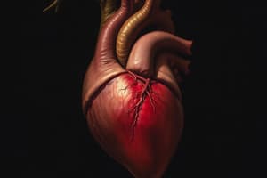
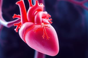
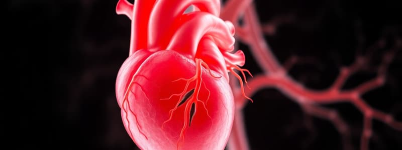
Development of the Heart
- The heart originates from a primitive heart tube derived from mesenchyme in the embryo's cardiogenic area.
- The primitive heart tube forms the endocardium, while surrounding splanchnic mesoderm differentiates into myocardium and epicardium.
- Initial heartbeat occurs around the 21st to 22nd day of embryonic development, with blood circulation starting by the 24th day.
Establishment of the Cardiogenic Area
- Cardiac progenitor cells emerge cranial to the primitive streak in the 3rd week of development.
- These cells migrate to form a horseshoe-shaped primitive heart field in the splanchnopleuric mesoderm by the end of the 3rd week.
- Endoderm of the primitive pharynx induces vasculogenesis in the primary heart field through the secretion of vascular endothelial growth factor (VEGF).
Formation of the Heart Tube
- Small vessels coalesce to form two endothelial heart tubes that later fuse into a single heart tube.
- The heart tube has a bifurcated structure, with a cranial (arterial) end and a caudal (venous) end.
- The heart tube differentiates into five dilations from cranial to caudal: truncus arteriosus, bulbus cordis, primitive ventricle, primitive atrium, and sinus venosus.
Arterial and Venous Ends of the Heart Tube
- The truncus arteriosus connects to the aortic sac, leading to the first pharyngeal arch artery and dorsal aorta.
- The venous end, or unfused sinus venosus, forms two horns which assist in receiving blood through three veins from the body wall.
Acquisition of Adult Heart Form
- Formation of an S-shaped cardiac loop occurs as the primitive atrium and sinus venosus shift position in the pericardial cavity relative to the primitive ventricle.
- Bulbus cordis and the primitive ventricle are initially separated by a sulcus which later disappears, resulting in a single chamber.
- The expansion of the primitive atrium leads to the development of auricles that project forward from the truncus arteriosus.
Clinical Correlations
- Cardiac looping defects can lead to Kartagener syndrome, characterized by situs inversus and dextrocardia.
- Ectopia cordis is a rare condition where the heart is exposed on the thoracic surface due to nonunion of the sternal plates.
Formation of Heart Chambers
- The single lumen of the primitive heart tube partitions into four definitive chambers via four septa: atrioventricular septum, interatrial septum, interventricular septum, and aorticopulmonary septum.
Atrioventricular Septum Formation
- Endocardial cushions arise from mesenchyme in the canal walls and fuse to create the atrioventricular septum around the 28th day of development.
Interatrial Septum Formation
- The interatrial septum separates the right and left atria and is formed by septum primum and septum secundum.
- Septum primum develops from the primitive atrium and grows towards the atrioventricular septum, creating the foramen primum.
- As septum primum fuses with the atrioventricular septum, the foramen secundum forms, facilitating communication between right and left atria through the foramen ovale.
Physiological Closure of Foramen Ovale
- The foramen ovale usually closes within the first year of life when pressure in the left atrium surpasses that in the right atrium, pushing the septum primum against the septum secundum.
Interventricular Septum Formation
- Development of the interventricular septum begins by the 4th week and is completed by the end of the 7th week.
- It consists of muscular, bulbar, and membranous parts, each evolving from different origins.
Aorticopulmonary Septum Formation
- Neural crest and endocardial cells form truncal and bulbar ridges that spiral and fuse to create the aorticopulmonary septum, dividing the outflow tract into the aorta and pulmonary trunk.
Congenital Heart Defects
- Congenital heart defects (CHDs) arise from abnormal cardiac morphogenesis, leading to shunts between heart chambers.
- Shunt types include left-to-right (causing pulmonary hypertension) and right-to-left (causing cyanosis).
Acyanotic and Cyanotic Heart Defects
- Acyanotic defects include conditions like ventricular septal defect (VSD) and atrial septal defect (ASD), characterized by left-to-right shunts.
- Cyanotic defects involve right-to-left shunting, typically manifesting as “blue baby” syndrome with symptoms like feeding problems and exertional dyspnea.
Learning Outcomes
- Understand the formation of hemangioblasts and blood islands.
- Explain similarities between heart tubes, dorsal aorta, and vitelline veins.
- Grasp how lateral folding affects developing cardiovascular structures.
- Identify components and derivatives of the heart tube.
- Describe cardiac looping and its clinical implications.
- Explain development and modification of atrioventricular canals.
- Analyze septation and modification of primitive atria and ventricles.
Overview of Cardiovascular Development
- Cardiovascular system (CVS) development initiates in the 3rd week of embryogenesis.
- All CVS components are derived from mesoderm.
- Key developmental processes include the formation of the heart and major blood vessels: umbilical and vitelline veins.
Positioning and Structure of Heart Tube
- Heart tube initially positioned caudal to the septum transversum and rotates 180° after head fold formation.
- Heart tube and pericardial cavity lie ventral to the foregut after rotation.
- Dorsal mesocardium connects the heart tube to the pericardial cavity but disintegrates, anchoring the heart tube at pericardial margins.
Formation of Cardiac Wall
- Endothelial heart tube originates from splanchnopleuric mesoderm and becomes the endocardium.
- Myoepicardial mantle forms myocardium, surrounded by cardiac jelly, which is replaced by the developing myocardium.
- Visceral pericardium arises from myoepicardial mantle; somatopleuric mesoderm gives rise to the parietal layer of pericardium.
Acquisition of Adult Heart Form
- Bulboventricular loop formation involves ventral growth of bulbus cordis and primitive ventricle.
- Transverse sinus develops from the breakdown of mesocardium connecting various segments of the heart.
- S-loop development results from positioning of the atrium and sinus venosus relative to the primitive ventricle.
Clinical Correlations
- Cartagener syndrome is linked to defects in cardiac looping, causing situs inversus and dextrocardia.
- Ectopia cordis occurs from failure of sternal plates to unite, leading to exposed heart and high neonatal mortality.
Development of Heart Chambers
- Primitive heart tube's single lumen partitions into four chambers: atrioventricular, interatrial, interventricular, and aorticopulmonary septa.
- Atrioventricular septum forms through the fusion of endocardial cushions, dividing it into right and left canals.
Interatrial Septum Formation
- Formed by the septum primum and septum secundum, developing from the primitive atrium.
- The foramen primum and foramen secundum are critical gaps during formation.
- The foramen ovale, a significant feature, allows temporary blood flow between atria.
Physiological Closure of Foramen Ovale
- Typically closes within 6 months to 1 year post-birth as left atrial pressure exceeds that of the right atrium, pushing septum primum against septum secundum.
Interventricular Septum Formation
- Development starts in week 4, completed by week 7; differs from atrial septation as it does not allow shunting.
- Comprised of muscular, bulbar, and membranous parts, each from distinct developmental origins.
Aorticopulmonary Septum Development
- Formed by migrating neural crest and endocardial cells, which create truncal and bulbar ridges that spiral and fuse.
- This division establishes distinct pathways for the aorta and pulmonary trunk.
Valve Formation and Clinical Correlations
- Valvular anomalies can present as stenosis, regurgitation, or complete atresia (e.g., tricuspid atresia).
- Congenital heart defects (CHDs) arise from disruptions in normal cardiac morphogenesis, causing abnormal circulatory connections.
Studying That Suits You
Use AI to generate personalized quizzes and flashcards to suit your learning preferences.