Podcast
Questions and Answers
What is the thickness of the junctional epithelium coronally?
What is the thickness of the junctional epithelium coronally?
- 30-50 cells
- 15-30 cells (correct)
- 1-3 cells
- 5-10 cells
What is the primary source of the junctional epithelium?
What is the primary source of the junctional epithelium?
- Oral epithelium
- Gingival epithelium
- Enamel epithelium
- Reduced dental epithelium (correct)
What is special about the interface between the epithelium and lamina propria in the gingival sulcus?
What is special about the interface between the epithelium and lamina propria in the gingival sulcus?
- It has epithelial ridges
- It has a rough interface
- It has a basement membrane
- It has a smooth interface (correct)
What is the characteristic of the cells in the junctional epithelium?
What is the characteristic of the cells in the junctional epithelium?
What is the function of the junctional epithelium?
What is the function of the junctional epithelium?
What is the rate of turnover of the junctional epithelium?
What is the rate of turnover of the junctional epithelium?
What is the characteristic of the lamina propria in the dento-gingival junction?
What is the characteristic of the lamina propria in the dento-gingival junction?
What is the function of hemidesmosomes in the dento-gingival junction?
What is the function of hemidesmosomes in the dento-gingival junction?
What can be seen in the intercellular spaces of healthy gingival tissue?
What can be seen in the intercellular spaces of healthy gingival tissue?
What is the term for the combination of hemidesmosomes and basal lamina in the dento-gingival junction?
What is the term for the combination of hemidesmosomes and basal lamina in the dento-gingival junction?
What is unique about the junctional epithelium?
What is unique about the junctional epithelium?
What is the term for the process of gingival recession due to disease?
What is the term for the process of gingival recession due to disease?
At what age does the second stage of passive eruption typically persist until?
At what age does the second stage of passive eruption typically persist until?
What is the anatomical landmark that marks the apical end of the clinical crown during the first stage of passive eruption?
What is the anatomical landmark that marks the apical end of the clinical crown during the first stage of passive eruption?
What is the term for the shift of the dento-gingival junction?
What is the term for the shift of the dento-gingival junction?
In primary teeth, until when does the first stage of passive eruption typically occur?
In primary teeth, until when does the first stage of passive eruption typically occur?
Flashcards are hidden until you start studying
Study Notes
Dento-Gingival Junction
- The dento-gingival junction is formed by the interaction of the tooth and the gingiva
- Histogenesis of dento-gingival junction involves the formation of a primary dentogingival junction (from reduced enamel epithelium) and a secondary dentogingival junction (from oral epithelium)
Histology of Dento-gingival Junction
- The junctional epithelium is 15-30 cells thick coronally and 1-3 cells thick apically
- It consists of two zones: a single cell layer of cuboidal cells (stratum germinativum) and several layers of flattened cells
- The junctional epithelium has a high rate of turnover (5-6 days) and is initially derived from the rapidly replaced reduced enamel epithelium
- The lamina propria shows many capillaries and appears to be more cellular than other parts of the gingiva
- The epithelial cells are separated from the lamina propria by a smooth basement membrane
- The junctional epithelium cells are joined by few desmosomes and have large intercellular spaces, allowing crevicular fluid and defense cells to pass through
Epithelial Attachment
- The cells of the junctional epithelium attach themselves to the enamel or cementum by hemidesmosomes within the cell and a basal lamina produced by the epithelial cells
- This combination of hemidesmosomes and basal lamina is known as the attached apparatus or epithelial attachment
- The basal lamina in contact with the tooth is termed the internal basal lamina, and the other surface of the junctional epithelium in contact with the lamina propria is the normal basal lamina (external basal lamina)
- The junctional epithelium is unique in having two basal laminae
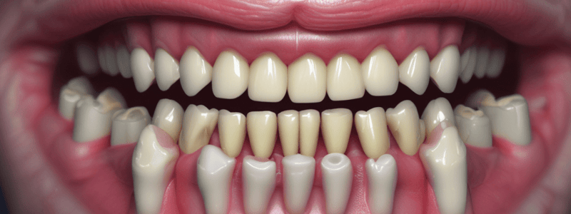
Gingival Sulcus and Dento-gingival Junction
- The gingival sulcus is lined by non-keratinized stratified squamous epithelium, which is thinner than the epithelium of the gingiva and lacks epithelial ridges
- The epithelium has a smooth interface with the lamina propria and is continuous with the gingival epithelium and attachment epithelium
- All three epithelia have a continuous basal lamina
Stages of Passive Eruption
- First stage: till 1 year before shedding in deciduous teeth and till 20-30 years in permanent teeth
- Second stage: persists to 40 years
- Third stage: transitory
- Fourth stage: gingival recession (pathologic) till loss
Studying That Suits You
Use AI to generate personalized quizzes and flashcards to suit your learning preferences.