Podcast
Questions and Answers
What is the primary function of the microscopic pores in enamel?
What is the primary function of the microscopic pores in enamel?
- To change the shape of the enamel rods
- To allow complete passage of ions and stains
- To selectively allow certain ions to pass through (correct)
- To block all ions and stains from passing through
What is the relationship between the shape of the enamel rods and the enamel prisms?
What is the relationship between the shape of the enamel rods and the enamel prisms?
- The shape of the enamel rods is independent of the enamel prisms
- The shape of the enamel prisms determines the shape of the enamel rods
- The shape of the enamel rods determines the shape of the enamel prisms (correct)
- The shape of the enamel prisms is irrelevant to the enamel rods
What is the significance of the inter-rod spaces or sheets in enamel?
What is the significance of the inter-rod spaces or sheets in enamel?
- They provide structural support to the enamel prisms
- They are irrelevant to enamel structure
- They contain organic material essential for enamel formation (correct)
- They allow for the passage of ions and stains
What is the significance of the Water+inter-rod spaces in enamel?
What is the significance of the Water+inter-rod spaces in enamel?
What is the term for the type of permeability exhibited by the microscopic pores in enamel?
What is the term for the type of permeability exhibited by the microscopic pores in enamel?
What is the primary reason why ground sections are not suitable for histological study?
What is the primary reason why ground sections are not suitable for histological study?
What is the approximate protein content in immature enamel?
What is the approximate protein content in immature enamel?
What is the term for the basic structural units of enamel?
What is the term for the basic structural units of enamel?
What is the arrangement of crystalites within enamel rods?
What is the arrangement of crystalites within enamel rods?
Why are demineralized sections used to study immature enamel?
Why are demineralized sections used to study immature enamel?
What term is used to describe the basic units of enamel composed of multiple crystals?
What term is used to describe the basic units of enamel composed of multiple crystals?
What is the shape of the enamel rods in the histological structure of enamel?
What is the shape of the enamel rods in the histological structure of enamel?
What is the percentage of mineralization in bone?
What is the percentage of mineralization in bone?
What is the term for the lines that appear on the surface of the enamel?
What is the term for the lines that appear on the surface of the enamel?
What is the junction between enamel and dentin called?
What is the junction between enamel and dentin called?
What is the term for the structures that appear as small, rounded, or oval bodies on the surface of the enamel?
What is the term for the structures that appear as small, rounded, or oval bodies on the surface of the enamel?
What is the type of mineralization that occurs in utero during embryological stages?
What is the type of mineralization that occurs in utero during embryological stages?
What is the rate of mineralization in enamel?
What is the rate of mineralization in enamel?
What is the term for the lines that appear as a result of the daily deposition of enamel?
What is the term for the lines that appear as a result of the daily deposition of enamel?
What is the type of microscopy used to study the structure of enamel in detail?
What is the type of microscopy used to study the structure of enamel in detail?
What is the term for the small, branching tubules that are sometimes seen in the enamel?
What is the term for the small, branching tubules that are sometimes seen in the enamel?
Flashcards
Function of Microscopic Pores in Enamel
Function of Microscopic Pores in Enamel
Selectively allow certain ions to pass through the enamel.
Rod and Prism Relationship
Rod and Prism Relationship
The shape of the enamel rods determines the shape of the enamel prisms.
Significance of Inter-rod Spaces
Significance of Inter-rod Spaces
Contain organic material essential for enamel formation.
Significance of Water+inter-rod Spaces
Significance of Water+inter-rod Spaces
Signup and view all the flashcards
Permeability of Enamel Pores
Permeability of Enamel Pores
Signup and view all the flashcards
Unsuitability of Ground Sections
Unsuitability of Ground Sections
Signup and view all the flashcards
Protein Content in Immature Enamel
Protein Content in Immature Enamel
Signup and view all the flashcards
Basic Structural Units of Enamel
Basic Structural Units of Enamel
Signup and view all the flashcards
Arrangement of Crystalites
Arrangement of Crystalites
Signup and view all the flashcards
Use of Demineralized Sections
Use of Demineralized Sections
Signup and view all the flashcards
Basic Units of Enamel
Basic Units of Enamel
Signup and view all the flashcards
Shape of Enamel Rods
Shape of Enamel Rods
Signup and view all the flashcards
Percentage of Bone Mineralization
Percentage of Bone Mineralization
Signup and view all the flashcards
Lines on Enamel Surface
Lines on Enamel Surface
Signup and view all the flashcards
Enamel-Dentin Border
Enamel-Dentin Border
Signup and view all the flashcards
Small bodies on enamel
Small bodies on enamel
Signup and view all the flashcards
Mineralization in Utero
Mineralization in Utero
Signup and view all the flashcards
Rate of Enamel Mineralization
Rate of Enamel Mineralization
Signup and view all the flashcards
Lines From Daily Deposition
Lines From Daily Deposition
Signup and view all the flashcards
Microscopy for Enamel Study
Microscopy for Enamel Study
Signup and view all the flashcards
Small tubules in enamel
Small tubules in enamel
Signup and view all the flashcards
Study Notes
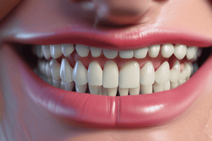
Enamel Structure Study
- Enamel structure is mainly studied in ground sections, but immature enamel can be studied in demineralized sections due to its high protein content (25-30%).
Enamel Rods (Prisms)
- Enamel rods (prisms) are the basic structural units of enamel, composed of a group of crystalites arranged on top of each other.
- Each rod has microscopic pores, making it semipermeable or selectively permeable, allowing some substances to pass through.
Enamel Formation
- Enamel formation begins in utero during embryological stages (primary mineralization) and continues after birth (secondary mineralization).
- The rate of mineralization is linear for enamel and logarithmic for dentin and bone.
- The process starts with the deposition of organic and inorganic material by ameloblasts, followed by mineralization, which reaches a plateau.
Histological Structure of Enamel
- Enamel rods, Hunter-Schreger bands, gnarled enamel, and aprismatic enamel are all part of the histological structure of enamel.
- Other features include incremental lines, cross striations, striae of Retzius, perikymata, and neonatal lines.
- The surface enamel has pits, caps, focal holes, and enamel brochs.
Studying Enamel
- Ground sections are used to study enamel structure, but they are not suitable for histological study.
- Demineralized sections can be used to study immature enamel, but not for studying enamel structure.
- Electron microscopes can be used to study enamel, especially when studying the organic structure.
Studying That Suits You
Use AI to generate personalized quizzes and flashcards to suit your learning preferences.