Podcast
Questions and Answers
Which cellular structure provides shape and support to the cell?
Which cellular structure provides shape and support to the cell?
- Motor Proteins
- Microfilaments
- Intermediate Filaments (correct)
- Microtubules
What is the main function of the cytoskeleton related to cell organelles?
What is the main function of the cytoskeleton related to cell organelles?
- Supporting intracellular movements (correct)
- Holding cell organelles in place
- Assisting in cell signalling
- Formation of vacuoles
Which model best describes the structure of the plasma membrane as a flexible, lipid bilayer?
Which model best describes the structure of the plasma membrane as a flexible, lipid bilayer?
- Cell Theory Model
- Fluid Mosaic Model (correct)
- Kinetic Molecular Theory Model
- Endosymbiotic Model
What is the primary function of the plasma membrane in a cell?
What is the primary function of the plasma membrane in a cell?
Which component of the plasma membrane creates a fluid appearance according to the Fluid Mosaic Model?
Which component of the plasma membrane creates a fluid appearance according to the Fluid Mosaic Model?
What is the diameter of microtubules?
What is the diameter of microtubules?
Which protein makes up microfilaments?
Which protein makes up microfilaments?
What is the main function of intermediate filaments?
What is the main function of intermediate filaments?
Which motor protein is responsible for muscle contractions?
Which motor protein is responsible for muscle contractions?
What type of proteins move along microtubules carrying cellular components?
What type of proteins move along microtubules carrying cellular components?
Which cytoskeleton fiber facilitates the division of chromosomes during cell division?
Which cytoskeleton fiber facilitates the division of chromosomes during cell division?
What is the main function of centrosomes in cell division?
What is the main function of centrosomes in cell division?
Which phase of the centrosome cycle involves centrosome maturation?
Which phase of the centrosome cycle involves centrosome maturation?
What role do motor proteins play in the cytoskeleton?
What role do motor proteins play in the cytoskeleton?
Which structure is composed of a network of fibrous structures?
Which structure is composed of a network of fibrous structures?
In what process do centrosomes help organize microtubules ensuring distribution to daughter cells?
In what process do centrosomes help organize microtubules ensuring distribution to daughter cells?
Flashcards are hidden until you start studying
Study Notes
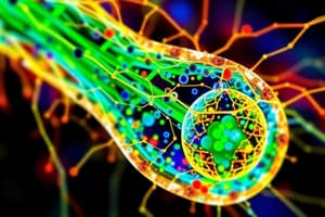
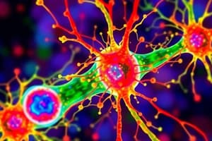
Cytoskeleton Structure
- Comprises three types of fibers: microfilaments, microtubules, and intermediate filaments.
Microtubules
- Small, hollow, round tubes with a diameter of about 24 nanometers.
- Composed of tubulin, a protein.
- Thirteen tubulins link together to form a single microtubule.
- Highly dynamic, capable of rapid changes.
- Can continuously grow or shrink.
- Transporting cellular materials.
- Facilitating the division of chromosomes during cell division.
Microfilaments
- Thread-like protein fibers, 3-6 nm in diameter.
- Composed of the protein actin.
- Responsible for muscle contraction, found in muscle cells.
- Play a role in various cellular movements, including cytokinesis, contraction, and gliding.
Intermediate Filaments
- Diameter: About 10 nm.
- Provide tensile strength to the cell.
- Facilitate the formation of keratins and neurofilaments.
Motor Proteins
- Include kinesin, dyneins, and myosin.
- Kinesin: move along the microtubules carrying the cellular components.
- Dyneins: pull the cell organelles towards the nucleus.
- Myosin: interact with actin protein, responsible for muscle contractions, and perform cytokinesis, exocytosis, and endocytosis.
Centrosome
- Consists of four phases: G1, G2, mitotic, and late mitotic phase.
- Helps in cell division.
- Maintains the chromosome number during cell division.
- Stimulates changes in the shape of the cell membrane by phagocytosis.
- In mitosis, helps in organizing the microtubules ensuring that the centrosomes are distributed to each daughter cell.
Cytoskeleton Function
- Maintains the shape and internal organization of the cell.
- Provides mechanical support.
- Present in eukaryotic cells, prokaryotic cells, and archaeans.
- Composed of a network of fibrous structures.
- Provides structural shape and support to the cell.
- Organizes organelles within the cell.
- Facilitates the intracellular transport of molecules.
- Essential for cell division processes.
- Participates in cell signaling pathways.
Plasma Membrane
- Outermost envelope-like membrane surrounding the cell and its organelles.
- Also known as the phospholipid bilayer.
- Present in both prokaryotic and eukaryotic cells.
- Functions as a boundary.
- Selectively permeable, regulating the entry and exit of specific substances.
- Acts as a connecting system, facilitating communication between the cell and its environment.
Plasma Membrane Structure
- Composed of carbohydrates, phospholipids, proteins, and conjugated molecules.
- Thickness ranges from 5 to 8 nanometers.
- Structure: flexible, lipid bilayer surrounding and containing the cell's cytoplasm.
- Described as the fluid mosaic model based on molecular arrangement.
Fluid Mosaic Model
- Proposed in 1972 by biologists Garth L. Nicolson and Seymour Jonathan Singer.
- Explains the detailed structure of the plasma membrane in eukaryotic cells.
- Components such as phospholipids, proteins, carbohydrates, and cholesterol create a fluid appearance in the plasma membrane.
Plasma Membrane Function
- Functions as a physical barrier between the external environment and the inner cell organelles.
Studying That Suits You
Use AI to generate personalized quizzes and flashcards to suit your learning preferences.