Podcast
Questions and Answers
Which of the following is NOT a major function of the cytoskeleton?
Which of the following is NOT a major function of the cytoskeleton?
- Enabling large-scale movements such as muscle contraction
- Providing structural support and determining cell shape
- Facilitating vesicle transport within the cell
- Generating the energy required for cell division (correct)
What distinguishes fluorescence microscopy from standard light microscopy in the study of the cytoskeleton?
What distinguishes fluorescence microscopy from standard light microscopy in the study of the cytoskeleton?
- Fluorescence microscopy can only be applied to fixed (non-living) cells.
- Fluorescence microscopy allows for the detection of specific proteins with fluorescent labels. (correct)
- Fluorescence microscopy has a lower resolution limit compared to light microscopy.
- Fluorescence microscopy uses beams of electrons for higher resolution.
Which of the following statements is TRUE regarding intermediate filaments?
Which of the following statements is TRUE regarding intermediate filaments?
- They are highly dynamic structures that constantly polymerize and depolymerize.
- They are primarily involved in cell division through chromosome segregation.
- They provide mechanical strength to cells and tissues. (correct)
- They are composed of actin monomers.
How does the use of antibodies enhance the specificity of fluorescence microscopy in studying the cytoskeleton?
How does the use of antibodies enhance the specificity of fluorescence microscopy in studying the cytoskeleton?
What is a key structural difference between actin filaments and microtubules?
What is a key structural difference between actin filaments and microtubules?
In the context of intermediate filaments, what is the role of lamin proteins?
In the context of intermediate filaments, what is the role of lamin proteins?
During microtubule formation, what is the primary role of GTP?
During microtubule formation, what is the primary role of GTP?
How does the polarity of actin filaments and microtubules contribute to their function?
How does the polarity of actin filaments and microtubules contribute to their function?
What is the significance of dynamic instability in microtubules?
What is the significance of dynamic instability in microtubules?
Which of the following best describes the function of microtubule-associated proteins (MAPs)?
Which of the following best describes the function of microtubule-associated proteins (MAPs)?
How do motor proteins utilize the cytoskeleton for intracellular transport?
How do motor proteins utilize the cytoskeleton for intracellular transport?
Which of the following statements accurately compares kinesins and dyneins?
Which of the following statements accurately compares kinesins and dyneins?
What is the role of ATP in actin filament dynamics?
What is the role of ATP in actin filament dynamics?
What does 'treadmilling' refer to in the context of actin filaments?
What does 'treadmilling' refer to in the context of actin filaments?
How do actin-binding proteins regulate actin filament behavior?
How do actin-binding proteins regulate actin filament behavior?
Which of the following is a shared characteristic of all three types of cytoskeletal filaments (actin filaments, microtubules, and intermediate filaments)?
Which of the following is a shared characteristic of all three types of cytoskeletal filaments (actin filaments, microtubules, and intermediate filaments)?
Which type of microscopy would be most appropriate to use in order to visualize the dynamic movement of microtubules in a LIVING cell?
Which type of microscopy would be most appropriate to use in order to visualize the dynamic movement of microtubules in a LIVING cell?
Which of the following cytoskeletal filaments is associated with providing mechanical strength to cells in animal tissues that are subject to mechanical stress?
Which of the following cytoskeletal filaments is associated with providing mechanical strength to cells in animal tissues that are subject to mechanical stress?
How does the centrosome contribute to the organization of microtubules?
How does the centrosome contribute to the organization of microtubules?
Which motor protein is primarily responsible for vesicle transport towards the plus end of microtubules, away from the cell body in a neuron?
Which motor protein is primarily responsible for vesicle transport towards the plus end of microtubules, away from the cell body in a neuron?
Which of the following statements accurately describes the movement of actin monomers during actin filament treadmilling?
Which of the following statements accurately describes the movement of actin monomers during actin filament treadmilling?
What type of cytoskeletal filament gives no polarity?
What type of cytoskeletal filament gives no polarity?
What is the name of the filament that is held together with noncovalent interactions?
What is the name of the filament that is held together with noncovalent interactions?
What type of filament diameter is about ~7 nm?
What type of filament diameter is about ~7 nm?
Which of the following filament functions are dependent on accessory proteins (select all that apply)?
Which of the following filament functions are dependent on accessory proteins (select all that apply)?
Which of the following are organizing functions in all eukaryotes?
Which of the following are organizing functions in all eukaryotes?
Which of the following are the subunits of the microtubules?
Which of the following are the subunits of the microtubules?
What direction does kinesin typically move relative to the microtubule structure within a cell, and how does it relate to the cell's center?
What direction does kinesin typically move relative to the microtubule structure within a cell, and how does it relate to the cell's center?
During actin filament growth, at which end are monomers added to create an ATP-actin?
During actin filament growth, at which end are monomers added to create an ATP-actin?
What cytoskeletal element is a long stiff hollow tube?
What cytoskeletal element is a long stiff hollow tube?
Which of the following proteins sequesters actin monomers?
Which of the following proteins sequesters actin monomers?
Myosins generally move towards what direction?
Myosins generally move towards what direction?
Which of the following subunits is hydrolysed to GDP?
Which of the following subunits is hydrolysed to GDP?
Which of the following accurately describes a function of myosins?
Which of the following accurately describes a function of myosins?
Cells are anchored to the intermediate filaments at what location?
Cells are anchored to the intermediate filaments at what location?
What is the role of GTP hydrolysis in the context of microtubule dynamics?
What is the role of GTP hydrolysis in the context of microtubule dynamics?
What is the name of the motor protein responsible for minus ends directed?
What is the name of the motor protein responsible for minus ends directed?
What type of movement can the cell do because of Cell Motility (Crawling)?
What type of movement can the cell do because of Cell Motility (Crawling)?
Below what size can light micoscopes NOT resolve cytoskeletal?
Below what size can light micoscopes NOT resolve cytoskeletal?
If a researcher observes that a cell's intermediate filaments are providing mechanical strength and are anchored to cell-cell junctions, which specific type of cell is MOST likely being observed?
If a researcher observes that a cell's intermediate filaments are providing mechanical strength and are anchored to cell-cell junctions, which specific type of cell is MOST likely being observed?
A researcher is studying microtubule dynamics in a cell and observes that the microtubules are rapidly disassembling at the plus ends. What is the MOST likely cause of this?
A researcher is studying microtubule dynamics in a cell and observes that the microtubules are rapidly disassembling at the plus ends. What is the MOST likely cause of this?
A cell biologist is investigating a cell that exhibits a high degree of dynamic instability in its microtubules. Which of the following scenarios would MOST likely promote microtubule growth in this cell?
A cell biologist is investigating a cell that exhibits a high degree of dynamic instability in its microtubules. Which of the following scenarios would MOST likely promote microtubule growth in this cell?
A researcher mutates a cell line such that kinesin motor proteins are non-functional. What is the MOST likely consequence of this mutation?
A researcher mutates a cell line such that kinesin motor proteins are non-functional. What is the MOST likely consequence of this mutation?
During actin filament treadmilling, if the rate of addition of actin monomers at the plus end is equal to the rate of dissociation at the minus end, what would be observed?
During actin filament treadmilling, if the rate of addition of actin monomers at the plus end is equal to the rate of dissociation at the minus end, what would be observed?
Flashcards
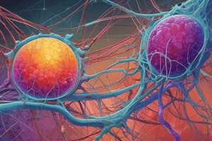
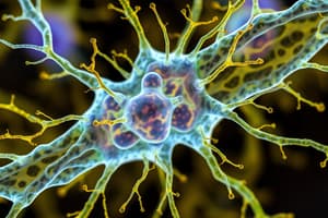
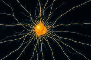
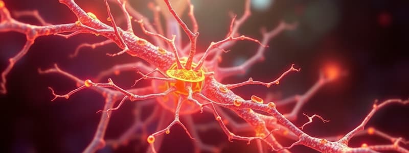
Cytoskeleton
Cytoskeleton
A network of protein filaments throughout the cytoplasm of eukaryotic cells.
Cytoskeleton's structural role
Cytoskeleton's structural role
Provide structural support and determine cell shape (Actin, MT, IF).
Cytoskeleton's organizational role
Cytoskeleton's organizational role
Internal organization of cell (MT) such as organelles and vesicle transport.
Cytoskeleton's role in cell division
Cytoskeleton's role in cell division
Signup and view all the flashcards
Cytoskeleton - Large Scale movements
Cytoskeleton - Large Scale movements
Signup and view all the flashcards
Cytoskeletal filaments
Cytoskeletal filaments
Signup and view all the flashcards
Fluorescence Microscopy
Fluorescence Microscopy
Signup and view all the flashcards
Transmission electron microscope
Transmission electron microscope
Signup and view all the flashcards
Immunofluorescence microscopy
Immunofluorescence microscopy
Signup and view all the flashcards
Three types of filaments
Three types of filaments
Signup and view all the flashcards
Actin Filaments
Actin Filaments
Signup and view all the flashcards
Intermediate Filaments
Intermediate Filaments
Signup and view all the flashcards
Microtubules
Microtubules
Signup and view all the flashcards
Cytoplasmic Intermediate Filaments
Cytoplasmic Intermediate Filaments
Signup and view all the flashcards
Cytoplasmic IF functions
Cytoplasmic IF functions
Signup and view all the flashcards
Nuclear IFs
Nuclear IFs
Signup and view all the flashcards
Cytoplasmic IF proteins
Cytoplasmic IF proteins
Signup and view all the flashcards
How Filaments held together
How Filaments held together
Signup and view all the flashcards
Keratin Filaments anchored
Keratin Filaments anchored
Signup and view all the flashcards
Microtubules - Role
Microtubules - Role
Signup and view all the flashcards
Microtubules involvement
Microtubules involvement
Signup and view all the flashcards
Microtubules
Microtubules
Signup and view all the flashcards
The structure of microtubules
The structure of microtubules
Signup and view all the flashcards
Microtubule polarity
Microtubule polarity
Signup and view all the flashcards
Microtubules growth
Microtubules growth
Signup and view all the flashcards
Microtubule Organizing Centers
Microtubule Organizing Centers
Signup and view all the flashcards
Microtubules in MTOCs
Microtubules in MTOCs
Signup and view all the flashcards
Microtubules are what?
Microtubules are what?
Signup and view all the flashcards
Microtubule organized
Microtubule organized
Signup and view all the flashcards
MAP
MAP
Signup and view all the flashcards
Microtubules can be stablilized if?
Microtubules can be stablilized if?
Signup and view all the flashcards
Motor proteins
Motor proteins
Signup and view all the flashcards
Kinesins Motor proteins
Kinesins Motor proteins
Signup and view all the flashcards
Dyneins Motor proteins
Dyneins Motor proteins
Signup and view all the flashcards
Motor proteins job
Motor proteins job
Signup and view all the flashcards
Microtubules job to help
Microtubules job to help
Signup and view all the flashcards
Actin filaments are,
Actin filaments are,
Signup and view all the flashcards
Actin monomers are,
Actin monomers are,
Signup and view all the flashcards
Actin filaments made of
Actin filaments made of
Signup and view all the flashcards
Actin filament has,
Actin filament has,
Signup and view all the flashcards
growth is what?
growth is what?
Signup and view all the flashcards
Actin monomers are what?
Actin monomers are what?
Signup and view all the flashcards
Plus
Plus
Signup and view all the flashcards
Cell crawling,
Cell crawling,
Signup and view all the flashcards
Motor proteins generally move
Motor proteins generally move
Signup and view all the flashcards
Study Notes
Cytoskeleton Overview
- The cytoskeleton consists of protein filaments within the cytoplasm.
- The cytoskeleton is highly dynamic and performs a number of important functions within eukaryotic cells
- The cytoskeleton is made up of Actin Filaments, Intermediate Filaments and Microtubules
Major Functions
- The cytoskeleton provides structural support, which helps to determine cell shape.
- The cytoskeleton provides internal organization by positioning organelles.
- The cytoskeleton facilitates in vesicle transport.
- Cell division relies on the cytoskeleton for chromosome segregation and overall cell division.
- The cytoskeleton contributes to large scale movements, like cell crawling and muscle contraction.
Microscopy Techniques
- Cytoskeletal filaments range from 7 nm to 25 nm in diameter.
- Light microscopes have a resolution limit of ~200 nm, constrained by the wavelength of visible light, making it impossible to resolve cytoskeletal filaments.
- Fluorescence microscopy uses a light microscope with the same resolution, however fluorescent labels are used to detect specific proteins, like cytoskeletal filaments.
- Transmission electron microscopes utilize beams of electrons to see structures with a short wavelength.
- This gives TEM microscopes a resolution of ~1 nm, capable of revealing detailed structures.
Immunofluorescence microscopy
- Immunofluorescence microscopy helps to specify the location of proteins.
- This type of microscopy is only compatible with cells that have been fixed, not living cells.
- This uses Primary and Secondary antibodies to bind to a specific protein
- Secondary antibodies are covalently tagged to a fluorescent marker.
- Fluorescence microscopes are used to excite the flourescent marker and visualise the light that has been emmitted.
Three Types of Filaments
- Actin filaments, with a diameter of ~7 nm, are one type of cytoskeletal filament.
- Intermediate filaments, with a diameter of ~10 nm, are another type of cytoskeletal filament.
- Microtubules, with a diameter of 25 nm, are a third type of cytoskeletal filament.
- All filaments are held together by noncovalent interactions.
Intermediate Filaments
- Intermediate filaments provide structural support, and have different types of IF proteins.
- Cytoplasmic IFs occur in animal cells subjected to mechanical stress and provide mechanical strength.
- Nuclear IFs include the nuclear lamina, which is a 2D meshwork consisting of lamins.
Cytoplasmic Intermediate Filaments
- Cytoplasmic IF proteins have a conserved α-helical central rod domain and N- and C-terminal domains that differ.
- Cytoplasmic IF pack together into rope-like filaments.
- 2 monomers form a coiled-coil dimer
- 2 dimers form staggered antiparallel tetramer
- 8 tetramers associate side by side and assemble into a filament
- Most interactions are noncovalent
- Cytoplasmic IF do not have filament polarity.
- Cytoplasmic IFs are tough, flexible, and have high tensile strength.
- Keratin filaments in epithelial cells create an interconnected network throughout the cytoplasm and are anchored at cell-cell junctions (desmosomes).
- Epithelium is a sheet of cells that cover external surfaces
Microtubules
- Microtubules' organizing function is present in all eukaryotes.
- Microtubules are involved in cell organization, including vesicle and organelle transport/positioning.
- Microtubules are involved in mitosis, and contribute to the structure of cells and structures like flagella.
- Microtubules are made of tubulin, forming long and stiff hollow tubes.
- They are inextensible (don't extend).
Microtubule Structure
- Microtubules are made of two related globular proteins called Alpha-tubulin and Beta-tubulin
- These form tubulin heterodimers that are bound to GTP.
- Alpha and Beta subunits are arranged for microtubule polarity, where the end (β) is different from the minus end (α).
- 13 parallel protofilaments make up a hollow tube.
Microtubule Protofilaments
- Protofilaments are bonded noncovalently
- Protofilament bonds are weaker than bonds within each protofilament.
- Microtubules can grow and disassemble at both ends, however this is faster at the plus end.
Microtubule Organising Centres (MTOC)
- Within the cell, microtubules grow out from MTOCs, for example the centrosome in animal cells.
- Ends are stabilised at the MTOC
- The plus ends of microtubules grow and shrink
- This continuous process needs remodeling
- These plus ends of microtubules can then grow or shrink
- MTOCs contribute to dynamic instability for microtubules
- The y-Tubulin Ring Complex (γ-TuRC) is an example of nucleation site.
- γ-TuRC is a protein complex of y-tubulin and accessory proteins, and ring of y-tubulin (gold) acts as an attachment site for αβ-tubulin dimers
Microtubule Dynamics
- The dynamic instability of microtubules is required in order to remodel.
- Microtubules in non-dividing animal cells radiate from one centrosome.
- In dividing animal cells, the centrosome duplicates to form two spindle poles which reorganise to form a bipolar mitotic spindle.
- Microtubule-associated proteins regulate the stability, dynamics, and organization of microtubules.
Microtubules & Transport
- Microtubules can be stabilized to prevent disassembly.
- Microtubules facilitate cargo transport in nerve cells (from the cell body to the axon terminal).
- Motor proteins on microtubules facilitate in axonal transport.
Motor Proteins and Intracellular Transport
- Kinesins move towards microtubules' plus end.
- Kinesin I transports organelles, vesicles, and macromolecules towards the axon terminus.
- Dyneins move towards microtubules' minus end.
- Cytoplasmic dynein transports worn-out mitochondria towards the cell body.
Organelle Positioning
- Microtubules are used to position organelles in cells.
- For example ER has kinesin-1 near to the nuclear envelope.
- Another example is the golgi which is positioned near to the centrosome by cytoplasmic dynein.
Actin Filaments
- Actin filaments are known as microfilaments and are present in all eukaryotes.
- Actin filaments are made of actin monomers, and are flexible but inextensible.
- Myosins are the motor proteins associated with actin filaments.
- Actin filaments contribute to stiff stable structures like microvilli or cell motility.
Actin Filaments Structure
- Actin filaments are usually 7nm in diameter
- Actin consists of:
- actin monomers
- noncovalent interactions
- two protofilaments twisted in a right direction
- Actin filament has polarity
- plus end is different from the minus end
- actin monomers are all in the same orientation in each protofillament
- growth is faster at the plus end of actin
Actin Monomers
- Free Monomers are bound to ATP
- Bound in the center of the protein
- Soon after the actin monomer has been added to the filament
- Actin hydrolyses ATP to ADP
- This reduces strength of binding between monomers in filament
- Rapid addition of Actin monomers
- faster than ATP hydrolysis in new ly added actin monomers
- Actin filament has an ATP cap
Actin Polymerisation in Test Tube
- This consists of
- Actin subunits and ATP added to a test tube
- In order to study actin filament polymeristaion
- Test tube also contains Nucleation - lag phase
- Small oligomers form, but are unstable
- Includes Elongation - growth phase
- some oligomers become more stable, leads to rapid filament elongation (faster at plus end)
- Can also consist of stabilised actin -Steady state (equilibrium phase) • decrease in [actin subunits] • rate of subunit addition = rate of subunit dissassociation Treadmilling is the rate of the sub unit
Actin Filaments and Treadmilling
- This can consist of ATP as a building block of actin
- addition of actin monomers - polymerization
- actin monomers quickly hydrolyse to ADP
- Then will lose an actin monomer to be depolymerised
- this needs a continual supply of ATP building blocks
- which can then produce the same size and looks stable
- This means that the continues exchange of monomers at each end
- Net addition at at the plus end
- net loss at the minus end
- this process moves throughout the filament
- This means that the continues exchange of monomers at each end
- which can then produce the same size and looks stable
- Actin filaments must rapidly assemble at the leading edge
- this pushes the cell forward
Comparing Actin Filaments and Microtubules
- Actin Filaments
- Composed of ATP
- Have subunits that are connected after hydrolysis
- Have a smaller rate of synthesis compared to microtubules
- Mictotubules
- Composed of GTP and a complex molecule chain
- Sub Units Connected After Hydrolisis
- Composed of GTP and a complex molecule chain
- Both Have a good rate of synthesis
Cellular Structures & The Environment;
- This includes the next set of lessons and studies
Successfully Learning
- The student should understand the differences in microscope,
- the difference in different filament properties
- Describe the properties in the cytoskeleton and if filaments that form
- Describe how they perform - Dynamic instabilty
- Describe and Understand them.
- Understand and describe motor proteins and if actin filaments and how they move.
Studying That Suits You
Use AI to generate personalized quizzes and flashcards to suit your learning preferences.