Podcast
Questions and Answers
Which type of reconstruction algorithm is not typically used in image reconstruction?
Which type of reconstruction algorithm is not typically used in image reconstruction?
- Iterative technique
- Forward projection (correct)
- Back projection
- Filtered back projection
What is a key characteristic of prospective reconstruction?
What is a key characteristic of prospective reconstruction?
- Creates lower quality images
- Produced during scanning (correct)
- Requires post-processing
- Uses previously acquired raw data
What does DFOV stand for in CT imaging?
What does DFOV stand for in CT imaging?
- Digital Field Of View (correct)
- Deferred Field Of View
- Diameter Field Of View
- Dynamic Field Of View
Which component is NOT part of the image display process?
Which component is NOT part of the image display process?
Which type of voxel has equal dimensions in all three axes?
Which type of voxel has equal dimensions in all three axes?
What image reformatting technique is specifically designed to display anatomy in various planes?
What image reformatting technique is specifically designed to display anatomy in various planes?
In which method is the geometric midpoint of the reconstructed CT image designated?
In which method is the geometric midpoint of the reconstructed CT image designated?
Which of the following 3D image formatting techniques focuses on displaying the maximum intensity across pixels?
Which of the following 3D image formatting techniques focuses on displaying the maximum intensity across pixels?
What is the first step in the computed tomography (CT) image formation process?
What is the first step in the computed tomography (CT) image formation process?
What does the term 'projection' refer to in CT imaging?
What does the term 'projection' refer to in CT imaging?
Which geometries are used in the projections of CT imaging?
Which geometries are used in the projections of CT imaging?
What is involved in the image reconstruction process in CT imaging?
What is involved in the image reconstruction process in CT imaging?
How are the matrix elements obtained during image reconstruction?
How are the matrix elements obtained during image reconstruction?
What is the role of the detector in the data acquisition step of CT imaging?
What is the role of the detector in the data acquisition step of CT imaging?
What transformation occurs to the X-ray beam as it passes through the body?
What transformation occurs to the X-ray beam as it passes through the body?
What type of processor is typically used for calculations in image reconstruction?
What type of processor is typically used for calculations in image reconstruction?
What happens to image quality when isotropic voxels are reformatted?
What happens to image quality when isotropic voxels are reformatted?
What is Curved Planar Reformation (cMPR) used for?
What is Curved Planar Reformation (cMPR) used for?
Which of the following is NOT one of the algorithms used in the Sliding Thin Slabs technique?
Which of the following is NOT one of the algorithms used in the Sliding Thin Slabs technique?
What do Maximum Intensity Projection (MIP) images primarily show?
What do Maximum Intensity Projection (MIP) images primarily show?
What is a significant limitation of the Maximum Intensity Projection technique?
What is a significant limitation of the Maximum Intensity Projection technique?
What does Average Intensity Projection (AIP) help in characterizing?
What does Average Intensity Projection (AIP) help in characterizing?
In which imaging scenario is Maximum Intensity Projection (MIP) particularly useful?
In which imaging scenario is Maximum Intensity Projection (MIP) particularly useful?
What characteristic does an AIP image represent?
What characteristic does an AIP image represent?
Flashcards
Iterative Reconstruction
Iterative Reconstruction
A type of image reconstruction that uses a mathematical approach to build an image from multiple projections taken from different angles.
Back Projection
Back Projection
A simple image reconstruction method that projects the data back onto the image matrix, resulting in a blurry image.
Filtered Back Projection
Filtered Back Projection
An improvement on back projection, where the projected data is filtered to reduce the blurriness, resulting in a sharper image.
Prospective Reconstruction
Prospective Reconstruction
Signup and view all the flashcards
Retrospective Reconstruction
Retrospective Reconstruction
Signup and view all the flashcards
Image Display
Image Display
Signup and view all the flashcards
Image Reformation
Image Reformation
Signup and view all the flashcards
Raw Data
Raw Data
Signup and view all the flashcards
Computed Tomography (CT) Image Formation
Computed Tomography (CT) Image Formation
Signup and view all the flashcards
CT Data Acquisition
CT Data Acquisition
Signup and view all the flashcards
CT Beam Geometry
CT Beam Geometry
Signup and view all the flashcards
CT Image Reconstruction
CT Image Reconstruction
Signup and view all the flashcards
Attenuation of X-rays
Attenuation of X-rays
Signup and view all the flashcards
X-ray Transmission and Detection
X-ray Transmission and Detection
Signup and view all the flashcards
Projection Data Set
Projection Data Set
Signup and view all the flashcards
Multiplanar Reformation (MPR)
Multiplanar Reformation (MPR)
Signup and view all the flashcards
Isotropic Voxels
Isotropic Voxels
Signup and view all the flashcards
Anisotropic Voxels
Anisotropic Voxels
Signup and view all the flashcards
Curved Planar Reformation (CPR)
Curved Planar Reformation (CPR)
Signup and view all the flashcards
Maximum Intensity Projection (MIP)
Maximum Intensity Projection (MIP)
Signup and view all the flashcards
Average Intensity Projection (AIP)
Average Intensity Projection (AIP)
Signup and view all the flashcards
Overlapping Images in MPR
Overlapping Images in MPR
Signup and view all the flashcards
Minimum Intensity Projection (MinIP)
Minimum Intensity Projection (MinIP)
Signup and view all the flashcards
Study Notes
Computed Tomography (CT)
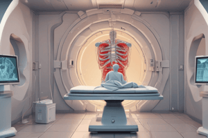
- CT is a medical imaging technique
- CT images are developed from multiple X-ray absorption measurements
- CT images show thin slices of the body
- CT image formation is a three-step process:
- Scan (or Data Acquisition): X-ray tube scans the patient systematically, collecting sufficient data for reconstruction. The sum of X-ray transmission through each detector yields a projection. Multiple projections are taken at various angles.
- Image Reconstruction: Using complex computer algorithms, mathematical equations, and physics, raw data is processed to generate a slice image.
- Image Display: Electrical signals from the image reconstruction are converted into a visual image on a monitor. Different shades of gray represent different CT numbers.
CT Image Formation: Geometries
- Two projection geometries are used in CT imaging:
- Parallel beam geometry: X-rays travel parallel to one another.
- Fan beam geometry: In this geometry, X-rays diverge from a source point at varying angles in a fan-like pattern.
- Cone beam geometry
Image Reconstruction
- The process uses raw data to create an image.
- The process uses complex computer algorithms, mathematical equations, and physics
- When the source-detector makes a sweep across the patient, X-ray beam attenuation varies based on tissue density and effective atomic number (Z), producing projections
- The intensity profile or projection is calculated; the values are stored digitally
- The computer processes projections to reconstruct an image of the anatomical structure.
- The individual values of the matrix elements are obtained by solving simultaneous equations.
- The matrix represents cross-sectional anatomy.
- Dedicated array processors are used for fast calculations and display.
- Common algorithms in image reconstruction include iterative techniques, back projection, and filtered back projection.
Image Reconstruction: Types
- Prospective reconstruction: Image generation during the scan
- Retrospective reconstruction: Data from previous scans analyzed to create a new image
Image Display
- Image display components convert digital data into electrical signals for the CT monitor.
- Images are displayed on a cathode ray tube (CRT) or flat-panel displays (e.g., TFT LCD)
- Each CT number is assigned a shade of gray (typically 256 shades).
Image Reformation (Post-Processing)
- Image post-processing technique for CT
- Raw Data: Initial measurements (projections) from the CT scanner during patient scan.
- Image Data: Processed, reconstructed form of raw data from applying mathematical transformations.
- DFOV (zoom/target): determines the portion of raw data used to create the image.
Image Reformation: Parameters
- Field of view (FOV): Geometric midpoint of the reconstructed CT image.
- Anisotropic voxel: Voxels with unequal dimensions (often with thinner slices in one dimension).
- Isotropic voxel: Voxels with equal dimensions in all three axes (x, y, z). Facilitates 3D reconstructions.
CT Image Reformatting Techniques
- Two-dimensional formats: Multiplanar reformation (MPR), Curved planar reformation (CPR)
- Three-dimensional formats: Surface shaded displays (SSD), Volume rendering (VR), Average intensity projection (AIP), Maximum intensity projection (MIP), Minimum intensity projection (MinIP), Endoluminal imaging.
Multiplanar Reformation (MPR)
- Reformats anatomical structures; generates multiplanar images from axial scans
- Ideal for 2D viewing of anatomy.
- Produces coronal, sagittal, and paraxial views from axial data.
- Using isotropic voxel, produces virtually identical image quality to original axial images.
- Anisotropic voxels can be improved with overlapping images.
Curved Planar Reformation(CPR)
- Reformats anatomical data along the centerline of tubular structures, including vessels.
- Used for visualizing tubular structures effectively.
Intensity Projection Rendering
- Thin-slice algorithms like AIP, MIP, and MinIP are used in this technique.
Average Intensity Projection (AIP)
- Combines average attenuation values within voxels
- Useful for structures with low contrast, such as a solid body or walls of hollow structures (blood vessels/intestines).
Maximum Intensity Projection (MIP)
- Shows only the highest attenuation values within voxels
- Enables detection of high-intensity structures
- Used in angiographic and urographic images
- Limited view of 3D relationships in a volume.
Minimum Intensity Projection (MinIP)
- Shows only lowest attenuation values
- Identifies low-density structures (e.g., air-containing structures)
- Aids in diagnosing diseases, like bronchiectasis and emphysema.
Studying That Suits You
Use AI to generate personalized quizzes and flashcards to suit your learning preferences.


