Podcast
Questions and Answers
In the context of computed tomography (CT), what does the term 'isotropic resolution' refer to, and what is its significance?
In the context of computed tomography (CT), what does the term 'isotropic resolution' refer to, and what is its significance?
- It refers to the ability to differentiate between tissues with similar densities, and its significance lies in improved contrast detail.
- It refers to the speed at which the CT scanner can acquire data, reducing the likelihood of motion artifacts.
- It refers to the intensity of radiation emitted during the scan, improving signal-to-noise ratio.
- It refers to the uniformity of voxel dimensions (x, y, and z) within the imaging volume, enhancing image detail in all planes. (correct)
According to Faraday's law of induction, what is the relationship between electronic current traveling along a wire loop and the generated magnetic field?
According to Faraday's law of induction, what is the relationship between electronic current traveling along a wire loop and the generated magnetic field?
- The magnetic field is generated perpendicularly to the current flow and is proportional to its strength. (correct)
- The magnetic field is generated parallel to the current flow and is proportional to the square of its strength.
- The magnetic field is generated parallel to the current flow and is inversely proportional to its strength.
- The magnetic field is generated randomly in relation to the current flow and its strength.
Which imaging sequence does not use inversion recovery?
Which imaging sequence does not use inversion recovery?
- STIR
- T1-weighted FLAIR
- T2-weighted FLAIR
- SPGR (correct)
For computed tomography and magnetic resonance imaging, which typically represents the limiting spatial resolution?
For computed tomography and magnetic resonance imaging, which typically represents the limiting spatial resolution?
During the image reconstruction process, which step determines the level of edge reinforcement?
During the image reconstruction process, which step determines the level of edge reinforcement?
What limits the appreciation of all shades of gray measured by the CT scanner?
What limits the appreciation of all shades of gray measured by the CT scanner?
Which one does not influence the magnitude of the equilibrium magnetization of tissues in the MR magnet?
Which one does not influence the magnitude of the equilibrium magnetization of tissues in the MR magnet?
What parameter settings would result in T2-weighting?
What parameter settings would result in T2-weighting?
T1 relaxation occurs when:
T1 relaxation occurs when:
What technological advance allowed helical scanning with computed tomography?
What technological advance allowed helical scanning with computed tomography?
What are the principal advantages of computed tomography over conventional radiography?
What are the principal advantages of computed tomography over conventional radiography?
Flashcards
Tomographic Imaging
Tomographic Imaging
Imaging technique using thin sections or slices, eliminating superimposition.
Contrast Resolution
Contrast Resolution
Ability to distinguish subtle differences in tissue characteristics, displayed as different shades of gray.
Spatial Resolution
Spatial Resolution
Minimum resolvable separation between high-contrast objects.
Volume Dataset Reformation
Volume Dataset Reformation
Signup and view all the flashcards
CT Advantages
CT Advantages
Signup and view all the flashcards
Elimination of Superimposition
Elimination of Superimposition
Signup and view all the flashcards
MR Imaging Contrast
MR Imaging Contrast
Signup and view all the flashcards
Tomographic Advantage
Tomographic Advantage
Signup and view all the flashcards
Isotropic Resolution
Isotropic Resolution
Signup and view all the flashcards
Image Detail
Image Detail
Signup and view all the flashcards
Attenuation Value
Attenuation Value
Signup and view all the flashcards
Multiplanar Reformatting (MPR)
Multiplanar Reformatting (MPR)
Signup and view all the flashcards
Voxel-to-Pixel Transmission
Voxel-to-Pixel Transmission
Signup and view all the flashcards
Contrast Resolution (CR)
Contrast Resolution (CR)
Signup and view all the flashcards
Spatial Resolution (SR)
Spatial Resolution (SR)
Signup and view all the flashcards
Radon's Principles in CT
Radon's Principles in CT
Signup and view all the flashcards
Spatial Resolution (CT vs. MR)
Spatial Resolution (CT vs. MR)
Signup and view all the flashcards
CT Imaging
CT Imaging
Signup and view all the flashcards
Thin section of the body.
Thin section of the body.
Signup and view all the flashcards
Voxels
Voxels
Signup and view all the flashcards
Pixels
Pixels
Signup and view all the flashcards
Pixel Brightness
Pixel Brightness
Signup and view all the flashcards
Voxel Value Display
Voxel Value Display
Signup and view all the flashcards
Slice Thickness
Slice Thickness
Signup and view all the flashcards
CT Angiography
CT Angiography
Signup and view all the flashcards
MR Imaging Purpose
MR Imaging Purpose
Signup and view all the flashcards
Magnet Role in MRI
Magnet Role in MRI
Signup and view all the flashcards
Magnet Performance Factors
Magnet Performance Factors
Signup and view all the flashcards
Faraday's Law in MRI
Faraday's Law in MRI
Signup and view all the flashcards
Contrast Media Role
Contrast Media Role
Signup and view all the flashcards
Phases in CT Angiography
Phases in CT Angiography
Signup and view all the flashcards
3D Volume Rendering
3D Volume Rendering
Signup and view all the flashcards
CT System Components
CT System Components
Signup and view all the flashcards
X-ray Attenuation
X-ray Attenuation
Signup and view all the flashcards
Hounsfield Units (HU)
Hounsfield Units (HU)
Signup and view all the flashcards
HU Values Above/Below Zero
HU Values Above/Below Zero
Signup and view all the flashcards
HU Measurement Range
HU Measurement Range
Signup and view all the flashcards
Multidetector CT
Multidetector CT
Signup and view all the flashcards
Larmor Equation
Larmor Equation
Signup and view all the flashcards
Reconstruction Algorithm
Reconstruction Algorithm
Signup and view all the flashcards
FLAIR
FLAIR
Signup and view all the flashcards
Susceptibility Artifact
Susceptibility Artifact
Signup and view all the flashcards
Fat Suppression
Fat Suppression
Signup and view all the flashcards
Magic Angle Effect
Magic Angle Effect
Signup and view all the flashcards
Motion Artifact
Motion Artifact
Signup and view all the flashcards
Pseudolesion
Pseudolesion
Signup and view all the flashcards
Pseudolayering Artifact
Pseudolayering Artifact
Signup and view all the flashcards
Parallel Imaging
Parallel Imaging
Signup and view all the flashcards
Study Notes
- CT and MR imaging are crucial for veterinary diagnoses.
- Understanding their principles helps optimize use and recognize limitations.
Computed Tomography and Magnetic Resonance Imaging in Veterinary Practice
- CT and MR offer superior diagnostics over radiography due to their tomographic nature, and increased contrast resolution.
- CT and MR images are thin sections or slices, eliminating superimposition.
- Volume datasets from CT and MR can be reformatted in any plane or as 3D projections for better anatomical representation.
- Contrast resolution is how well a system represents differences in tissue characteristics.
- Better contrast resolution means more subtle tissue variations are displayed as different shades of gray pixels.
- CT can detect smaller differences in x-ray attenuation compared to radiographs, explained by scatter reduction, and more sensitive x-ray detectors.
- CT differentiates pure liquids from soft tissues, unlike radiographs.
- MR excels in contrast resolution, enhanced by combining different imaging sequences.
- Spatial resolution, is the minimum separation between high-contrast objects, and is more limited in CT and MR compared to radiography.
- CT and MR spatial resolution is linked to slice thickness, approximately 0.3 and 1.0 mm respectively.
- Technical advances have contributed to reducing the value of the magnetic resolution.
Image Formation: General Concepts
- CT and MR both produce images of thin sections or slices composed of voxels, or volume elements.
- Images are displayed on a flat monitor as a matrix of pixels, or picture elements.
- A voxel with greater tissue signal intensity in MR or x-ray attenuation in CT is shown as a brighter pixel.
- The mean value within each voxel is converted to pixel brightness, even if multiple values coexist.
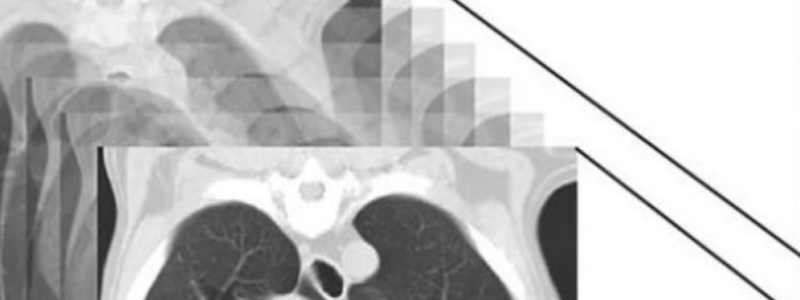
- Slice thickness limits CT and MR imaging, and is typically the largest dimension of each voxel.
- Scanners aim for isotropic resolution, imaging tissue sections with perfectly cubical voxels.
- Multidetector row scanners nearly achieve isotropic resolution, allowing submillimeter slice generation.
- Image detail is enhanced in all planes with the thinner resolution.
- Image sections can be acquired in any plane with MR imaging, whereas CT images are typically transverse due to system gantry limitations.
- CT sections are scanned contiguously, forming a volume dataset of millions of tiny cubes, each with an attenuation value.
- CT images can be rearranged so voxels along a different plane are displayed as a new pixel matrix, known as multiplanar reformatting (MPR).
- MPR images help recognize structures especially tubular or tortuous ones, as well as assess normal and abnormal tissues.
- Imaging software generates 3D representations through volume or surface rendering for understanding morphologic changes and planning surgery.
Computed Tomography
- G. N. Hounsfield and A. M. Cormack won the 1979 Nobel Prize in medicine for developing CT imaging.
- CT imaging has significantly expanded diagnostic applications, while improving comfort and reducing motion artifacts.
- CT technology is based on Radon's mathematic principles developed in 1917.
- The principle shows an image of an object can be produced using an infinite number of projections.
- Thousands of radiographic projections can be performed at precise angles.
- X-ray transmission through tissues can be quantified for each projection.
- A sophisticated computer geometrically reconstructs data and assigns x-ray transmission values to body regions.
- Thinly collimated x-ray beams rotate around a patient.
- Slices of tissue are computer-generated, each with a voxel matrix and a definite attenuation value.
- Modern systems can acquire slices in fractions of a second.
Computed Tomography System Geometry
- A CT system includes:
- A scanning unit (gantry) with a rotating x-ray emitting tube and a detector system
- A patient table
- A console with a computer for adjusting acquisition parameters and image reconstruction.
- Rotating x-ray tubes are powerful enough to operate for long periods without overheating, enabling the acquisition of large data volumes.
- Helical scanning systems rapidly image a large body region without interruption.
- Rapid tube rotation and table advancement trace a helical path around the patient's body.
- Flat slices of cubical voxels can be reconstructed from any part of the dataset using interpolation methods.
- Fast scanners reduce motion artifact and allow precise angiographic procedures with contrast-medium bolus tracking.
- Advanced scanners rapidly acquire sub-millimeter slices, and produce smoother images in multiplanar (MP) and 3D reformatting.
- Detector systems convert incident x-rays into electronic signals, which are key to image quality.
- Ceramic, solid-state detectors offer better x-ray absorption and are used in current scanners.
- Each detector has a scintillation crystal that reacts with incident x-rays, generating light converted into digital pulses.
- Detectors are positioned over semicircular or annular arrays, depending on system geometry.
- Annular arrays are stationary, semicircular arrays move with the opposing x-ray tube.
- CT scanners with multidetector row configurations are standard in veterinary hospitals.
- These units acquire multiple thin contiguous slices simultaneously, which is a important upgrade over previous single-row detectors.
- 1-mm-thick slices of a 30-cm thorax may require only a few seconds with a 64-slice scanner, compared to minutes with a single-row scanner.
- Recent systems have faster computers that rapidly reconstruct and provide images.
- Multidetector row detectors decrease motion artifacts and tissue misregistration, and use radiation from the x-ray tube more efficiently.
- Interscan delays are reduced with multidetector row systems, allowing angiographic procedures with multiple series in a short period, such as for portosystemic shunt detection.
- The system reconstructs data into images as the tube rotates around the patient, it begins with understanding x-ray attenuation, which depends on electron density.
- X-ray attenuation is quantified measuring the portion of radiation removed in passing through a given thickness of material and is based on the linear attenuation coefficient (µ).
- For image purposes, x-ray attenuation values are expressed as Hounsfield units (HU), normalized to the x-ray attenuation of water (HU = zero).
- Tissues causing more x-ray attenuation have a HU value above zero, whereas tissues causing less attenuation have a HU value less than zero (negative).
- Liquids and soft tissues can be discriminated using their HU values, displaying greater contrast resolution of CT over radiography.
- Pre-set parameters before image reconstruction include the adjustment of overlap between adjacent slices of tissues used and the reconstruction filter.
- Reconstruction filters determine edge reinforcement.
- A low-pass filter, recommended when soft tissue contrast must be emphasized, creating a blurrier image.
- "Bone filters" maximize spatial resolution, but reduce contrast resolution, creating sharper but grainier images.
- Intermediate pass filtration is for smaller soft tissue areas to prevent blurring.
- Reconstruction filters must be selected based on the size of the body part and the content to be emphasized.
- HU values from typical scanners range from -1000 to +3095 HU, which is a total of 4096 shades of gray (12 bits of grayscale).
- The human cannot differentiate these values.
- Use window width (W) and level (L) of the image grayscale, per the median and range composing the area of interest.
- Digital Imaging and Communications in Medicine (DICOM) viewing software adjusts the displayed gray shades and the HU at the window center or contrast.
- W/L must be adapted to tissues and usually requires several adjustments.
- Understanding radiographic opacities and anatomy aids in interpreting CT images.
- Tissue attenuation characteristics are assessed qualitatively and quantitatively using DICOM viewing software to precisely measure a selected number of pixels.
Contrast-Enhanced Procedures
- Iodinated contrast medium is standard for CT procedures.
- Hyperattenuating contrast distribution provides data on vascular perfusion as well as the integrity of natural barriers.
- For nonangiographic postcontrast scans, they happen a few minutes following bolus injection.
- Tissue HU content will increase in proportion to the concentration of the contrast medium.
Cone-Beam Computed Tomography
- Cone-beam CT is increasingly used due to low cost equipment and installation.
- Cone-beam CT includes an x-ray source, image detector, a computer and an image.
- The system utilizes a cone-shaped x-ray beam instead of a collimated fan beam.
- Using a cone-beam reduced the radiation dose, but increases scatter radiation.
- Instead of aligned row detectors like flat panels or image intensifiers in cone beam CT technology are used and rotate synchronously through 360 degrees.
- Helpful in the assessment of heads and dental structures in dogs and cats with high spatial revolution.
Magnetic Resonance Imaging
- MR imaging benefits the veterinary industry for it's high contrast resolution quality.
- Centers around the electromagnetic properties of hydrogen nuclei (protons), for it is abundant in tissues.
- Energy transfer is localized spatially and is the source of image formation.
- Map the distribution of H protons in water (H2O) and lipid (CH2/CH3).
- MR imaging is mainly used to investigate neurologic structure and the horse, can be imaged.
Instrumentation
- MR combines electronics, RF generators, coils and gradients that interact with a computer.
- Provides proper tissue excitation, signal localization, and recognition.
- A magnet provides the external magnetic field.
- Performances increase through its strength, stability, and uniformity.
- Illustrated components of the MR system are shown.
Main Magnetic Field and Radiofrequency Energy
- Per Faraday's law, whenever electronic current travels through a loop of wire, a magnetic field is generated.
- Fundamentally how MR scanners work mainly with high field strength system by maintaining high magnetic field strength and superconducting wires which uses a cooling agent agent such as helium.
Spins, Excitation, and Relaxation
- Each hydrogen proton is positively charged (H+) and spins, similarly to a spinning top.
- Individually, the tiny magnets are oriented randomly.
- Placed in a strong magnetic field, spinning protons forces protons to either be parallel or antiparallel.
- Protons cancel out, but a excess proton amounts, which is proportional that's parallel, and magnetic flow that's along the axis.
- size depends on he photon density and the external Bo
- Wobbles around behavior called pression and this motion is controlled by application of an external force.
- The frequency proportional constant specific 42.6 MHz/T
- Each protons moment has an component whether aligned or longitudinal to the xy-plane in positive magnetization.
- Apply RF pulse by matching the Larmor frequency of the protons by causing a state of imbalance.
- causes a stream of energy (parallel to antiparallel) and away from the xyz.
- Angle is called the flip angle.
- change results by a difference.
Tissue Contrast
- Manipulate tissue for images through diagnostic potential of MR imaging.
- To improve the contrast, one must enhance anatomic by adjusting TE, so that anatomical differences can be measured among structures.
- One can adjust the imaging to identify tissue structures from T1 and T2.
- Optimizing the image helps determine how the image will appear in a particular.
- T1 weighted requires short TR
- T2 Weighted images requires log TE if both results were limited
- PD effect signal intensity depending of the density of the weight of the tissue which are displayed though varying images.
Magic Angle Effect
- Magic angle effect are able to alter what intensity appears for ligaments.
Studying That Suits You
Use AI to generate personalized quizzes and flashcards to suit your learning preferences.