Podcast
Questions and Answers
Which cranial nerve is associated with the mnemonic 'Once'?
Which cranial nerve is associated with the mnemonic 'Once'?
- Olfactory
- Oculomotor
- Abducens
- Optic (correct)
What is the mnemonic device used for the cranial nerve VII?
What is the mnemonic device used for the cranial nerve VII?
- Final (correct)
- Very
- Anatomy
- The
Which cranial nerve is indicated by the mnemonic 'Good'?
Which cranial nerve is indicated by the mnemonic 'Good'?
- Optic
- Abducens
- Glossopharyngeal (correct)
- Vagus
What is the primary function of the afferent neurons in the PNS?
What is the primary function of the afferent neurons in the PNS?
Which mnemonic device corresponds to cranial nerve XI?
Which mnemonic device corresponds to cranial nerve XI?
Which part of the nervous system is responsible for the fight/flight/freeze response?
Which part of the nervous system is responsible for the fight/flight/freeze response?
What cranial nerve is represented by the phrase 'Very'?
What cranial nerve is represented by the phrase 'Very'?
In the context of the motor division of the PNS, what role do efferent neurons play?
In the context of the motor division of the PNS, what role do efferent neurons play?
What is the primary function of the parasympathetic nervous system?
What is the primary function of the parasympathetic nervous system?
Which of the following accurately describes the relationship between the PNS and CNS?
Which of the following accurately describes the relationship between the PNS and CNS?
Upon touching something hot, what is the immediate response driven by sensory receptors?
Upon touching something hot, what is the immediate response driven by sensory receptors?
What are the two main divisions of the peripheral nervous system?
What are the two main divisions of the peripheral nervous system?
What distinguishes the sympathetic nervous system's responses from those of the parasympathetic nervous system?
What distinguishes the sympathetic nervous system's responses from those of the parasympathetic nervous system?
What is the main role of the association fibers in the brain?
What is the main role of the association fibers in the brain?
Which area of the cerebral cortex is primarily responsible for conscious intellect, personality, and judgment?
Which area of the cerebral cortex is primarily responsible for conscious intellect, personality, and judgment?
What type of fibers connect gyri in opposite hemispheres?
What type of fibers connect gyri in opposite hemispheres?
What are the three layers of the cerebrum?
What are the three layers of the cerebrum?
Which lobe of the brain is primarily associated with memory?
Which lobe of the brain is primarily associated with memory?
What is the primary function of the cerebral cortex?
What is the primary function of the cerebral cortex?
In which area of the brain does sensory information recognition primarily occur?
In which area of the brain does sensory information recognition primarily occur?
Which type of fibers are responsible for transmitting nerve signals along the same hemisphere?
Which type of fibers are responsible for transmitting nerve signals along the same hemisphere?
What is the primary role of the central nervous system (CNS)?
What is the primary role of the central nervous system (CNS)?
Which division of the peripheral nervous system is responsible for voluntary motor control?
Which division of the peripheral nervous system is responsible for voluntary motor control?
What type of nerve fibers are involved in conducting impulses from receptors to the CNS?
What type of nerve fibers are involved in conducting impulses from receptors to the CNS?
What is the primary function of the cerebellum?
What is the primary function of the cerebellum?
Which anatomical structure is known as the 'horse's tail'?
Which anatomical structure is known as the 'horse's tail'?
The autonomic nervous system (ANS) can be divided into what two divisions?
The autonomic nervous system (ANS) can be divided into what two divisions?
What part of the central nervous system is primarily involved in regulating alertness and attention?
What part of the central nervous system is primarily involved in regulating alertness and attention?
Which of the following statements about the somatic nervous system is true?
Which of the following statements about the somatic nervous system is true?
The gray matter in the spinal cord is shaped like which letter?
The gray matter in the spinal cord is shaped like which letter?
What function does the sympathetic division of the ANS serve?
What function does the sympathetic division of the ANS serve?
What occurs when the reticular formation is inhibited?
What occurs when the reticular formation is inhibited?
Which type of fiber is responsible for conducting impulses from the CNS to smooth muscles?
Which type of fiber is responsible for conducting impulses from the CNS to smooth muscles?
What structures separate the spinal cord into right and left halves?
What structures separate the spinal cord into right and left halves?
What role do cranial and spinal nerves play in the peripheral nervous system?
What role do cranial and spinal nerves play in the peripheral nervous system?
The limbic system is primarily associated with which function?
The limbic system is primarily associated with which function?
The cerebrum is primarily associated with what function in the nervous system?
The cerebrum is primarily associated with what function in the nervous system?
Which part of the nervous system is involved in reflex actions?
Which part of the nervous system is involved in reflex actions?
What is the role of the central canal in the spinal cord?
What is the role of the central canal in the spinal cord?
Which function is NOT associated with the autonomic nervous system?
Which function is NOT associated with the autonomic nervous system?
What does damage to the reticular formation potentially lead to?
What does damage to the reticular formation potentially lead to?
What is the primary function of the sensory division of the PNS?
What is the primary function of the sensory division of the PNS?
What is the primary purpose of the folia in the cerebellum?
What is the primary purpose of the folia in the cerebellum?
Which structure anchors the conus medullaris of the spinal cord to the coccyx?
Which structure anchors the conus medullaris of the spinal cord to the coccyx?
Which structure is considered part of the central nervous system?
Which structure is considered part of the central nervous system?
Which of the following statements about the motor (efferent) division of the PNS is true?
Which of the following statements about the motor (efferent) division of the PNS is true?
Which of the following best describes the location of the cerebellum?
Which of the following best describes the location of the cerebellum?
What is a significant component of the Reticular Activating System (RAS)?
What is a significant component of the Reticular Activating System (RAS)?
Which cranial nerve is mainly associated with the forebrain?
Which cranial nerve is mainly associated with the forebrain?
Which cranial nerves are classified as purely sensory?
Which cranial nerves are classified as purely sensory?
What type of cranial nerves comprise the majority, being classified as mixed nerves?
What type of cranial nerves comprise the majority, being classified as mixed nerves?
Which of the following cranial nerves is associated with the midbrain?
Which of the following cranial nerves is associated with the midbrain?
Where are the cell bodies of motor neurons located in the cranial nerves?
Where are the cell bodies of motor neurons located in the cranial nerves?
Which cranial nerve is associated with the pons?
Which cranial nerve is associated with the pons?
Which cranial nerve is known for being mainly sensory, yet also has a motor component?
Which cranial nerve is known for being mainly sensory, yet also has a motor component?
What is the function of cranial nerves classified as 'mixed nerves'?
What is the function of cranial nerves classified as 'mixed nerves'?
Flashcards
Cerebral Cortex
Cerebral Cortex
The outermost layer of the cerebrum, responsible for higher cognitive functions like conscious thought, memory, and language.
Association Areas
Association Areas
Regions in the cerebral cortex that integrate and process information from different sensory areas, forming complex perceptions and memories.
Prefrontal Cortex
Prefrontal Cortex
The frontmost part of the frontal lobe, responsible for executive functions like planning, decision-making, and personality.
Tracts (White Matter)
Tracts (White Matter)
Signup and view all the flashcards
Association Tracts
Association Tracts
Signup and view all the flashcards
Commissural Tracts
Commissural Tracts
Signup and view all the flashcards
Gyrus
Gyrus
Signup and view all the flashcards
What connects corresponding gyri in opposite hemispheres?
What connects corresponding gyri in opposite hemispheres?
Signup and view all the flashcards
Nervous System (NS)
Nervous System (NS)
Signup and view all the flashcards
Peripheral NS (PNS)
Peripheral NS (PNS)
Signup and view all the flashcards
Afferent division
Afferent division
Signup and view all the flashcards
Efferent division
Efferent division
Signup and view all the flashcards
Autonomic NS - Sympathetic
Autonomic NS - Sympathetic
Signup and view all the flashcards
Autonomic NS - Parasympathetic
Autonomic NS - Parasympathetic
Signup and view all the flashcards
Receptors
Receptors
Signup and view all the flashcards
Effectors
Effectors
Signup and view all the flashcards
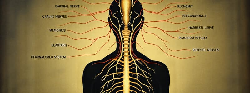
Cranial Nerves
Cranial Nerves
Signup and view all the flashcards
Olfactory Nerve
Olfactory Nerve
Signup and view all the flashcards
Optic Nerve
Optic Nerve
Signup and view all the flashcards
Oculomotor Nerve
Oculomotor Nerve
Signup and view all the flashcards
Trochlear Nerve
Trochlear Nerve
Signup and view all the flashcards
Cranial Nerves: Sensory Only
Cranial Nerves: Sensory Only
Signup and view all the flashcards
Cranial Nerves: Mainly Sensory
Cranial Nerves: Mainly Sensory
Signup and view all the flashcards
Cranial Nerves: Mixed
Cranial Nerves: Mixed
Signup and view all the flashcards
Motor Neuron Cell Bodies
Motor Neuron Cell Bodies
Signup and view all the flashcards
Sensory Neuron Cell Bodies
Sensory Neuron Cell Bodies
Signup and view all the flashcards
What are cranial nerve I and II?
What are cranial nerve I and II?
Signup and view all the flashcards
What is cranial nerve VIII?
What is cranial nerve VIII?
Signup and view all the flashcards
What are examples of mixed cranial nerves?
What are examples of mixed cranial nerves?
Signup and view all the flashcards
What is the nervous system?
What is the nervous system?
Signup and view all the flashcards
What are the two main divisions of the nervous system?
What are the two main divisions of the nervous system?
Signup and view all the flashcards
What does the central nervous system (CNS) consist of?
What does the central nervous system (CNS) consist of?
Signup and view all the flashcards
What is the role of the CNS?
What is the role of the CNS?
Signup and view all the flashcards
What does the peripheral nervous system (PNS) consist of?
What does the peripheral nervous system (PNS) consist of?
Signup and view all the flashcards
What is the role of the PNS?
What is the role of the PNS?
Signup and view all the flashcards
What are the two divisions of the PNS?
What are the two divisions of the PNS?
Signup and view all the flashcards
What is the role of the sensory/afferent division?
What is the role of the sensory/afferent division?
Signup and view all the flashcards
What is the role of the motor/efferent division?
What is the role of the motor/efferent division?
Signup and view all the flashcards
What are the two divisions of the motor/efferent division?
What are the two divisions of the motor/efferent division?
Signup and view all the flashcards
What is the role of the somatic nervous system?
What is the role of the somatic nervous system?
Signup and view all the flashcards
What is the role of the autonomic nervous system?
What is the role of the autonomic nervous system?
Signup and view all the flashcards
What are the two divisions of the autonomic nervous system?
What are the two divisions of the autonomic nervous system?
Signup and view all the flashcards
What is the role of the sympathetic division?
What is the role of the sympathetic division?
Signup and view all the flashcards
What is the role of the parasympathetic division?
What is the role of the parasympathetic division?
Signup and view all the flashcards
Cerebellum
Cerebellum
Signup and view all the flashcards
Folia
Folia
Signup and view all the flashcards
Arbor Vitae
Arbor Vitae
Signup and view all the flashcards
Limbic System
Limbic System
Signup and view all the flashcards
Reticular Formation
Reticular Formation
Signup and view all the flashcards
Reticular Activating System (RAS)
Reticular Activating System (RAS)
Signup and view all the flashcards
Conus Medullaris
Conus Medullaris
Signup and view all the flashcards
Cauda Equina
Cauda Equina
Signup and view all the flashcards
Filum Terminale
Filum Terminale
Signup and view all the flashcards
Central Canal
Central Canal
Signup and view all the flashcards
Gray Matter in Spinal Cord
Gray Matter in Spinal Cord
Signup and view all the flashcards
Gray Commissure
Gray Commissure
Signup and view all the flashcards
Anterior Median Fissure
Anterior Median Fissure
Signup and view all the flashcards
Posterior Median Sulcus
Posterior Median Sulcus
Signup and view all the flashcards
Spinal Nerves
Spinal Nerves
Signup and view all the flashcards
Study Notes
Nervous System Overview
- The nervous system is a network of cells and tissues that communicate information, control body functions, and coordinate thoughts, actions, and emotions.
- It has two main divisions: the Central Nervous System (CNS) and the Peripheral Nervous System (PNS).
Central Nervous System (CNS)
- The CNS acts as a "command center" and includes the brain and spinal cord.
- It receives sensory information and integrates it, forming responses.
Peripheral Nervous System (PNS)
- The PNS consists of cranial and spinal nerves.
- It connects the CNS to the rest of the body.
- The PNS has two functional divisions: sensory and motor.
- Sensory (afferent) division brings impulses to the CNS.
- Motor (efferent) division carries impulses from the CNS to effectors, such as muscles and glands.
PNS Sensory Division
- Receptors detect and respond to changes in the internal or external environment.
- Location-based:
- Exteroceptors – stimuli from outside the body, e.g., touch, vision, hearing, smell, taste
- Interoceptors – stimuli from the internal environment, e.g., blood vessels, visceral organs
- Proprioceptors – stimuli from musculoskeletal system, e.g., body position
- Stimulus-based:
- Mechanoreceptors -- respond to mechanical stimuli, examples including pressure, touch, and hearing
- Thermoreceptors -- detect temperature
- Chemoreceptors -- detect chemicals
- Photoreceptors – detect light
- Nociceptors – detect pain
- Location-based:
- Consist of first order neurons with cell bodies in sensory ganglia of cranial nerves (or sensory ganglia in the spinal ganglia).
PNS Motor Division
- The motor division carries information from the CNS to effectors, causing a response.
- Two subdivisions:
- Somatic Nervous System (SNS) – voluntary; Skeletal muscles, single multipolar neuron from cell bodies in ventral horn of spinal cord to effector.
- Autonomic Nervous System (ANS) – involuntary (internal environment), two successive multipolar neurons from CNS to effector:
- Preganglionic neuron – starts in CNS (myelinated)
- Postganglionic neuron – communicates with effector (unmyelinated).
CNS Protective Features
- Bone: skull and vertebral column
- Meninges: three layers (dura mater, arachnoid mater, pia mater)
- Dura mater – tough outer layer, brain has two fused layers, spinal cord has one layer. Deep to dura mater is the subdural space, filled with interstitial fluid (ISF). Superficial to dura mater is the epidural space, filled with fat, blood vessels, etc.
- Arachnoid mater – middle layer, avascular, contains cerebrospinal fluid (CSF) . Has arachnoid granulations (in brain only) which project into dural venous sinuses allowing CSF to return to blood.
- Pia mater – inner layer, vascular (surrounds CNS directly).
- Cerebrospinal Fluid (CSF): cushions and protects CNS structures; circulates through ventricles of brain + central spinal canal; formed from blood plasma, produced by choroid plexuses.
Brain Regions Summary
- Forebrain
- Cerebrum – right and left cerebral hemispheres; three layers:
- Cerebral cortex – gray matter (outer layer), 2-4 mm thick; functional areas for motor, sensory, and association functions.
- Tracts (white matter) – beneath cortex, includes association tracts, commissural tracts, projection tracts (projection fibers.)
- Basal Nuclei – paired masses of gray matter within white matter; control muscle movement
- Cerebrum – right and left cerebral hemispheres; three layers:
- Diencephalon – all gray matter:
- Thalamus – 2 lobes connected by intermediate mass; relay station for impulses coming to cortex (from spinal cord)
- Hypothalamus – below thalamus, above pituitary, major regulator of internal environment including e.g., blood pressure, heart rate
- Midbrain – connects pons + diencephalon, contains cerebral aqueduct; anterior portion = cerebral peduncles (projection tracts.)
- Hindbrain –
- Pons – anterior to cerebellum; tracts between brain + spinal cord, tracts to/from cerebellum, pontine respiratory centers
- Medulla oblongata – inferior to pons; ends at foramen magnum; two bulges (pyramids) = large motor tracts (= part of corticospinal tracts), vital centers in medulla: cardiac, vasomotor (blood vessels), respiratory; several non-vital centers for swallowing, sneezing, vomiting
- Cerebellum – posterior to pons/medulla; has folds similar to gyri = folia; cortex = gray matter; arbor vitae (deep to cortex, anterior cerebellum) = white matter, coordinates skeletal muscle contraction – balance, posture
- Functional Systems:
- Limbic System – nuclei in cerebral hemisphere + diencephalon; regulates emotions (laughing, crying, etc.); areas involved in memory.
- Reticular Formation – nuclei in brain stem; filters stimuli + only sends new/unusual signals to other brain areas, sleep results when inhibited; If damaged = coma
Spinal Cord
- Structure: foramen magnum to L1-L2 (conus medullaris); nerves continue down through vertebral foramina as cauda equina (horse's tail), exits via intervertebral foramina.
- Filum terminale – CT extension of pia mater – anchors conus medullaris of spinal cord to coccyx.
- Cross-sectional structures a) anterior and posterior median fissure and sulcus, separate cord into halves, b) central canal contains CSF , c) gray matter – H shaped; dorsal horn = sensory, lateral horn = motor, ventral horn = motor, and d) white matter - myelinated axons carrying sensory or motor tracts.
- The different columns within the spinal cord conduct different types of impulses
Blood Flow to Brain
- Anteriorly, flow from internal carotids, splitting to form the anterior cerebral arteries, middle cerebral arteries, and anterior communicating artery.
- Posteriorly, vertebral arteries form the basilar artery, which feeds into the posterior cerebral arteries.
Studying That Suits You
Use AI to generate personalized quizzes and flashcards to suit your learning preferences.


