Podcast
Questions and Answers
What is the primary purpose of delivering an electrical current to the heart during countershock?
What is the primary purpose of delivering an electrical current to the heart during countershock?
- To increase the heart's pumping efficiency.
- To reset the heart rhythm. (correct)
- To induce ventricular fibrillation.
- To enhance the heart's natural rhythm.
During elective cardioversion, the electric current is synchronized with what part of the patient's heart rhythm?
During elective cardioversion, the electric current is synchronized with what part of the patient's heart rhythm?
- The R wave. (correct)
- The T wave.
- The P wave.
- The PR interval.
What range of energy is typically used initially for elective cardioversion?
What range of energy is typically used initially for elective cardioversion?
- 50-70 J
- 300-360 J
- 250-300 J
- 100-120 J (correct)
Which of the following rhythms would most likely require unsynchronized defibrillation?
Which of the following rhythms would most likely require unsynchronized defibrillation?
Why is it critical to push the 'sync' button on a cardioverter/defibrillator before performing elective cardioversion?
Why is it critical to push the 'sync' button on a cardioverter/defibrillator before performing elective cardioversion?
What is a key difference in the delivery of electrical shock between cardioversion and defibrillation?
What is a key difference in the delivery of electrical shock between cardioversion and defibrillation?
What should be done immediately after delivering an electrical shock during a resuscitation attempt?
What should be done immediately after delivering an electrical shock during a resuscitation attempt?
Why is it crucial to confirm intravenous (IV) access and address electrolyte imbalances prior to performing cardioversion?
Why is it crucial to confirm intravenous (IV) access and address electrolyte imbalances prior to performing cardioversion?
Why is it important to remove oxygen and metallic objects from the patient before performing cardioversion or defibrillation?
Why is it important to remove oxygen and metallic objects from the patient before performing cardioversion or defibrillation?
What safety precaution is emphasized before delivering a countershock?
What safety precaution is emphasized before delivering a countershock?
What is the primary function of a cardiac pacemaker?
What is the primary function of a cardiac pacemaker?
What type of pacemaker lead placement involves wires being inserted through the subclavian or jugular veins?
What type of pacemaker lead placement involves wires being inserted through the subclavian or jugular veins?
In a patient with complete heart block, which type of pacing is most likely to be used?
In a patient with complete heart block, which type of pacing is most likely to be used?
What does the term 'triggered' mean in the context of pacemaker modes?
What does the term 'triggered' mean in the context of pacemaker modes?
What should a nurse ensure regarding a temporary pacemaker's settings?
What should a nurse ensure regarding a temporary pacemaker's settings?
What does 'failure to capture' refer to in the context of pacemaker malfunction?
What does 'failure to capture' refer to in the context of pacemaker malfunction?
Which intervention is unique to ICDs that can prevent the need for a defibrillation shock?
Which intervention is unique to ICDs that can prevent the need for a defibrillation shock?
What is the primary goal of inducing hypothermia after cardiac arrest?
What is the primary goal of inducing hypothermia after cardiac arrest?
What electrolyte imbalance is a major concern during the rewarming phase of hypothermia therapy?
What electrolyte imbalance is a major concern during the rewarming phase of hypothermia therapy?
According to the information provided, which coronary artery supplies blood to the anterior wall of the heart?
According to the information provided, which coronary artery supplies blood to the anterior wall of the heart?
In elective cardioversion, what is the significance of synchronizing the electrical current with the patient's heart rhythm?
In elective cardioversion, what is the significance of synchronizing the electrical current with the patient's heart rhythm?
Why is an unsynchronized shock used during defibrillation?
Why is an unsynchronized shock used during defibrillation?
Before elective cardioversion, a patient is often assessed using a transesophageal echocardiogram (TEE). What is the primary reason for this?
Before elective cardioversion, a patient is often assessed using a transesophageal echocardiogram (TEE). What is the primary reason for this?
What is the primary reason for removing metallic objects from a patient before performing cardioversion or defibrillation?
What is the primary reason for removing metallic objects from a patient before performing cardioversion or defibrillation?
Why is it important for the nurse to report electrolyte imbalances, particularly potassium, calcium, and magnesium, to the physician before cardioversion?
Why is it important for the nurse to report electrolyte imbalances, particularly potassium, calcium, and magnesium, to the physician before cardioversion?
In the context of cardiac pacing, what does 'inhibition' refer to?
In the context of cardiac pacing, what does 'inhibition' refer to?
What is the significance of ensuring that the temporary pacemaker is kept insulated with a sterile glove and close to the patient's chest when it is not in use?
What is the significance of ensuring that the temporary pacemaker is kept insulated with a sterile glove and close to the patient's chest when it is not in use?
What is the implication of a pacemaker 'competing' with the patient's own impulse generation?
What is the implication of a pacemaker 'competing' with the patient's own impulse generation?
Which of the following best describes how an ICD’s anti-tachycardia pacing (ATP) works to prevent ventricular tachycardia from escalating to ventricular fibrillation?
Which of the following best describes how an ICD’s anti-tachycardia pacing (ATP) works to prevent ventricular tachycardia from escalating to ventricular fibrillation?
What is the primary rationale behind using cooling techniques, such as cold saline infusion or cooling blankets, in post-cardiac arrest care?
What is the primary rationale behind using cooling techniques, such as cold saline infusion or cooling blankets, in post-cardiac arrest care?
During the rewarming phase of hypothermia therapy, what electrolyte imbalance is of greatest concern and requires careful monitoring?
During the rewarming phase of hypothermia therapy, what electrolyte imbalance is of greatest concern and requires careful monitoring?
In a patient experiencing a myocardial infarction (MI) with ST segment elevation (STEMI) in leads II, III, and aVF, which coronary artery is most likely occluded?
In a patient experiencing a myocardial infarction (MI) with ST segment elevation (STEMI) in leads II, III, and aVF, which coronary artery is most likely occluded?
What ECG change is indicative of myocardial ischemia?
What ECG change is indicative of myocardial ischemia?
On an ECG, what abnormality suggests there is myocardial injury?
On an ECG, what abnormality suggests there is myocardial injury?
What ECG change is characteristic of an established myocardial infarction (MI) with necrosis?
What ECG change is characteristic of an established myocardial infarction (MI) with necrosis?
In a patient with an anteroseptal MI, which ECG leads would most likely show indicative changes?
In a patient with an anteroseptal MI, which ECG leads would most likely show indicative changes?
What is the primary concern when a patient with a temporary pacemaker develops symptomatic bradycardia postoperatively, and the pacemaker is set to a fixed rate?
What is the primary concern when a patient with a temporary pacemaker develops symptomatic bradycardia postoperatively, and the pacemaker is set to a fixed rate?
A patient with known coronary artery disease is admitted with chest pain. The 12-lead ECG shows ST depression in multiple leads. What does this finding suggest?
A patient with known coronary artery disease is admitted with chest pain. The 12-lead ECG shows ST depression in multiple leads. What does this finding suggest?
A patient undergoes cardioversion for atrial fibrillation and converts to sinus rhythm. Post-procedure, the ECG shows new T wave inversions in several leads. What is the most likely explanation for this finding?
A patient undergoes cardioversion for atrial fibrillation and converts to sinus rhythm. Post-procedure, the ECG shows new T wave inversions in several leads. What is the most likely explanation for this finding?
When setting up transcutaneous pacing, placing the pads in which configuration will likely provide the most effective pacing?
When setting up transcutaneous pacing, placing the pads in which configuration will likely provide the most effective pacing?
Why is it important to deliver an electrical current that is 'synchronized' in elective cardioversion?
Why is it important to deliver an electrical current that is 'synchronized' in elective cardioversion?
For which of the following rhythms is elective cardioversion the MOST appropriate intervention?
For which of the following rhythms is elective cardioversion the MOST appropriate intervention?
What is the significance of the 'sync' button on a cardioverter/defibrillator?
What is the significance of the 'sync' button on a cardioverter/defibrillator?
A patient is scheduled for elective cardioversion. Which of the following pre-procedure interventions is critical for preventing potential complications?
A patient is scheduled for elective cardioversion. Which of the following pre-procedure interventions is critical for preventing potential complications?
What is the MOST critical action to take immediately before delivering an electrical shock during cardioversion?
What is the MOST critical action to take immediately before delivering an electrical shock during cardioversion?
In the context of temporary cardiac pacing, what does the term 'triggered' signify?
In the context of temporary cardiac pacing, what does the term 'triggered' signify?
What is the MOST appropriate action if a patient with a temporary pacemaker exhibits a 'failure to capture'?
What is the MOST appropriate action if a patient with a temporary pacemaker exhibits a 'failure to capture'?
What is the primary advantage of anti-tachycardia pacing (ATP) in an implantable cardioverter-defibrillator (ICD)?
What is the primary advantage of anti-tachycardia pacing (ATP) in an implantable cardioverter-defibrillator (ICD)?
Why is it crucial to closely monitor for electrolyte imbalances, particularly potassium levels, during the rewarming phase of hypothermia therapy after cardiac arrest?
Why is it crucial to closely monitor for electrolyte imbalances, particularly potassium levels, during the rewarming phase of hypothermia therapy after cardiac arrest?
A patient post-cardiac arrest is undergoing targeted temperature management (TTM). What is the primary rationale for maintaining the patient's temperature between 90-93°F (32-34°C)?
A patient post-cardiac arrest is undergoing targeted temperature management (TTM). What is the primary rationale for maintaining the patient's temperature between 90-93°F (32-34°C)?
Which of the following ECG changes is MOST indicative of myocardial ischemia?
Which of the following ECG changes is MOST indicative of myocardial ischemia?
On an ECG, which finding is MOST suggestive of myocardial injury?
On an ECG, which finding is MOST suggestive of myocardial injury?
What ECG characteristic is MOST indicative of an established myocardial infarction (MI) with necrosis?
What ECG characteristic is MOST indicative of an established myocardial infarction (MI) with necrosis?
A patient is experiencing an acute myocardial infarction (MI). If the infarct is located in the inferior wall of the left ventricle, which coronary artery is MOST likely occluded?
A patient is experiencing an acute myocardial infarction (MI). If the infarct is located in the inferior wall of the left ventricle, which coronary artery is MOST likely occluded?
After open-heart surgery, a patient's temporary epicardial pacemaker is set to a fixed rate. Postoperatively, the patient develops symptomatic bradycardia, and the ECG shows pacing spikes that are not consistently followed by a QRS complex. What is the primary concern?
After open-heart surgery, a patient's temporary epicardial pacemaker is set to a fixed rate. Postoperatively, the patient develops symptomatic bradycardia, and the ECG shows pacing spikes that are not consistently followed by a QRS complex. What is the primary concern?
A patient with known coronary artery disease is admitted to the emergency department with complaints of chest pain. The 12-lead ECG reveals ST-segment depression in multiple leads. Which of the following is the MOST likely interpretation of this finding?
A patient with known coronary artery disease is admitted to the emergency department with complaints of chest pain. The 12-lead ECG reveals ST-segment depression in multiple leads. Which of the following is the MOST likely interpretation of this finding?
A patient undergoes cardioversion for atrial fibrillation. Post-procedure, the ECG shows new T wave inversions in several leads. What is the MOST likely explanation for this finding?
A patient undergoes cardioversion for atrial fibrillation. Post-procedure, the ECG shows new T wave inversions in several leads. What is the MOST likely explanation for this finding?
A patient is receiving transcutaneous pacing. Which pad placement is MOST likely to provide effective cardiac pacing?
A patient is receiving transcutaneous pacing. Which pad placement is MOST likely to provide effective cardiac pacing?
A patient with a dual-chamber pacemaker is being evaluated. What does it mean if the pacemaker is classified as DDD?
A patient with a dual-chamber pacemaker is being evaluated. What does it mean if the pacemaker is classified as DDD?
A nurse is caring for a patient with a newly inserted transvenous pacemaker. Which nursing intervention is MOST important to prevent infection at the insertion site?
A nurse is caring for a patient with a newly inserted transvenous pacemaker. Which nursing intervention is MOST important to prevent infection at the insertion site?
Flashcards
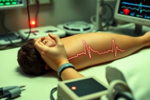
Countershock
Countershock
The use of electric current to reset a heart rhythm.
Elective Cardioversion
Elective Cardioversion
A synchronized countershock to convert dysrhythmias.
Defibrillation
Defibrillation
An unsynchronized, high-energy shock to convert dysrhythmias.
AED
AED
Signup and view all the flashcards
Pacemaker
Pacemaker
Signup and view all the flashcards
Permanent Pacemaker
Permanent Pacemaker
Signup and view all the flashcards
External Pacemakers
External Pacemakers
Signup and view all the flashcards
Output
Output
Signup and view all the flashcards
Sensitivity
Sensitivity
Signup and view all the flashcards
Triggered
Triggered
Signup and view all the flashcards
electrical activity
electrical activity
Signup and view all the flashcards
Inhibited
Inhibited
Signup and view all the flashcards
Capture
Capture
Signup and view all the flashcards
Sensing
Sensing
Signup and view all the flashcards
Failure to sense
Failure to sense
Signup and view all the flashcards
Failure to capture
Failure to capture
Signup and view all the flashcards
Hypothermia Therapy
Hypothermia Therapy
Signup and view all the flashcards
Ischemia
Ischemia
Signup and view all the flashcards
Injury
Injury
Signup and view all the flashcards
Infarction
Infarction
Signup and view all the flashcards
Elective Cardioversion Energy
Elective Cardioversion Energy
Signup and view all the flashcards
Defibrillation Energy
Defibrillation Energy
Signup and view all the flashcards
Cardioversion Indications
Cardioversion Indications
Signup and view all the flashcards
Defibrillation Indications
Defibrillation Indications
Signup and view all the flashcards
Cardioversion Synchronization
Cardioversion Synchronization
Signup and view all the flashcards
Pacemaker: Primary Function
Pacemaker: Primary Function
Signup and view all the flashcards
Standard Pad Placement
Standard Pad Placement
Signup and view all the flashcards
Epicardial pacemakers
Epicardial pacemakers
Signup and view all the flashcards
Transvenous Pacemakers
Transvenous Pacemakers
Signup and view all the flashcards
Pacing Rate
Pacing Rate
Signup and view all the flashcards
Ischemia EKG Change
Ischemia EKG Change
Signup and view all the flashcards
Injury EKG Change
Injury EKG Change
Signup and view all the flashcards
Infarction: EKG Change
Infarction: EKG Change
Signup and view all the flashcards
Anterior MI Artery
Anterior MI Artery
Signup and view all the flashcards
Anteroseptal MI Changes
Anteroseptal MI Changes
Signup and view all the flashcards
Inferior MI Artery
Inferior MI Artery
Signup and view all the flashcards
Synchronized Cardioversion
Synchronized Cardioversion
Signup and view all the flashcards
Post-shock actions
Post-shock actions
Signup and view all the flashcards
Epicardial Pacing
Epicardial Pacing
Signup and view all the flashcards
Failure to fire
Failure to fire
Signup and view all the flashcards
Therapeutic Hypothermia
Therapeutic Hypothermia
Signup and view all the flashcards
Cooling Concerns
Cooling Concerns
Signup and view all the flashcards
Anterior Wall Artery
Anterior Wall Artery
Signup and view all the flashcards
ST depression
ST depression
Signup and view all the flashcards
Concave ST elevation
Concave ST elevation
Signup and view all the flashcards
Convex ST elevation
Convex ST elevation
Signup and view all the flashcards
Study Notes
- Study notes on circulatory assist devices, pacemakers, myocardial infarction (MI), and electrocardiogram (EKG) changes
Countershock
- Electric current to reset the heart rhythm
- Includes cardioversion and defibrillation
Elective Cardioversion vs. Defibrillation
- Elective Cardioversion: Delivers synchronized electric current to convert dysrhythmias, initially at 100-120 J, up to 200 J
- Defibrillation: Delivers unsynchronized electric shock, administering a larger number of joules up to 360 J
- In defibrillation there is no discernible R wave to sync with
Indications for Countershocks
- Elective Cardioversion: Used when SVT is resistant to medication, in atrial fibrillation or flutter and for unstable patients with ventricular tachycardia,
- Symptoms for unstable ventricular tachycardia in patients can include hypotension, dyspnea, or chest pain
- May also be used if the person has symptoms suggestive of heart failure, myocardial infarction, or myocardial ischemia
- Defibrillation: Used for pulseless patients with ventricular tachycardia or ventricular fibrillation
Interventions for Countershocks
- Once patient is attached, push "sync" button before each cardioversion attempt
- Syncs with the R wave and the patient MUST HAVE an R or QS wave to sync with
- The machine discharges after the R wave and before the downstroke of the T wave
- During the absolute refractory period
- If performed during relative refractory period, ventricular fibrillation may result
- Apply conductive paste or gel pads on the chest wall at the apex and base of the heart
- Obtain continuous ECGs during the procedure
- Automatic External Defibrillator (AED) can be used by medical/lay persons
- AED uses IECG to detect patterns through large patches on the patient's chest
- If lethal dysrhythmia is detected, AED defibrillates
- AED will instruct the user on how to operate
Nursing Role in Preparation for Countershocks
- Start at low frequency, then increase
- Obtain informed consent and educate on the procedure
- Ensure an MD, RN, RT and RPh are present
- Obtain an ECG strip
- Check for atrium clots with a transesophageal echocardiogram
- Confirm IV access and meds are given
- If hemodynamically unstable then sedative meds are given
- Light sedative
- Conscious
- Moderate sedation
- Hypnotic sedative and narcotic analgesic: Etomidate, fentanyl
- Report any electrolyte imbalance (Ca, Mg, K)
- Remove oxygen and metallic objects
- Apply conductive pads to the right chest and midaxillary line
- Can also be placed on anterior and posterior chest walls, which is better for people with atrial fibrillation
- Ensure that "All clear!" is called before the shock, and no one touches the bed or patient
Nursing Role after Countershocks
- Resume CPR and ACLS immediately after the shock
- Administer pharmacological interventions intravenously
- Every Q2 minutes (~5 cycles of CPR), check pulse and rhythm, continue if indicated
- Assess for Emboli(especially cerebral), respiratory depression, skin burns and dysrhythmias after the procedure
Pacemakers - Primary Function
- Pulse generator to provide electrical stimulus to the heart when the heart's conduction fails or doesn’t produce sufficient cardiac output
- Connected to wires that carry an electrical stimulus to the myocardial cells
- Used in addition to drug therapy when the conduction system fails or cannot initiate an impulse spontaneously, or is unable to maintain primary pacing control
- Can be programmed to pace when an intrinsic beat is not sensed as a backup trigger
Permanent Pacemakers
- Internally implants pulse generator in subcutaneous tissue of the chest wall
- Leads are passed transvenously to the heart and rest on the endocardium
- Pacer wires are inserted through subclavian or jugular veins into the atria and/or ventricles
Ventricle Pacing
- Most common type of pacemakers
- Appear as a spike before wide and bizarre QRS
- Impulses from the atria are blocked, such as in a complete heart block
Atrial Pacing
- Appears as a spike before the P wave
- Common with Sinus node disease leading to bradycardia
- The SA node fires improperly as a result of ischemic damage to SA node which leads to bradycardia
Single Chamber Pacemakers
- Can be in the atrium or the ventricle
Dual Chamber/Atrioventricular Pacemakers
- Requires 2 wires for atrioventricular pacing
- Used to synchronize heart depolarizations in order to maintain cardiac output
Temporary Pacemaker Types
- External Pacemakers
- Pads are placed on anterior and posterior walls of the chest
- Delivers direct electrical impulses through the skin and body tissues via transcutaneous pacemakers
- Can be painful and requires medication with sedatives and analgesics
- Analyze ECG strip before and after
Epicardial Pacemakers
- Temporary pacing used during open heart surgery
- 2 wires stitched in at the atrial level and 2 wires stitched in at the ventricular level
- Wires must be brought under the skin just under the sternum for access
- Lightly sutured to easily remove when unneeded
- Implanted to treat symptomatic bradycardia that arises post-surgery
- When not in use must be kept insulated by a sterile glove near the chest
- Nurse must keep sterile technique during the insertion site
Transvenous Pacemakers
- Electrical stimulation of the right ventricular or R atrial endocardium via electrode-tipped catheter
- Uses direct insertion of the pacing wire of a pulmonary artery catheter with embedded pacing port
- Temporary, sterile, bedside procedure, requires x-ray to confirm placement
Pacemaker Box/Generator
- Essential to ensure the pacemaker is set correctly
- Pacing rate: Top dial in beats per minute (BPM)
- Output: Middle dial, Energy amount in milliamperes
- Sensitivity: Bottom dial, amount of electrical activity pacer senses to fire
- Blue caps underneath connect wires
Pacemaker Terms
- Know how to identify on an ECG
- Triggered: Fires an impulse in response to sensing
- Inhibited: Only initiates pacing, will not activate if the pacemaker senses an intrinsic beat
- Double: Pacemaker reacts to both inhibition and triggering
- Firing: Sends out an electrical impulse
- Capture: A stimulus strong enough to produce depolarization
- Sensing: Pacemaker "senses" the patient's own impulse generation
Three Letter Codes for Pacemakers
- Chamber Paced: represents the chamber that will be paced - Atrium (A), Ventricle (V), or Dual (D)
- Chamber Sensed: represents the chamber that the pacemaker will react to - Atrium (A), Ventricle (V), or Dual (D)
- Pacemaker Response: represents what the pacemaker will do upon reaction - Triggered, Inhibited, or Double
- Example for VVI: Ventricle, Ventricle, Inhibited - the ventricles will be paced only if necessary, if no intrinsic beat
Pacemaker Malfunctions
- Failure to sense: Pacemaker competes with the patient's own impulses, it may discharge an impulse during the refractory period
- Failure to capture: Initiates an impulse, but not strong enough for depolarization
- Failure to fire: Unable to send an impulse, indicated by absence of a pacemaker spike
ICDs
- Function in "Countershock" area
- Used to prevent sudden cardiac arrest as well as sustained ventricular tachycardia
- Prophylactically used in high risk groups; cardiomyopathy.
- Pacemaker capabilities: To defibrillate or cardiovert and anti-tachycardia pacing to avoid conversion, treats bradycardia
Hypothermia Therapy and Cardiac Arrest
- Cooling technique for those who have already experienced cardiac arrest to 90-93 degrees F
- Slows body metabolism to protect the brain and reduce mortality
- 80% of patients who received hypothermia resulted in a more favorable neurological outcome than normothermia,
- Externally cools via cooling blanket device, cool pads, or blankets
- Internally cools via cold intravenous saline infusion which circulates fluid percutaneously through jugular catheters
- Measures skin for cold burns
- Hypothermia leads to Atrial and ventricular dysrhythmias with PR, QRS, QT elongations
- Also leads to common hypokalemia
Nursing Care when Rewarming from Hypothermia
- Occurs after 12-24 hours of therapy
- Can lead to hyperkalemia and vasodilation, electrolyte shifts
- Closely follow CVP, ScVO2, and urine output
- Fluid shifts are an indicator, if CVP and UO declines, may need fluid therapy
MI Patterns on a 12 Lead ECG
- Anterior MI: Front wall, supplied by the left anterior descending artery
- Inferior MI: Bottom and right side wall, supplied by the right coronary artery
- Lateral MI: Left side wall, supplied by the circumflex artery
- Posterior MI: Back wall, supplied by the right coronary artery
- Anterior leads have ST elevation in V2-V4 and reciprocal changes in II, III, aVF
- Inferior leads have ST elevations in II, III, aVF and reciprocal changes in 1, aVL, and V1-V6
ECG Changes with Ischemia, Injury and Infarction
- Ischemia: Heart starves for blood and oxygen, appears as an inverted T wave and pale or whitish tissue
- Injury: Tissue injured by lack of perfusion, appears as ST segment elevation and blueish tissue
- Infarction: Tissue necrosis, appears as significant Q wave and black tissue
ST Segment Abnormalities
- Normal ST: At the same baseline as PR
- ST Depression: ST below baseline indicates ischemia or reciprocal changes of an infarct
- Concave ST elevation: ST above the baseline with rounded edges, may indicate pericarditis or myocardial infarction (STEMIs)
- Convex ST elevation: ST above the baseline with coved edges, typically associated with myocardial injury
MI Locations + CA distribution
- Anterior MI
- ST elevation in V2-V4
- Reciprocal changes II, III, aVF
- CA: left anterior descending (LAD)
- Inferior ΜΙ
- ST elevations in II, III, avF
- Reciprocal changes in I, aVL, and V1-V6
- CA: right coronary artery (RCA)
- Lateral MI
- ST changes in I, aVL, V5, V6
- Reciprocal changes II, III, aVF
- CA: circumflex
- Posterior MI
- ST depression with tall R wave and upright T waves
- Reciprocal changes in V1 & V2
- CA: RCA or circumflex
- Extensive Anterior MI
- Indicative changes in I, aVL, V1 to V6
- Reciprocal changes in II, III, aVF
- CA: LAD or left main
- Anteroseptal MI
- Indicative changes in V1 plus any anterior leads
- Usually no reciprocal changes
- CA: LAD
- MIs can extend across into other walls, besides the left ventricle
Studying That Suits You
Use AI to generate personalized quizzes and flashcards to suit your learning preferences.