Podcast
Questions and Answers
What is the primary protein found in the center of elastic fibers?
What is the primary protein found in the center of elastic fibers?
- Elastin (correct)
- Collagen
- Reticulin
- Fibrillin
Which of the following structures is NOT a site where elastic fibers are typically found?
Which of the following structures is NOT a site where elastic fibers are typically found?
- Ligamentum flavum
- Bronchioles
- Trachea
- Spleen (correct)
Which type of fiber is characterized by its ability to form a branching network?
Which type of fiber is characterized by its ability to form a branching network?
- Fibroelastic fibers
- Reticular fibers (correct)
- Collagen fibers
- Elastic fibers
What color can reticular fibers appear when stained with silver?
What color can reticular fibers appear when stained with silver?
Which type of glycosaminoglycan is linked to a core protein to form proteoglycans?
Which type of glycosaminoglycan is linked to a core protein to form proteoglycans?
Which component of connective tissue acts as adhesive material and is coated by glycoproteins?
Which component of connective tissue acts as adhesive material and is coated by glycoproteins?
In which type of connective tissue can you find synovial fluid?
In which type of connective tissue can you find synovial fluid?
What does metachromasia refer to when staining tissue?
What does metachromasia refer to when staining tissue?
Which cell type is primarily responsible for the synthesis of connective tissue matrix and fibers?
Which cell type is primarily responsible for the synthesis of connective tissue matrix and fibers?
What is a unique characteristic of pericytes?
What is a unique characteristic of pericytes?
In which context are mesenchymal cells most commonly found?
In which context are mesenchymal cells most commonly found?
What is the primary function of connective tissue?
What is the primary function of connective tissue?
What characterizes the appearance of fibroblasts compared to adipose cells?
What characterizes the appearance of fibroblasts compared to adipose cells?
Which of the following best describes white collagenous fibers?
Which of the following best describes white collagenous fibers?
What is the primary function of macrophage cells derived from migrating blood monocytes?
What is the primary function of macrophage cells derived from migrating blood monocytes?
How are tropocollagen molecules structured?
How are tropocollagen molecules structured?
Which of the following statements about reticular cells is true?
Which of the following statements about reticular cells is true?
What color do white collagenous fibers appear when viewed microscopically?
What color do white collagenous fibers appear when viewed microscopically?
What types of tissues can collagenous fibers be found in?
What types of tissues can collagenous fibers be found in?
What aspect of fibroblasts contributes to the healing of wounds?
What aspect of fibroblasts contributes to the healing of wounds?
Which characteristic sets plasma cells apart from other connective tissue cells?
Which characteristic sets plasma cells apart from other connective tissue cells?
Which of the following characteristics is true of yellow elastic fibers?
Which of the following characteristics is true of yellow elastic fibers?
Which type of connective tissue is primarily characterized by the presence of reticular fibers?
Which type of connective tissue is primarily characterized by the presence of reticular fibers?
What enzymatic action can digest yellow elastic fibers?
What enzymatic action can digest yellow elastic fibers?
What characteristic defines the M1 'killer' macrophages?
What characteristic defines the M1 'killer' macrophages?
Which statement most accurately describes the reticular fibers?
Which statement most accurately describes the reticular fibers?
What is a major function of the M2 'repair' macrophages?
What is a major function of the M2 'repair' macrophages?
What type of cell acts as a multipotent stem cell in the context of connective tissue?
What type of cell acts as a multipotent stem cell in the context of connective tissue?
Which of the following is NOT a characteristic of the macrophages described?
Which of the following is NOT a characteristic of the macrophages described?
How are the M1 macrophages primarily identified histologically?
How are the M1 macrophages primarily identified histologically?
What is the main difference in the types of adipocytes derived from lipoblasts?
What is the main difference in the types of adipocytes derived from lipoblasts?
Which staining technique is typically used to visualize reticular fibers?
Which staining technique is typically used to visualize reticular fibers?
Which of the following is a life-threatening condition associated with Marfan syndrome?
Which of the following is a life-threatening condition associated with Marfan syndrome?
What component of connective tissue is typically synthesized by a class of integral membrane proteins?
What component of connective tissue is typically synthesized by a class of integral membrane proteins?
In which type of connective tissue can you typically find reticular fibers that form a network?
In which type of connective tissue can you typically find reticular fibers that form a network?
What is the primary role of glycoproteins in connective tissue?
What is the primary role of glycoproteins in connective tissue?
Which staining technique would result in a reddish-purple color due to the presence of glycosaminoglycans?
Which staining technique would result in a reddish-purple color due to the presence of glycosaminoglycans?
What role do pericytes play in connective tissue?
What role do pericytes play in connective tissue?
What is the primary cell type responsible for producing elastic fibers in connective tissue?
What is the primary cell type responsible for producing elastic fibers in connective tissue?
Which statement accurately reflects the characteristics of fibroblasts?
Which statement accurately reflects the characteristics of fibroblasts?
What characteristic distinguishes metachromasia in histological staining?
What characteristic distinguishes metachromasia in histological staining?
Which feature is associated with white collagenous fibers?
Which feature is associated with white collagenous fibers?
What is a key feature of adipose cells compared to fibroblasts?
What is a key feature of adipose cells compared to fibroblasts?
Which type of glycosaminoglycan is commonly found in cartilage and bone?
Which type of glycosaminoglycan is commonly found in cartilage and bone?
What is a defining characteristic of yellow elastic fibers?
What is a defining characteristic of yellow elastic fibers?
What is the approximate length of a tropocollagen molecule?
What is the approximate length of a tropocollagen molecule?
What distinguishes plasma cells from other cells within connective tissue?
What distinguishes plasma cells from other cells within connective tissue?
Which connective tissue type is NOT directly mentioned as containing collagenous fibers?
Which connective tissue type is NOT directly mentioned as containing collagenous fibers?
Which type of cell is most numerous in the loose connective tissue?
Which type of cell is most numerous in the loose connective tissue?
Which cell type is described as having a central, large, oval, and vesicular nucleus?
Which cell type is described as having a central, large, oval, and vesicular nucleus?
What unique property distinguishes white collagenous fibers from yellow elastic fibers?
What unique property distinguishes white collagenous fibers from yellow elastic fibers?
Which statement is true regarding the structural arrangement of fibroblasts?
Which statement is true regarding the structural arrangement of fibroblasts?
What type of connective tissue is identified by the presence of branching spindle-shaped cells?
What type of connective tissue is identified by the presence of branching spindle-shaped cells?
What is the origin of macrophages that reside in connective tissue?
What is the origin of macrophages that reside in connective tissue?
How do the branches of yellow elastic fibers contribute to their function?
How do the branches of yellow elastic fibers contribute to their function?
Which of the following is a primary characteristic of reticular fibers?
Which of the following is a primary characteristic of reticular fibers?
What is a primary characteristic of M1 'killer' macrophages?
What is a primary characteristic of M1 'killer' macrophages?
Which of the following statements about M2 'repair' macrophages is accurate?
Which of the following statements about M2 'repair' macrophages is accurate?
What is the main feature of reticular cells in loose connective tissue?
What is the main feature of reticular cells in loose connective tissue?
Which staining technique is often utilized to visualize reticular fibers?
Which staining technique is often utilized to visualize reticular fibers?
What type of cells derive from lipoblasts in the context of adipose tissue?
What type of cells derive from lipoblasts in the context of adipose tissue?
What cellular feature distinguishes M1 macrophages from M2 macrophages?
What cellular feature distinguishes M1 macrophages from M2 macrophages?
What type of organelles are typically abundant in macrophages, especially the M1 type?
What type of organelles are typically abundant in macrophages, especially the M1 type?
Which type of connective tissue is primarily characterized by the presence of a reticular stroma?
Which type of connective tissue is primarily characterized by the presence of a reticular stroma?
Flashcards
Fibroblast function
Fibroblast function
Fibroblasts are the most common cells in connective tissue and synthesize connective tissue matrix and fibers. They are responsible for tissue growth and wound healing.
Fibroblast characteristics
Fibroblast characteristics
Fibroblasts are spindle-shaped cells with branching processes, numerous ribosomes, and prominent endoplasmic reticulum(rER) and Golgi apparatus, which are all vital for producing large amounts of protein fibers, such as collagen and elastin.
Fibroblast origin
Fibroblast origin
Fibroblasts originate from mesenchymal cells, which are undifferentiated cells of the mesoderm. These undifferentiated mesenchymal cells are extremely common in embryonic connective tissue. They also reside in the pulp of deciduous teeth.
Mesenchymal cell function
Mesenchymal cell function
Signup and view all the flashcards
Macrophage origin
Macrophage origin
Signup and view all the flashcards
Mast cell
Mast cell
Signup and view all the flashcards
Plasma cells
Plasma cells
Signup and view all the flashcards
Reticular cell
Reticular cell
Signup and view all the flashcards
White Collagenous Fibers
White Collagenous Fibers
Signup and view all the flashcards
Tropocollagen Molecules
Tropocollagen Molecules
Signup and view all the flashcards
Collagen Fibrils
Collagen Fibrils
Signup and view all the flashcards
Yellow Elastic Fibers
Yellow Elastic Fibers
Signup and view all the flashcards
Fibroblast
Fibroblast
Signup and view all the flashcards
Connective Tissue
Connective Tissue
Signup and view all the flashcards
Acidophilic Staining
Acidophilic Staining
Signup and view all the flashcards
Elastic membrane
Elastic membrane
Signup and view all the flashcards
Acidophilic fibers
Acidophilic fibers
Signup and view all the flashcards
Elastic fibers location
Elastic fibers location
Signup and view all the flashcards
Marfan syndrome
Marfan syndrome
Signup and view all the flashcards
Reticular fibers function
Reticular fibers function
Signup and view all the flashcards
Reticular fiber structure
Reticular fiber structure
Signup and view all the flashcards
Glycosaminoglycans (GAGs)
Glycosaminoglycans (GAGs)
Signup and view all the flashcards
Proteoglycans
Proteoglycans
Signup and view all the flashcards
Ground substance components
Ground substance components
Signup and view all the flashcards
Macrophage types
Macrophage types
Signup and view all the flashcards
M1 Macrophage function
M1 Macrophage function
Signup and view all the flashcards
M2 Macrophage function
M2 Macrophage function
Signup and view all the flashcards
Reticular Fibers
Reticular Fibers
Signup and view all the flashcards
UMCs
UMCs
Signup and view all the flashcards
Adipocytes: Division
Adipocytes: Division
Signup and view all the flashcards
Adipocytes: Location
Adipocytes: Location
Signup and view all the flashcards
Reticular stroma location
Reticular stroma location
Signup and view all the flashcards
Fibroblast function
Fibroblast function
Signup and view all the flashcards
Mesenchymal cell origin
Mesenchymal cell origin
Signup and view all the flashcards
Macrophage origin
Macrophage origin
Signup and view all the flashcards
Mast cell role
Mast cell role
Signup and view all the flashcards
Adipocyte characteristic
Adipocyte characteristic
Signup and view all the flashcards
Reticular cell function
Reticular cell function
Signup and view all the flashcards
Fibroblast characteristics (brief)
Fibroblast characteristics (brief)
Signup and view all the flashcards
UMCs definition
UMCs definition
Signup and view all the flashcards
White Collagenous Fibers
White Collagenous Fibers
Signup and view all the flashcards
Tropocollagen Molecules
Tropocollagen Molecules
Signup and view all the flashcards
Collagen Fibrils
Collagen Fibrils
Signup and view all the flashcards
Yellow Elastic Fibers
Yellow Elastic Fibers
Signup and view all the flashcards
Collagen Fiber Function
Collagen Fiber Function
Signup and view all the flashcards
Elastic Fiber Function
Elastic Fiber Function
Signup and view all the flashcards
White Collagenous Fiber Stain
White Collagenous Fiber Stain
Signup and view all the flashcards
Connective Tissue Function
Connective Tissue Function
Signup and view all the flashcards
Acidophilic Fibers
Acidophilic Fibers
Signup and view all the flashcards
Elastic Fibers Location
Elastic Fibers Location
Signup and view all the flashcards
Reticular Fibers Function
Reticular Fibers Function
Signup and view all the flashcards
Reticular Fibers Structure
Reticular Fibers Structure
Signup and view all the flashcards
GAGs (Glycosaminoglycans)
GAGs (Glycosaminoglycans)
Signup and view all the flashcards
Proteoglycans
Proteoglycans
Signup and view all the flashcards
Marfan Syndrome
Marfan Syndrome
Signup and view all the flashcards
Ground Substance Components
Ground Substance Components
Signup and view all the flashcards
Adipocyte Division
Adipocyte Division
Signup and view all the flashcards
Adipocyte Location
Adipocyte Location
Signup and view all the flashcards
Macrophage Types
Macrophage Types
Signup and view all the flashcards
M1 Macrophage Function
M1 Macrophage Function
Signup and view all the flashcards
M2 Macrophage Function
M2 Macrophage Function
Signup and view all the flashcards
Reticular Stroma Location
Reticular Stroma Location
Signup and view all the flashcards
Reticular Cell Feature
Reticular Cell Feature
Signup and view all the flashcards
UMCs
UMCs
Signup and view all the flashcards
Study Notes
Connective Tissue (CT)
- CT supports, binds, and separates various tissues and organs
- Two types:
- Connective tissue proper
- Special types: cartilage, bone, blood, hematopoietic tissue, lymphoid tissue
Connective Tissue Proper
-
Fibers:
- White collagenous fibers: white, strong, resist stretch, form wavy bundles (branching bundles, single fibers don't branch), affected by boiling (converted to gelatin), destroyed by acids/alkalis, digested by pepsin and collagenase enzymes
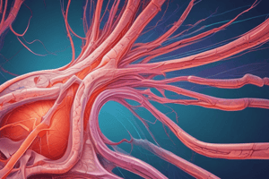
- Yellow elastic fibers: yellow, thin, long, highly refractile (able to reflect light), elastic, recoil after stretch, branch, found in aorta walls, digested by pancreatic elastase
- Reticular fibers: very thin, form network, argyrophilic (affinity for silver), found in stroma of parenchymatous organs, reticular lamina of basement membranes
-
Matrix (Ground Substance):
- Macromolecules: multi-adhesive glycoproteins
- Hyaluronic acid, soft, jelly-like, not linked to protein core, synthesized by hyaluronan synthases
- Chondroitin sulfate: linked to protein core, forming proteoglycans; found in cartilage and bone; heparan sulfate in basal lamina
- Glycoproteins: examples: chondronectin (cartilage), osteonectin (bone), fibronectin (CT proper). Act like adhesives
-
Cells:
- Fibroblasts: responsible for synthesizing CT matrix and fibers; spindle-shaped, branched, central nucleus
- Macrophages: phagocytize foreign bodies like bacteria; important in wound healing; large, rounded cell with pseudopodia (processes); nucleus is eccentric, small, oval and pale
- Mast cells: found in connective tissue, involved in inflammatory response.
- Plasma cells: produce antibodies; small, round nuclei
- Lymphocytes (important parts of the immune system): important in responding to infectious pathogens.
- Adipose: fat cells; store energy; spherical shaped
- Leukocytes: white blood cells
- Other cells: mesenchymal cells, pericytes, reticular cells
Connective Tissue Fibers - Characterization
- White collagenous fibers: color: white; resistant to stretch; wave-like bundles; branching bundles are found; single fiber does not branch; affect by boiling, converted in gelatin; destroyed by acids and alkalis; digested by pepsin and collagenase
- Yellow elastic fibers: color: yellow; thin, long, highly refractive meaning able to reflect light; elastic in nature; recoils after release of stretch; branch and may form elastic membranes e.g. in the aorta walls; not affected by boiling; digested by pancreatic elastase enzyme
Types and Sites of Collagen
- Type I: Bone, tendon, teeth, dermis
- Type II: Hyaline and elastic cartilage
- Type III: Reticular stroma of liver, spleen, kidney, lymph nodes
- Type IV: Basal lamina of basement membranes
- Type V: Basal lamina of the placenta
Staining
- H&E: Acidophilic
- Mallory Trichrome Stain: Collagen fibers (blue)
- Van Gieson's Stain: Collagen fibers (red)
- Verhoeff's Stain: Elastic fibers (black)
- Silver stain: reticular fibers (black)
- PAS: magenta colored (polysaccharides)
Fixed vs. Free CT Cells
-
Fixed: Undifferentiated mesenchymal, fibroblasts, adipose, reticular, macrophages (originate locally, remain in the CT).
-
Free: Mast cells, free macrophages, plasma, pigment cells, blood leukocytes (originate elsewhere, temporarily in CT)
Undifferentiated Mesenchymal Cells (UMCs)
- Mesodermal cells of the embryo
- Can divide
- Differentiate into various CT cells (fibroblasts, chondrocytes, etc).
- Found in embryonic connective tissue (mesenchymal and mucoid CT), pulp of deciduous teeth
Fibroblasts
- The most common CT cells; responsible for synthesizing CT matrix and fibers
- Responsible for growth, repair, and healing of wounds
- Commonly found in loose CT; spindle shaped, contain central nuclei, pale basophilic cytoplasm containing free ribosomes
Macrophages
- Originate from blood monocytes (originally developing from UMCs in the bone marrow)
- Cannot divide, fully differentiated
- Primarily in loose CT
- Large, rounded with pseudopodia (processes)
- Second most common cell in loose CT
- Eccentric, small, kidney-shaped/irregular, single, and pale
Reticular cells
- UMC (undifferentiated mesenchymal cell).
- Cannot divide, fully differentiated.
- Reticular stroma of parenchymal organs.
Adipocytes
-
Brown Adipocytes:
- Multi-nucleated
- Many fat droplets
- In sites of brown adipose CT
- 10 times smaller than white adipocytes
- Oval shaped cells, with single central nucleus
- Cytoplasm is acidophilic (with many vacuoles due to dissolved fat droplets)
-
White Adipocytes:
- Multi-nucleated
- One fat globule
- In sites of white adipose CT
- Also in loose CT
- 10 times larger than brown adipocytes
- Oval shaped cells, with peripheral nucleus (signet ring appearance)
- Cytoplasm contains a large fat globule
Studying That Suits You
Use AI to generate personalized quizzes and flashcards to suit your learning preferences.