Podcast
Questions and Answers
Which of the following mechanisms is NOT considered a primary cause of direct injury in cases of globe trauma from a missile?
Which of the following mechanisms is NOT considered a primary cause of direct injury in cases of globe trauma from a missile?
- Shock waves transmitted through the globe
- Direct impact of the missile on ocular structures
- Damage adjacent to the missile's pathway
- Simultaneous retraction of the choroid and retina (correct)
Why are the Bruch's membrane and RPE (Retinal Pigment Epithelium) more prone to rupture compared to the sclera in cases of ocular trauma?
Why are the Bruch's membrane and RPE (Retinal Pigment Epithelium) more prone to rupture compared to the sclera in cases of ocular trauma?
- The sclera is inelastic and brittle.
- Bruch's membrane and RPE are more elastic than the sclera.
- The sclera contains more collagen fibers providing improved tensile strength.
- Bruch's membrane and RPE are inelastic, making them more susceptible to rupture. (correct)
A young male presents with limited visual acuity, vitreous hemorrhage, and a relative afferent pupillary defect after a BB gun injury. What additional acute finding is MOST likely to be observed?
A young male presents with limited visual acuity, vitreous hemorrhage, and a relative afferent pupillary defect after a BB gun injury. What additional acute finding is MOST likely to be observed?
- Surrounding commotio retinae (correct)
- Phthisis bulbi
- Optic nerve atrophy
- Cataract formation
After the initial hemorrhage clears following a choroidal rupture, what characteristic features are MOST likely to become visible?
After the initial hemorrhage clears following a choroidal rupture, what characteristic features are MOST likely to become visible?
What diagnostic procedure is MOST important in evaluating the extent of vitreous hemorrhage and ruling out retinal detachment in a patient with ocular trauma?
What diagnostic procedure is MOST important in evaluating the extent of vitreous hemorrhage and ruling out retinal detachment in a patient with ocular trauma?
Which systemic condition is least likely to be associated with Purtscher-like retinopathy?
Which systemic condition is least likely to be associated with Purtscher-like retinopathy?
A patient presents with Purtscher retinopathy following a traumatic injury. What is the most appropriate initial treatment approach?
A patient presents with Purtscher retinopathy following a traumatic injury. What is the most appropriate initial treatment approach?
A patient with significant vision loss secondary to choroidal rupture should be monitored closely for the development of which secondary complication?
A patient with significant vision loss secondary to choroidal rupture should be monitored closely for the development of which secondary complication?
What is the MOST likely underlying cause of Purtscher retinopathy?
What is the MOST likely underlying cause of Purtscher retinopathy?
Valsalva retinopathy is primarily caused by which of the following mechanisms?
Valsalva retinopathy is primarily caused by which of the following mechanisms?
What is the pathognomonic finding in Purtscher retinopathy that helps distinguish it from other causes of retinal ischemia?
What is the pathognomonic finding in Purtscher retinopathy that helps distinguish it from other causes of retinal ischemia?
A patient presents with a subhyaloid hemorrhage following a bout of severe coughing. What is the most likely underlying condition?
A patient presents with a subhyaloid hemorrhage following a bout of severe coughing. What is the most likely underlying condition?
A patient is diagnosed with Purtscher retinopathy. What fluorescein angiography (FA) finding would be LEAST likely to be observed?
A patient is diagnosed with Purtscher retinopathy. What fluorescein angiography (FA) finding would be LEAST likely to be observed?
Which of the following activities is least likely to cause Valsalva retinopathy?
Which of the following activities is least likely to cause Valsalva retinopathy?
Optical coherence tomography (OCT) imaging of Purtscher flecken will show hyperreflectivity in which retinal layer?
Optical coherence tomography (OCT) imaging of Purtscher flecken will show hyperreflectivity in which retinal layer?
What is the primary benefit of performing a YAG laser membranotomy in a patient with a non-clearing subhyaloid hemorrhage?
What is the primary benefit of performing a YAG laser membranotomy in a patient with a non-clearing subhyaloid hemorrhage?
Prior to considering laser treatment for Valsalva retinopathy with subhyaloid hemorrhage, what diagnostic test is most important to perform?
Prior to considering laser treatment for Valsalva retinopathy with subhyaloid hemorrhage, what diagnostic test is most important to perform?
A college student visits his eye doctor after noticing blurry vision in his right eye after an intense weightlifting session at the gym. Fundus examination reveals a pre-retinal hemorrhage. What should the doctor advise?
A college student visits his eye doctor after noticing blurry vision in his right eye after an intense weightlifting session at the gym. Fundus examination reveals a pre-retinal hemorrhage. What should the doctor advise?
In which of the following scenarios would steroid therapy be most theoretically beneficial for treating PAMM?
In which of the following scenarios would steroid therapy be most theoretically beneficial for treating PAMM?
Laser membranotomy for Valsalva retinopathy aims to disrupt which structure to facilitate drainage of subhyaloid hemorrhage?
Laser membranotomy for Valsalva retinopathy aims to disrupt which structure to facilitate drainage of subhyaloid hemorrhage?
A patient presents with blurry vision after blunt trauma to the eye, and an examination reveals gray-white opacification of the neurosensory retina. What is the most likely diagnosis?
A patient presents with blurry vision after blunt trauma to the eye, and an examination reveals gray-white opacification of the neurosensory retina. What is the most likely diagnosis?
What is the primary mechanism behind the retinal changes observed in commotio retinae?
What is the primary mechanism behind the retinal changes observed in commotio retinae?
In the context of commotio retinae, which of the following complications is most likely to lead to permanent vision loss?
In the context of commotio retinae, which of the following complications is most likely to lead to permanent vision loss?
What is the typical prognosis for visual recovery in cases of commotio retinae without complications?
What is the typical prognosis for visual recovery in cases of commotio retinae without complications?
A patient who sustained a high-velocity projectile injury near the orbit presents with a full-thickness chorioretinal defect. Which condition is most likely?
A patient who sustained a high-velocity projectile injury near the orbit presents with a full-thickness chorioretinal defect. Which condition is most likely?
Which of the following mechanisms is most directly associated with the pathology observed in chorioretinitis sclopetaria?
Which of the following mechanisms is most directly associated with the pathology observed in chorioretinitis sclopetaria?
What is a key differentiating factor between commotio retinae and chorioretinitis sclopetaria in terms of mechanism of injury?
What is a key differentiating factor between commotio retinae and chorioretinitis sclopetaria in terms of mechanism of injury?
A patient with chorioretinitis sclopetaria develops significant fibrotic scarring and atrophy in the affected area. What is the most likely long-term visual outcome?
A patient with chorioretinitis sclopetaria develops significant fibrotic scarring and atrophy in the affected area. What is the most likely long-term visual outcome?
In cases of commotio retinae, why might visual acuity be diminished?
In cases of commotio retinae, why might visual acuity be diminished?
What is the most likely progression of visual symptoms and retinal changes following blunt trauma that causes commotio retinae?
What is the most likely progression of visual symptoms and retinal changes following blunt trauma that causes commotio retinae?
Flashcards
Commotio Retinae
Commotio Retinae
Traumatic retinopathy from direct trauma to the globe, known as Berlin’s edema.
Traumatic Retinopathy
Traumatic Retinopathy
Retinal damage due to direct trauma, leading to vision loss.
Symptoms of Commotio Retinae
Symptoms of Commotio Retinae
Gray-white opacification of the retina with blurry vision or vision loss.
Visual Recovery Prognosis
Visual Recovery Prognosis
Signup and view all the flashcards
Management of Commotio Retinae
Management of Commotio Retinae
Signup and view all the flashcards
Chorioretinitis Sclopetaria
Chorioretinitis Sclopetaria
Signup and view all the flashcards
Damage from High-Velocity Projectiles
Damage from High-Velocity Projectiles
Signup and view all the flashcards
Visual Loss Mechanism
Visual Loss Mechanism
Signup and view all the flashcards
Macular Hole Risk
Macular Hole Risk
Signup and view all the flashcards
Annular Opacification
Annular Opacification
Signup and view all the flashcards
Choroidal rupture
Choroidal rupture
Signup and view all the flashcards
Purtscher Retinopathy
Purtscher Retinopathy
Signup and view all the flashcards
Relative Afferent Pupillary Defect (RAPD)
Relative Afferent Pupillary Defect (RAPD)
Signup and view all the flashcards
Cotton-wool spots
Cotton-wool spots
Signup and view all the flashcards
Bruch's membrane
Bruch's membrane
Signup and view all the flashcards
Intra-retinal hemorrhage
Intra-retinal hemorrhage
Signup and view all the flashcards
Vitreous hemorrhage
Vitreous hemorrhage
Signup and view all the flashcards
Significant vision loss
Significant vision loss
Signup and view all the flashcards
Diagnostic imaging
Diagnostic imaging
Signup and view all the flashcards
Purtscher-like retinopathy
Purtscher-like retinopathy
Signup and view all the flashcards
Traumatic Purtscher retinopathy
Traumatic Purtscher retinopathy
Signup and view all the flashcards
Valsalva retinopathy
Valsalva retinopathy
Signup and view all the flashcards
Symptoms of Purtscher retinopathy
Symptoms of Purtscher retinopathy
Signup and view all the flashcards
Non-traumatic causes of Purtscher
Non-traumatic causes of Purtscher
Signup and view all the flashcards
Treatment for Purtscher retinopathy
Treatment for Purtscher retinopathy
Signup and view all the flashcards
Ocular massage
Ocular massage
Signup and view all the flashcards
Subhyaloidal hemorrhage
Subhyaloidal hemorrhage
Signup and view all the flashcards
YAG laser treatment
YAG laser treatment
Signup and view all the flashcards
Conservative management
Conservative management
Signup and view all the flashcards
Study Notes
Commotio Retinae
- Definition: Traumatic retinopathy resulting from direct blunt trauma to the eye.
- Causes: High-impact sports, violence, motor vehicle accidents.
- Mechanism: Shock waves from the impact damage outer retinal layers.
- Appearance: Sheen-like retinal whitening appears hours after injury, often in the posterior pole.
- Associated Complications: Macular pigment epitheliopathy, choroidal rupture, cystoid macular edema, macular hole (often due to vitreous detachment).
- Signs and Symptoms: Gray-white opacification of neurosensory retina (with or without internal hemorrhages), blurry vision or vision loss post-trauma, potential decrease in visual acuity down to 20/200.
- Complete Commotio Retinae (peripheral): Swelling resolves, vision returns to 20/20 as no fibrovascular tissue formed.
- Annular Opacification: Possible annular opacification around the macula after blunt trauma.
- Management and Prognosis: Good prognosis for visual recovery; typically clears within 3-4 weeks. No acute treatment, IV steroids sometimes used.
- Late Complications: Possible late choroidal atrophy.
Chorioretinitis Sclopetaria
- Definition: (AKA traumatic chorioretinal rupture, chorioretinitis proliferans) A rare injury to the choroid and overlying retina.
- Cause: High-velocity projectiles (bullets, BBs, explosions) passing adjacent to or through the orbit without penetrating the globe.
- Mechanism: The injury causes a full-thickness chorioretinal defect. Damage may be direct (adjacent to missile pathway) or indirect (shock waves). The "split and retract" mechanism is key.
- Signs and Symptoms: Often significant vision loss (NLP to 20/1000). Vitreous hemorrhage common, with hemorrhages potentially extending to all choroidal and retinal layers. Surrounding commotio often present. Claw-like breaks in Bruch membrane, and choriocapillaris can be seen as hemorrhage clears.
- Prognosis: Poor prognosis if macular involvement. Extensive scarring and atrophy can lead to permanent vision loss.
- Risk Factors: Young males; Pellets, bullets, paintballs, corks, tree branches more prone.
Purtscher Retinopathy
- Definition: Described in 1910, caused by microvascular damage/occlusion, specifically ischemia, usually from severe head trauma (or chest compression)
- Causes: Various conditions (head trauma, chest compression, long bone fractures, orthopedic surgery, weight lifting, etc.) leading to retinal ischemia and damage. Non-traumatic causes include acute pancreatitis, fat embolism, amniotic fluid embolism, preeclampsia, HELLP syndrome, vasculitic disorders like lupus.
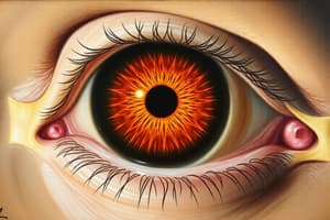
- Signs and Symptoms: Bilateral or unilateral, superficial white retinal patches (like cotton-wool spots or edema). Peripapillary hemorrhages common, also associated with Purtscher flecken (pathognomonic finding - a polygonal area of retinal whitening with a clear demarcation adjacent to retinal vessels).
- Appearance: Cotton-wool spots, retinal hemorrhages, Purtscher flecken.
- Diagnostic Findings: Fluorescein angiograms often show variable findings including retinal arteriolar obstruction, capillary non-perfusion, venous staining, and disk leakage. OCT may reveal hyperreflectivity in inner retinal layers (corresponding to cotton-wool spots), and variable macular edema. Chronic cases may also have outer retinal atrophy and photoreceptor loss.
- Management: No proven treatment, but IV steroids are sometimes used in cases of vasculitis.
Valsalva Retinopathy
- Definition: Pre-retinal and subhyaloidal hemorrhages due to sudden increase in intrathoracic/intrabdominal pressure.
- Cause: Increased intraocular venous pressure leading to retinal capillary rupture and bleeding. Clinically, this presents as a hemorrhagic detachment of the inner limiting membrane (ILM)
- Causes: Ocular massage, CPR, sexual activity, heavy exercise, coughing, vomiting, straining, lifting, labor, bowel movements, blowing instruments
- Appearance: Subhyaloidal hemorrhage (often clears spontaneously).
- Management: Observation and conservative measures. Spontaneous resolution common within weeks to months. Avoid anticoagulants and strenuous activities. Laser treatment may accelerate resolution in cases of non-clearing hemorrhage.
Studying That Suits You
Use AI to generate personalized quizzes and flashcards to suit your learning preferences.