Podcast
Questions and Answers
Which of the following best describes the central nervous system (CNS)?
Which of the following best describes the central nervous system (CNS)?
- A network of peripheral nerves that relay signals to the brain.
- A series of endocrine glands that regulate hormone production.
- The primary communication hub of the body, responsible for processing sensory information and directing responses. (correct)
- A simple structure that primarily handles reflex actions.
The dorsal structures of the spinal cord are primarily involved in which function?
The dorsal structures of the spinal cord are primarily involved in which function?
- Sensory input (correct)
- Regulating blood pressure
- Autonomic functions
- Motor output
What is the role of the meninges in protecting the brain?
What is the role of the meninges in protecting the brain?
- Providing a protective membrane layer and shock absorption. (correct)
- Attaching directly to the brain tissue to provide nourishment.
- Secreting cerebrospinal fluid (CSF)
- Producing myelin to insulate brain tissue.
Which of the following is NOT a function of cerebrospinal fluid (CSF)?
Which of the following is NOT a function of cerebrospinal fluid (CSF)?
What is the primary function of the blood-brain barrier (BBB)?
What is the primary function of the blood-brain barrier (BBB)?
Which cells are responsible for forming the myelin sheath in the central nervous system (CNS)?
Which cells are responsible for forming the myelin sheath in the central nervous system (CNS)?
Nodes of Ranvier boost the amplitude of the action potential to prevent what?
Nodes of Ranvier boost the amplitude of the action potential to prevent what?
The influx of which ion is primarily responsible for the depolarization phase of an action potential?
The influx of which ion is primarily responsible for the depolarization phase of an action potential?
What is the primary role of astrocytes in the neurovascular unit?
What is the primary role of astrocytes in the neurovascular unit?
Hyperpolarization makes the membrane potential more negative and less likely to trigger an action potential by allowing what?
Hyperpolarization makes the membrane potential more negative and less likely to trigger an action potential by allowing what?
Which glial cells act as the primary immune cells within the CNS?
Which glial cells act as the primary immune cells within the CNS?
What is the role of the sodium-potassium pump in maintaining the resting membrane potential of a neuron?
What is the role of the sodium-potassium pump in maintaining the resting membrane potential of a neuron?
What is the significance of 'tight junctions' in the context of the blood-brain barrier (BBB)?
What is the significance of 'tight junctions' in the context of the blood-brain barrier (BBB)?
Which of the following best describes 'synaptic plasticity'?
Which of the following best describes 'synaptic plasticity'?
Which cerebral structure is primarily responsible for motor control and balance?
Which cerebral structure is primarily responsible for motor control and balance?
What is the role of interneurons in the spinal cord?
What is the role of interneurons in the spinal cord?
What is the main role of the pia mater?
What is the main role of the pia mater?
Which of the following transport mechanisms across the blood-brain barrier (BBB) involves the binding of molecules to specific receptors on endothelial cells?
Which of the following transport mechanisms across the blood-brain barrier (BBB) involves the binding of molecules to specific receptors on endothelial cells?
What is meant by the term 'resting membrane potential' in a neuron?
What is meant by the term 'resting membrane potential' in a neuron?
Which primary function is associated with the cerebrum?
Which primary function is associated with the cerebrum?
What is a primary function of the brainstem?
What is a primary function of the brainstem?
What is the primary function of afferent neurons?
What is the primary function of afferent neurons?
What is the main role of ependymal cells in the CNS?
What is the main role of ependymal cells in the CNS?
What is the primary role of myelin sheath around an axon?
What is the primary role of myelin sheath around an axon?
Which of the following is an accurate comparison between electrical and chemical synapses?
Which of the following is an accurate comparison between electrical and chemical synapses?
Which event characterizes the repolarization phase of an action potential?
Which event characterizes the repolarization phase of an action potential?
What is the role of mechanosensitive ion channels in myogenic autoregulation?
What is the role of mechanosensitive ion channels in myogenic autoregulation?
A decrease in PaCO2 (partial pressure of carbon dioxide in arterial blood) typically leads to which vascular response in the brain?
A decrease in PaCO2 (partial pressure of carbon dioxide in arterial blood) typically leads to which vascular response in the brain?
Which mechanism helps maintain constant cerebral blood flow despite changes in blood pressure?
Which mechanism helps maintain constant cerebral blood flow despite changes in blood pressure?
Which of the following is NOT a component of the neurovascular unit?
Which of the following is NOT a component of the neurovascular unit?
Flashcards
What is the CNS?
What is the CNS?
The body's control center, processing sensory information and directing responses.
What is the Cerebrum?
What is the Cerebrum?
Initiates voluntary activity and controls reasoning.
What does the Brainstem Control?
What does the Brainstem Control?
Controls heart rate and blood pressure.
What is the Cerebellum responsible for?
What is the Cerebellum responsible for?
Signup and view all the flashcards
What is the Spinal Cord?
What is the Spinal Cord?
Signup and view all the flashcards
Function of Dorsal Structures (Spinal Cord)
Function of Dorsal Structures (Spinal Cord)
Signup and view all the flashcards
Ventral Structures
Ventral Structures
Signup and view all the flashcards
Lateral Horn
Lateral Horn
Signup and view all the flashcards
Efferent Neurons
Efferent Neurons
Signup and view all the flashcards
Afferent Neurons
Afferent Neurons
Signup and view all the flashcards
What are the Meninges?
What are the Meninges?
Signup and view all the flashcards
Dura Mater
Dura Mater
Signup and view all the flashcards
Arachnoid Mater
Arachnoid Mater
Signup and view all the flashcards
Pia Mater
Pia Mater
Signup and view all the flashcards
What is CSF?
What is CSF?
Signup and view all the flashcards
Blood-Brain Barrier (BBB)
Blood-Brain Barrier (BBB)
Signup and view all the flashcards
Blood-brain barrier (cerebral vasculature)
Blood-brain barrier (cerebral vasculature)
Signup and view all the flashcards
Blood-CSF barrier (choroid plexus)
Blood-CSF barrier (choroid plexus)
Signup and view all the flashcards
Outer brain-CSF barrier (pia arachnoid)
Outer brain-CSF barrier (pia arachnoid)
Signup and view all the flashcards
Inner CSF-brain barrier (neuroependyma)
Inner CSF-brain barrier (neuroependyma)
Signup and view all the flashcards
Endothelial Cells
Endothelial Cells
Signup and view all the flashcards
Astrocyte Foot Processes
Astrocyte Foot Processes
Signup and view all the flashcards
What are Neurons?
What are Neurons?
Signup and view all the flashcards
Cell Body (Neuron)
Cell Body (Neuron)
Signup and view all the flashcards
What are Dendrites?
What are Dendrites?
Signup and view all the flashcards
What do Axons do?
What do Axons do?
Signup and view all the flashcards
What is Myelin
What is Myelin
Signup and view all the flashcards
Node of Ranvier
Node of Ranvier
Signup and view all the flashcards
Sensory Neurons
Sensory Neurons
Signup and view all the flashcards
Interneurons
Interneurons
Signup and view all the flashcards
Study Notes
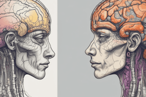
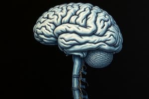
Structure and Function of the CNS
- The central nervous system (CNS) is a complex and rewarding organ to study
- This material serves as a refresher on the CNS's basic structure, function, support, and protection as a communication hub
Structure and Functions of the CNS
- The CNS comprises the brain and spinal cord
- The CNS acts as the body's control center, processing sensory information and directing responses
- The CNS coordinates voluntary and involuntary functions
- Knowing how different structures impact functions helps in understanding how disordered functions result in disease
- Divisions of the human nervous system communicate within the CNS and the body
- Impacts of CNS disorders inherently involve signaling to the periphery and receiving signals from it
- Life-sustaining functions are typically located deeper within the brain
Structure and Function of the Spinal Cord
- The spinal cord runs from the brain stem through the spine to the lower back, protected by vertebrae
- The spinal cord carries nerve signals between the CNS and the rest of the body
- Spinal cord cross-sections display a 'butterfly' pattern with internal grey matter surrounded by white matter
- Dorsal structures like the dorsal root and ganglion are involved in sensory input
- Ventral structures are responsible for motor output
- The lateral horn is involved in autonomic functions
- Motor neurons transmit signals from the spinal cord to muscles and they are called efferent neurons
- Sensory neurons in the dorsal root ganglia, known as afferent neurons, carry information from sensory receptors in the periphery to the CNS
- Sensory information goes to the spinal cord and is processed by interneurons, then translated into motor commands for muscle movement
- This translation is essential for coordinating bodily functions and responding to stimuli
- Different levels of the spinal cord interact with different organs
- Outcomes from spinal cord damage depend on the amount of damage and the level of the spine impacted
Protection and Support of the CNS
- The brain relies on several structures for protection and support
- The first layer of defense is the skin
- This is followed by the skull, which is a thick layer of bone
- Meninges are three protective membranes beneath the brain
- The dura mater is a thick, strong, fibrous membrane that attaches to the skull as a shock absorber
- The arachnoid mater is the middle layer, which attaches to the innermost layer and forms the subarachnoid space filled with cerebrospinal fluid
- The pia mater is closest to the brain tissue, providing support and nourishment
- The brain filters blood into CSF, which circulates through the subarachnoid space and ventricles
- CSF provides cushioning and blood brain barrier
- The blood-brain barrier is a collective term for four sites preventing unwanted substances and pathogens from entering the brain
Cerebrospinal Fluid (CSF)
- CSF is found within the brain and spinal cord
- The role of the CSF is to:
- Cushion the brain and spinal cord from mechanical shocks
- Exchange nutrients and waste between blood and tissue
- Provide a constant chemical environment for neuronal signaling
- There are four fluid-filled cavities within the brain called ventricles
- The ventricles' choroid plexus (formed by ependymal cells) filters blood to form the CSF secreted into the ventricles
- The CSF circulates from the lateral, third, and fourth ventricles to the cisterns of the brain
- It then flows into the subarachnoid space, enveloping the cortical convexities of the brain
- From here, CSF can enter the spinal cord's central canal or circulate through the subarachnoid space to the arachnoid granulations
- Arachnoid granulations are small protrusions of the arachnoid mater that extend into the dural venous sinuses, where CSF returns to venous circulation
- The rate of CSF production normally equals the rate of CSF absorption
- A retrograde influx of CSF occurs from the subarachnoid space into the parenchyma, where CSF and interstitial fluid interact in the perivascular space alongside blood vessels
- Astrocytes lining the perivascular space transport fluid to remove inflammatory waste proteins secreted by neurons.
- Fluid carrying the inflammatory waste returns to the subarachnoid space and drains into meningeal lymphatic vessels and arachnoid granulations
- CSF consists of glucose, salts, enzymes, and a few white blood cells (lymphocytes)
- Its constituents are tightly regulated to impact neuronal functioning:
- Concentrations of positively and negatively charged ions are highly regulated, acting as key signaling molecules for neuronal resting membrane potential, action potentials, and neurotransmission
- Large molecules (immunoglobulins and other proteins) cannot cross the blood-brain barrier
- Glutamate is a neurotransmitter in the mammalian CNS which has tightly controlled concentrations.
- Increased protein in the CSF may indicate health conditions and Increased plasma levels of glutamate will not impact CSF concentrations
Blood-Brain Barrier (BBB)
- The blood-brain barrier (BBB) is a collective term for four main interfaces between the blood and the central nervous system
- Blood-brain barrier (cerebral vasculature) regulates molecule passage from cerebral blood vessels into cerebral tissue
- The Blood-CSF barrier (choroid plexus) regulates molecule passage from the blood as it forms the CSF at the choroid plexus epithelium
- The Outer brain-CSF barrier (pia arachnoid) regulates molecule passage between blood and CSF at the arachnoid epithelium
- The Inner CSF-brain barrier (neuroependyma) is only present during early development and is formed by strap junctions of neuroependymal cells lining ventricular surfaces
- Barriers maintain the neural environment by regulating molecule passage into and out of the brain
- The BBB protects the brain from microorganisms and toxins
- A neurovascular unit is a complex network of cells ensuring proper brain function
- It regulates blood flow, maintains the BBB, provides nutrients, and removes waste
- Vascular endothelial cells, astrocytes, and pericytes form the neurovascular unit of the BBB
- Endothelial cells forming capillary walls are joined by tight junctions which prevent large molecules/pathogens from passing through
- Endothelial cells are surrounded by astrocyte foot processes enhancing the barrier and maintaining tight junctions
- Astrocytes exchange nutrients and waste between blood vessels and neurons
- Pericytes surround blood vessels and regulate blood flow by contracting or relaxing
- Microglia are immune cells that continuously scan the brain environment for damage/pathogens
- The BBB selectively controls what enters the brain
- Lipid-soluble molecules pass through lipid membranes, water-soluble molecules cannot, and larger molecules cannot pass through tight junctions.
- Specific transport systems allow larger or water-soluble molecules to cross the BBB
- Carrier-mediated transport uses protein carriers embedded in the cell membrane to move molecules such as glucose and amino acids
- Adsorptive-mediated transport involves transporting positively charged molecules that adhere to the negatively charged cell membrane
- Receptor-mediated transport involves molecules binding to specific receptors on endothelial cells, which is internalized and transported across the barrier, and is often used for large molecules like insulin and transferrin
Cells of the CNS
- There are two main types of cells in the CNS
- Neurons are the functional units transmitting information through electrical and chemical signaling
- Glial cells support and protect the CNS
- Neurons make up 50% of the CNS space but are outnumbered 3:1 by glial cells
- Neuron structure has different roles for receiving and transmitting information
- The cell body is responsible for the metabolism, genetic information and ptotein synthesis
- Dendrites receive incoming signals
- Axons convey electrical signals (0.1–3 mm)
- Myelin increases action potential speed
- Nodes of Ranvier boost action potential amplitude
- Glial cells are non-neuronal cells in the CNS and PNS providing structural and functional support to neurons
- Glial cells assist with various support functions to ensure efficient neuron operation
- Astrocytes:
- Provide physical support
- Clean up debris through phagocytosis
- Uptake neurotransmitters and provide necessary ions for chemical environment regulation
- Supply nourishment
- Repair and maintain tissues
- Isolate the CNS as BBB cells
- Ependymal cells:
- Line the ventricles and central canal of the spinal cord
- Form the choroid plexus, which produces cerebrospinal fluid (CSF)
- Use cilia to circulate CSF
- Use microvilli to reabsorb CSF
- Play an essential role in brain homeostasis
- Microglia:
- Immune cells that act as macrophages
- Protect against microorganisms
- Act as phagocytes
- Oligodendrocytes:
- Support axons
- Produce myelin sheath in the CNS
- One oligodendrocyte supports multiple axons
Signaling Within the CNS
- Neurons are excitable cells with the ability to generate + transmit electrical signals
- Neurons at rest have a resting membrane potential, which is the uneven distribution of charged ions
- Neurons respond to stimuli with voltage changes that increase or decrease the likelihood of an electrical signal
Action Potentials
- An action potential is a brief, rapid change in a neuron’s cell membrane electrical potential
- Voltage change can transmit electrical impulses
- Action potentials are long-range signals actively propagated in one direction without amplitude change
- Neurons either triggers an action potential or do not, its an "all or nothing" principle
- Action potential generation depends on ion movement
- If the incoming signal allows more negative ions into the cell, the membrane becomes hyperpolarized
- If the signal results in the influx of positive ions, the membrane is depolarized
- Synaptic transmission occurs once an action potential reaches its terminus, relaying the signal to adjacent cells at a synapse
- An average neuron forms about 1,000 synaptic connections and receives up to 10,000
- Synapses are either electrical or chemical
- Electrical synapses transmit quick depolarizing (excitatory) signals through gap junctions
- Chemical synapses, which are more common, release chemicals to exert influence on adjacent cells
- Diversity in signaling relies on neurotransmitters present and receptors in adjacent cell membranes
Diversity in Signaling
- Signaling diversity in the CNS arises from the neuron network and individual properties
- The CNS contains specialized neurons that exhibit structural differences
- Neurons express specific genes that produce proteins
- Neurons have varying electrical properties which affects how they respond to incoming signals
- There are over 100 neurotransmitters, each acting as different receptors and releasing specific neurotransmitters
- Many neurotransmitters act at both ion channel-coupled receptors and G protein-coupled receptors
- Binding at an ion channel-coupled receptor opens an ion channel,selective for cations or anions
- Binding to G protein-coupled receptors activates G proteins, then activates specific enzyme systems resulting in intracellular signaling
- Neuropeptides, acting as neurotransmitters, work slowly but have longer-lasting effects
- Synaptic plasticity is the ability of synapses to change in strength, key to learning and memory.
- Long-term potentiation and depression enhance or weaken signaling pathways and are associated with cognitive functions
- Neurons are structured in intricate networks allowing complex processing through the integration of signals
Metabolic Needs of the CNS
- The human brain has a high metabolic rate and oxygen requirement
- It is over three times larger than the brains of other animals and possess over one hundred billion neurons, with over 16,800 GHz of processing power and 1 million GB of memory
- The brain receives 15% of cardiac output, despite representing only 2% of body weight
- Neurons rely on ATP produced in the mitochondria (30–36 ATP molecules per glucose molecule) and a high oxygen requirement
- The brain consumes ca. 20% of O₂ and requires 100g of glucose which is about 50% of the the individuals daily intake of Carbohydrates
- The brain receives 750–1000 ml/min blood flow and grey matter blood flow is four times that of white matter
- Constant cerebral blood flow is critical for optimum function and thus is autoregulated in blood gas concentration and preassure levels
- Myogenic autoregulation allows smooth muscle cells in cerebral arteries and arterioles to maintain steady blood flow
- Blood pressure increases leads to vasoconstriction due to mechanosensitive ion channel activation, activates MLCK which Reduces blood flow
- Conversely, when pressure decreases, smooth muscle relaxes (vasodilation) which allow Increased blood flow is compensating for the pressure
Responses to Blood Pressure
- Impaired myogenic tone can lead to recurrent ischaemic strokes from poor blood vessel responsiveness
- Parenchymal arteries buffer rapid blood pressure changes and protect the brain’s microcirculation
- Neurogenic regulation of cerebral blood flow involves vessel diameter control through neurotransmitters released from neurons.
- Vasoactive neurotransmitters include nitric oxide and acetylcholine promote vasodilation, while others like serotonin and neuropeptide Y cause vasoconstriction
- Increased neuron firing enhances O& Glucose which localized blood flow changes is tightly coupled with neuronal activity
- Increases in oxygen can be visualized by functional MRI and and this is blood oxygen level-dependent (BOLD) signal, which detects metabolic demand induced fluctuations
- The metabolic mechanism adjusts blood flow based on local tissue demands, which creates byproducts e.g. carbon dioxide accumulation
- For cerebral blood flow is highly sensitive at 1 mmHg increase
- Cerebral blood flow for PaCO2 increases by 4%, allowing for rapid adjustments in blood flow.
- Maintaining the homeostasis in response to carbon dioxide/ Oxygen levels and Meeting the brains metabolic needs
- When PaCO2 decreases H+ concentration decreases vasoconstriction caused due to lower O&G levels + Metabolic Demand
- The vasocontriction/ blood-flow blockage can result in dizziness or fainting.
- Endothelial cells lining cerebral blood vessels regulate vascular tone through vasoactive substance release
- Nitric oxide (NO) is a vasodilator released in response to metabolic signals and stress. and and enhances blood flow to given Regions
- Endothelial cells release vasoconstrictors to fine-tune blood vessel diameter
- Statins enhance endothelial function by upregulating nitric oxide synthase
Studying That Suits You
Use AI to generate personalized quizzes and flashcards to suit your learning preferences.