Podcast
Questions and Answers
The circulatory system is split into two circuits to maximize what?
The circulatory system is split into two circuits to maximize what?
- oxygenation of blood (correct)
- movement of blood
- coagulation of blood
- filtration of blood
The pulmonary circuit carries deoxygenated blood to the lungs and oxygenated blood back to the heart.
The pulmonary circuit carries deoxygenated blood to the lungs and oxygenated blood back to the heart.
True (A)
The systemic circuit carries oxygenated blood to the body and deoxygenated blood back to the heart.
The systemic circuit carries oxygenated blood to the body and deoxygenated blood back to the heart.
True (A)
The heart consists of two pumps that are separated by the ___________.
The heart consists of two pumps that are separated by the ___________.
What are the two chambers that each heart pump contains?
What are the two chambers that each heart pump contains?
The direction of blood flow is ALWAYS from the atrium to the ventricle.
The direction of blood flow is ALWAYS from the atrium to the ventricle.
Which of these is NOT a specific type of valve found in the heart?
Which of these is NOT a specific type of valve found in the heart?
Atrioventricular (AV) valves prevent the backflow of blood from a ventricle into an atrium.
Atrioventricular (AV) valves prevent the backflow of blood from a ventricle into an atrium.
Semilunar valves prevent the backflow of blood from an artery into a ventricle.
Semilunar valves prevent the backflow of blood from an artery into a ventricle.
In heart diagrams, the left side of the heart is actually on the right, and the right side of the heart is on the left.
In heart diagrams, the left side of the heart is actually on the right, and the right side of the heart is on the left.
What is the purpose of the right pump in the heart?
What is the purpose of the right pump in the heart?
What are the two vessels that carry deoxygenated blood to the right atrium?
What are the two vessels that carry deoxygenated blood to the right atrium?
What is the name of the valve between the right atrium and right ventricle?
What is the name of the valve between the right atrium and right ventricle?
Blood is pumped from the right ventricle into the pulmonary arteries.
Blood is pumped from the right ventricle into the pulmonary arteries.
What is the valve between the left atrium and left ventricle?
What is the valve between the left atrium and left ventricle?
Blood is pumped from the left ventricle into the aorta.
Blood is pumped from the left ventricle into the aorta.
What is the purpose of the coronary pathway?
What is the purpose of the coronary pathway?
The heart can stop to rest if blood flow is cut off in the coronary arteries.
The heart can stop to rest if blood flow is cut off in the coronary arteries.
What type of muscle tissue is found in the heart?
What type of muscle tissue is found in the heart?
Cardiac muscle is a type of myogenic muscle, which means it contracts only when stimulated by external nerve impulses.
Cardiac muscle is a type of myogenic muscle, which means it contracts only when stimulated by external nerve impulses.
The heart will still continue to beat if removed from the body.
The heart will still continue to beat if removed from the body.
What is the name of the bundle of specialized nerves in the upper right atrium that sets the tempo of the heart?
What is the name of the bundle of specialized nerves in the upper right atrium that sets the tempo of the heart?
What is the function of the atrioventricular (AV) node?
What is the function of the atrioventricular (AV) node?
The AV node delays the impulse for a split second so that the ventricles contract before the atria.
The AV node delays the impulse for a split second so that the ventricles contract before the atria.
What is the function of the Purkinje fibers?
What is the function of the Purkinje fibers?
Electrocardiograms are used to record the chemical activity of the heart.
Electrocardiograms are used to record the chemical activity of the heart.
Which part of the ECG represents the contraction of the atria?
Which part of the ECG represents the contraction of the atria?
Which part of the ECG represents the contraction of the ventricles?
Which part of the ECG represents the contraction of the ventricles?
Which part of the ECG represents the relaxation phase of the ventricles?
Which part of the ECG represents the relaxation phase of the ventricles?
The rate of heartbeat is influenced by the nervous system.
The rate of heartbeat is influenced by the nervous system.
Which nervous system prepares the body for stress and increases heart rate?
Which nervous system prepares the body for stress and increases heart rate?
Which nervous system returns the body to resting levels after stress?
Which nervous system returns the body to resting levels after stress?
The 'lub-dub' sound of the heart is caused by the closing of the valves.
The 'lub-dub' sound of the heart is caused by the closing of the valves.
The 'lub' sound of the heart is produced by the closing of the semilunar valves.
The 'lub' sound of the heart is produced by the closing of the semilunar valves.
The 'dub' sound of the heart is produced by the closing of the AV valves.
The 'dub' sound of the heart is produced by the closing of the AV valves.
Flashcards
Pulmonary System
Pulmonary System
The circulatory pathway that moves blood between the heart and lungs. It carries deoxygenated blood to the lungs for oxygenation and then returns oxygenated blood to the heart.
Systemic System
Systemic System
The circulatory pathway that moves blood between the heart and the rest of the body. It carries oxygenated blood to the body's cells and returns deoxygenated blood to the heart.
Septum
Septum
The wall that divides the left and right sides of the heart, preventing oxygenated and deoxygenated blood from mixing.
Atria
Atria
Signup and view all the flashcards
Ventricles
Ventricles
Signup and view all the flashcards
Vena Cava
Vena Cava
Signup and view all the flashcards
Tricuspid Valve
Tricuspid Valve
Signup and view all the flashcards
Semilunar Valve
Semilunar Valve
Signup and view all the flashcards
Bicuspid Valve
Bicuspid Valve
Signup and view all the flashcards
Aorta
Aorta
Signup and view all the flashcards
Coronary Pathway
Coronary Pathway
Signup and view all the flashcards
Heart Attack
Heart Attack
Signup and view all the flashcards
Myogenic Muscle
Myogenic Muscle
Signup and view all the flashcards
SA Node
SA Node
Signup and view all the flashcards
AV Node
AV Node
Signup and view all the flashcards
Purkinje Fibers
Purkinje Fibers
Signup and view all the flashcards
Sympathetic Nervous System
Sympathetic Nervous System
Signup and view all the flashcards
Parasympathetic Nervous System
Parasympathetic Nervous System
Signup and view all the flashcards
What is the function of the tricuspid valve?
What is the function of the tricuspid valve?
Signup and view all the flashcards
Explain the role of the SA node in the heart.
Explain the role of the SA node in the heart.
Signup and view all the flashcards
What is the difference between the systemic and pulmonary circuits?
What is the difference between the systemic and pulmonary circuits?
Signup and view all the flashcards
How does a heart attack occur?
How does a heart attack occur?
Signup and view all the flashcards
What is the function of the Purkinje fibers?
What is the function of the Purkinje fibers?
Signup and view all the flashcards
How do the sympathetic and parasympathetic nervous systems affect heart rate?
How do the sympathetic and parasympathetic nervous systems affect heart rate?
Signup and view all the flashcards
What are the 'lub' and 'dub' sounds of the heart?
What are the 'lub' and 'dub' sounds of the heart?
Signup and view all the flashcards
Which side of the heart receives deoxygenated blood?
Which side of the heart receives deoxygenated blood?
Signup and view all the flashcards
Which side of the heart pumps oxygenated blood?
Which side of the heart pumps oxygenated blood?
Signup and view all the flashcards
What is the function of the septum in the heart?
What is the function of the septum in the heart?
Signup and view all the flashcards
Study Notes
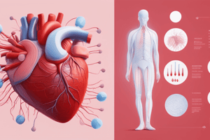
Circulatory System Part 2: The Heart
- The heart is composed of two pumps separated by the septum.
- Each pump has two chambers: an atrium and a ventricle.
- Blood always flows from the atrium to the ventricle.
- The right atrium receives deoxygenated blood from the body (superior and inferior vena cava).
- The right ventricle pumps this deoxygenated blood to the lungs via the pulmonary artery.
- The left atrium receives oxygenated blood from the lungs via the pulmonary veins.
- The left ventricle pumps this oxygenated blood to the body via the aorta.
- Valves (tricuspid, bicuspid/mitral, and semilunar) prevent backflow of blood.
- Atrioventricular (AV) valves prevent backflow from the ventricles to the atria.
- Semilunar valves prevent backflow from the arteries to the ventricles.
- Red text indicates oxygenated blood; blue text indicates deoxygenated blood.
- The pulmonary circuit carries deoxygenated blood to the lungs and oxygenated blood back to the heart.
- The systemic circuit carries oxygenated blood to the body and deoxygenated blood back to the heart.
- The heart's muscle is called cardiac muscle, a type of myogenic muscle that contracts without external nerve stimulation.
- The sinoatrial (SA) node is the heart's pacemaker, setting the heart's tempo. It's a bundle of specialized nerves in the upper right atrium.
- The atrioventricular (AV) node delays the impulse from the SA node, allowing the atria to contract before the ventricles.
- Purkinje fibers carry the nerve signal to the ventricles, causing them to contract.
- The coronary pathway supplies oxygen to the heart muscle. Blockage can lead to a heart attack.
- Diagrams of the heart are typically viewed from the front, meaning the left side of the heart is on the right of the diagram.
Lesson 9 Goals
- Definitions: Includes terms such as pulmonary system, systemic system, septum, atria, ventricle, vena cava, valves (tricuspid, semilunar, bicuspid), aorta, coronary pathway, heart attack, myogenic muscle, SA node, AV node, Purkinje fibers, sympathetic nervous system, and parasympathetic nervous system.
- Curriculum: Outlines learning objectives, including identifying heart structures, blood vessels, and the action of the heart.
Blood Flow Summary
- Blood flow through the heart is summarized in a simplified diagram illustrating the pulmonary and systemic circuits.
- These circuits take blood around the body (systemic) and to the lungs (pulmonary).
Electrocardiograms (ECG/EKG)
- ECGs record the electrical activity of the heart.
- Key components include the P wave (atrial contraction), QRS complex (ventricular contraction), and T wave (ventricular relaxation).
- The graphic representation of the electrical activity of the heart.
Setting the Heart's Tempo
- Heart rate is regulated by the autonomic (involuntary) nervous system, which has two parts:
- Sympathetic nervous system: Prepares the body for stress; increases heart rate
- Parasympathetic nervous system: Returns the body to resting levels after stress; decreases heart rate
What Part of the Heart Makes the "Lub-Dub" Noise?
- The "lub-dub" sound is caused by the closing of heart valves.
- "Lub" is produced by the closing of the AV valves when the ventricles contract.
- "Dub" is produced by the closing of the semilunar valves when the ventricles relax
- These are important sounds for a healthy heart.
Studying That Suits You
Use AI to generate personalized quizzes and flashcards to suit your learning preferences.