Podcast
Questions and Answers
What is the primary function of the corpus callosum?
What is the primary function of the corpus callosum?
- Initiating muscle tone
- Producing cerebrospinal fluid
- Connecting the cerebral hemispheres (correct)
- Processing sensory information
The grey matter is located on the outer surface of the cerebrum with the white matter beneath it.
The grey matter is located on the outer surface of the cerebrum with the white matter beneath it.
True (A)
What are the infoldings or furrows in the cerebral cortex called, and what is their purpose?
What are the infoldings or furrows in the cerebral cortex called, and what is their purpose?
convolutions, increase the surface area
The boundaries of the lobes of the cerebrum are defined by deep ______.
The boundaries of the lobes of the cerebrum are defined by deep ______.
Match the lobes of the cerebrum with their corresponding location under the cranial bones:
Match the lobes of the cerebrum with their corresponding location under the cranial bones:
Which of the following is NOT a primary function associated with the cerebral cortex?
Which of the following is NOT a primary function associated with the cerebral cortex?
The internal capsule connects the cerebral cortex with grey matter of lower parts of the brain and with the spinal cord.
The internal capsule connects the cerebral cortex with grey matter of lower parts of the brain and with the spinal cord.
Name two important masses of grey matter located deep within the cerebral hemispheres.
Name two important masses of grey matter located deep within the cerebral hemispheres.
The basal nuclei are involved in initiating muscle tone in slow and ______ activities.
The basal nuclei are involved in initiating muscle tone in slow and ______ activities.
Which of the following best describes the internal capsule's location?
Which of the following best describes the internal capsule's location?
Which of the following is the primary function of the thalamus?
Which of the following is the primary function of the thalamus?
The hypothalamus is directly connected to the anterior lobe of the pituitary gland via nerve fibers.
The hypothalamus is directly connected to the anterior lobe of the pituitary gland via nerve fibers.
What are the three main components of the brainstem?
What are the three main components of the brainstem?
The ______ is located in front of the cerebellum, below the midbrain and above the medulla oblongata, and acts as a bridge between the two hemispheres of the cerebellum.
The ______ is located in front of the cerebellum, below the midbrain and above the medulla oblongata, and acts as a bridge between the two hemispheres of the cerebellum.
Match the center located in the medulla oblongata with its primary function:
Match the center located in the medulla oblongata with its primary function:
Which of the following stimuli directly influences the respiratory center in the medulla oblongata?
Which of the following stimuli directly influences the respiratory center in the medulla oblongata?
The cerebellum's primary function is initiating voluntary muscle movements.
The cerebellum's primary function is initiating voluntary muscle movements.
What are the sensory inputs that provide information to the cerebellum for maintaining balance and equilibrium?
What are the sensory inputs that provide information to the cerebellum for maintaining balance and equilibrium?
The ______ nervous system transmits vasomotor impulses from the vasomotor center to the blood vessels.
The ______ nervous system transmits vasomotor impulses from the vasomotor center to the blood vessels.
Damage to the cerebellum would most likely result in which of the following?
Damage to the cerebellum would most likely result in which of the following?
Flashcards
Thalamus
Thalamus
Relays sensory information to the cerebrum.
Hypothalamus
Hypothalamus
Controls hormone output, appetite, thirst, body temperature, and emotional reactions.
Brainstem
Brainstem
Connects the cerebrum to the spinal cord; controls basic life-sustaining functions.
Midbrain
Midbrain
Signup and view all the flashcards
Pons
Pons
Signup and view all the flashcards
Medulla Oblongata
Medulla Oblongata
Signup and view all the flashcards
Cardiovascular Center
Cardiovascular Center
Signup and view all the flashcards
Respiratory Center
Respiratory Center
Signup and view all the flashcards
Vasomotor Center
Vasomotor Center
Signup and view all the flashcards
Cerebellum Function
Cerebellum Function
Signup and view all the flashcards
Cerebrum
Cerebrum
Signup and view all the flashcards
Longitudinal Cerebral Fissure
Longitudinal Cerebral Fissure
Signup and view all the flashcards
Corpus Callosum
Corpus Callosum
Signup and view all the flashcards
Cerebral Cortex
Cerebral Cortex
Signup and view all the flashcards
Convolutions
Convolutions
Signup and view all the flashcards
Cerebral Lobes
Cerebral Lobes
Signup and view all the flashcards
Internal Capsule
Internal Capsule
Signup and view all the flashcards
Higher Cerebral Functions
Higher Cerebral Functions
Signup and view all the flashcards
Basal Nuclei Function
Basal Nuclei Function
Signup and view all the flashcards
Basal Nuclei
Basal Nuclei
Signup and view all the flashcards
Study Notes
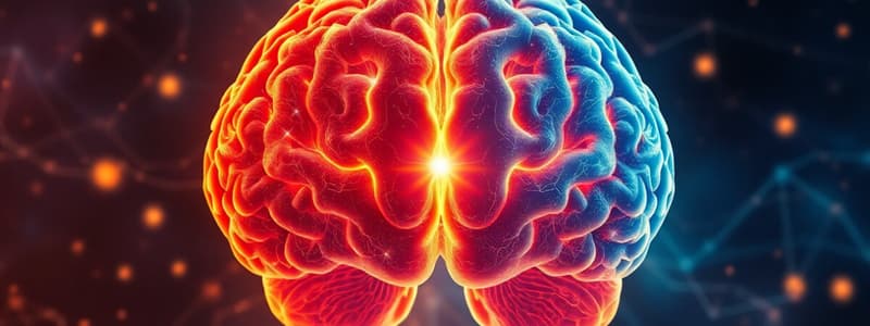
- The Cerebrum is the largest part of the brain, occupying the anterior and middle portion of the brain.
- A deep cleft, the longitudinal cerebral fissure, divides it into right and left cerebral hemispheres.
- Each hemisphere contains one lateral ventricle, which produces and circulates cerebrospinal fluid
- The cerebrospinal fluid cushions and protects the brain and removes toxins and wastes.
- The hemispheres are connected deep within the brain by the corpus callosum, a mass of white matter with myelinated nerve fibres.
- The cerebrum's peripheral part is composed of nerve cell bodies, or grey matter, forming the cerebral cortex.
- The grey matter is located on the outer surface with the white matter beneath it, opposite the arrangement in the spinal cord.
- The cerebral cortex shows many infoldings or furrows called convolutions
- Convolutions greatly increase the surface area of the cerebrum.
- Each hemisphere is divided into lobes named after the cranial bones they lie under: Frontal, Parietal, Temporal, Occipital.
- Deep sulci (fissures) define the boundaries of the lobes.
Interior of the Cerebrum
- Lobes are connected by nerve fibre masses, or tracts, that make up the brain's white matter.
- The internal capsule is an area with projection fibres that connect the cerebral cortex with grey matter and the spinal cord.
- The internal capsule lies deep within the brain between the basal nuclei (ganglia) and the thalamus.
- Many nerve impulses to and from the cerebral cortex are carried by the internal capsule.
Functions of the Cerebrum
- Mental activities such as memory, intelligence, thinking, reasoning, moral sense, and learning.
- Sensory perception, including pain, temperature, touch, sight, hearing, taste, and smell.
- Initiation and control of skeletal (voluntary) muscle contraction.
Other areas of the Cerebrum
- Deep within the cerebral hemispheres are masses of grey matter: Basal nuclei, Thalamus, and Hypothalamus.
- Basal nuclei are areas of grey matter connecting to the cerebral cortex and thalamus, and are involved in initiating muscle tone in slow, coordinated activities
- Inadequate or absent control of the basal nuclei results in jerky, clumsy, and uncoordinated movements.
- The thalamus consists of two masses of nerve cells and fibres within the cerebral hemispheres, below the corpus callosum.
- Sensory input from the skin, viscera, and special sense organs is transmitted to the thalamus before being redistributed to the cerebrum.
- The hypothalamus is composed of groups of nerve cells below and in front of the thalamus, immediately above the pituitary gland.
- The hypothalamus is linked to the posterior pituitary lobe by nerve fibres and to the anterior lobe by a complex system of blood vessels to controls hormone output from both lobes.
Hypothalamus Functions Include Control of
- The autonomic nervous system
- Appetite, thirst, and water balance
- Body temperature and circadian rhythms
- Emotional reactions, such as pleasure, fear, rage, and sexual behaviour
Brain Stem
- The brain stem is a vital structure connecting the cerebrum to the spinal cord, controlling basic life-sustaining functions and reflexes.
- The brainstem consists of three parts: Midbrain, Pons, and Medulla Oblongata.
Midbrain
- Located around the cerebral aqueduct between the cerebrum and the pons.
- It consists of cell bodies and nerve fibres connecting the cerebrum with lower brain parts and the spinal cord.
- The cell bodies act as relay stations for ascending and descending nerve fibres.
Pons
- Situated in front of the cerebellum, below the midbrain and above the medulla oblongata.
- Consists mainly of nerve fibres forming a bridge between the two hemispheres of the cerebellum and fibres passing between the brain and the spinal cord.
- There are groups of cells within the pons which act as relay stations and some of these are associated with the cranial nerves.
- Pons has grey matter lying deeply and white matter lying on the surface.
Medulla Oblongata
- The medulla oblongata connects the brainstem and the spinal cord.
- It extends from the pons and is continuous with the spinal cord.
- It is about 2.5 cm long, with white matter on the outer aspect and grey matter centrally.
- Vital centres, consisting of groups of cells associated with autonomic reflex activity, lie in its deeper structure.
Medulla Oblongata Vital Centers
- Cardiac centre.
- Respiratory centre.
- Vasomotor centre.
- Reflex centres of vomiting, coughing, sneezing, and swallowing.
- The cardiovascular centre controls the rate and force of cardiac contraction.
- Sympathetic stimulation increases the rate and force of the heartbeat, and parasympathetic stimulation has the opposite effect.
- The respiratory centre controls the rate and depth of respiration, stimulating contraction of the diaphragm and intercostal muscles to initiate inspiration.
- The respiratory centre is stimulated by excess carbon dioxide, deficiency of oxygen, and nerve impulses from chemoreceptors in the carotid bodies.
- The vasomotor centre controls the diameter of blood vessels, especially small arteries and arterioles, through the autonomic nervous system, and stimulation may cause constriction or dilatation.
- Sources of stimulation of the vasomotor centre include body temperature and emotions.
- Reflex centres are stimulated by irritating substances in the stomach or respiratory tract, initiating reflex actions to expel the irritant.
Cerebellum
- Situated behind the pons and immediately below the posterior portion of the cerebrum.
- Ovoid in shape with two hemispheres, separated by the vermis.
- Grey matter forms the surface of the cerebellum, and the white matter lies deeply.
Cerebellum Functions
- Coordination of voluntary muscular movement, posture, and balance.
- The cerebellum controls and coordinates the movements of various muscle groups, ensuring smooth, even, and precise actions.
- The cerebellum coordinates activities associated with balance and equilibrium, with sensory input from the muscles, joints, eyes, and ears.
- Impulses from the cerebellum influence the contraction of skeletal muscle to maintain balance and posture.
- Damage to the cerebellum results in clumsy, uncoordinated muscular movement, staggering gait, and inability to carry out smooth, steady, precise movements.
Studying That Suits You
Use AI to generate personalized quizzes and flashcards to suit your learning preferences.


