Podcast
Questions and Answers
What major structures comprise the central nervous system (CNS)?
What major structures comprise the central nervous system (CNS)?
- Brain and spinal cord (correct)
- Spinal cord and sensory organs
- Brain and peripheral nerves
- Nerves and ganglia
From which structure does the brain develop during embryonic development?
From which structure does the brain develop during embryonic development?
- Neural crest
- Ectoderm
- Mesoderm
- Neural tube (correct)
What is the process by which the brain develops folds?
What is the process by which the brain develops folds?
- Gyrification (correct)
- Growth migration
- Neurogenesis
- Neural differentiation
Which lobe of the cerebral hemisphere is primarily responsible for processing visual information?
Which lobe of the cerebral hemisphere is primarily responsible for processing visual information?
Which of the following is NOT a function of the cerebral cortex?
Which of the following is NOT a function of the cerebral cortex?
How many lobes are present in each cerebral hemisphere?
How many lobes are present in each cerebral hemisphere?
Which area of the cerebral cortex is responsible for voluntary movement?
Which area of the cerebral cortex is responsible for voluntary movement?
What type of brain structure does the telencephalon develop into?
What type of brain structure does the telencephalon develop into?
What are the cavities within the brain that contain cerebrospinal fluid called?
What are the cavities within the brain that contain cerebrospinal fluid called?
Which type of neuroglia is responsible for producing cerebrospinal fluid?
Which type of neuroglia is responsible for producing cerebrospinal fluid?
What does the motor homunculus represent?
What does the motor homunculus represent?
Which area of the brain is responsible for planning and sequencing movements?
Which area of the brain is responsible for planning and sequencing movements?
What is the function of Broca's area?
What is the function of Broca's area?
Which part of the brain is involved in regulating temperature and hunger?
Which part of the brain is involved in regulating temperature and hunger?
What does the primary auditory cortex do?
What does the primary auditory cortex do?
What role does the cerebellum play in the body?
What role does the cerebellum play in the body?
Which layer of the meninges is the outermost?
Which layer of the meninges is the outermost?
What connects the medulla oblongata to the spinal cord?
What connects the medulla oblongata to the spinal cord?
What is the main function of the thalamus?
What is the main function of the thalamus?
What type of neurons primarily compose the gray matter of the spinal cord?
What type of neurons primarily compose the gray matter of the spinal cord?
What are the three primary brain vesicles formed from the expanding neural tube?
What are the three primary brain vesicles formed from the expanding neural tube?
Which layer of the cerebral hemispheres contains nerve cell bodies and nonmyelinated fibers?
Which layer of the cerebral hemispheres contains nerve cell bodies and nonmyelinated fibers?
Which brain structure is involved in regulating sleep cycles and emotional responses?
Which brain structure is involved in regulating sleep cycles and emotional responses?
What is the primary role of astrocytes in the central nervous system?
What is the primary role of astrocytes in the central nervous system?
Which part of the brain is composed of the anterior and posterior association areas?
Which part of the brain is composed of the anterior and posterior association areas?
Which structure serves as a protective barrier that separates the brain from the skull?
Which structure serves as a protective barrier that separates the brain from the skull?
What type of cells form myelin sheaths around neurons in the central nervous system?
What type of cells form myelin sheaths around neurons in the central nervous system?
Which area processes sensory information and is involved in creating a rational understanding of objects?
Which area processes sensory information and is involved in creating a rational understanding of objects?
Where is cerebrospinal fluid produced within the brain?
Where is cerebrospinal fluid produced within the brain?
From which embryonic structure does the spinal cord develop?
From which embryonic structure does the spinal cord develop?
What is the role of oligodendrocytes in the central nervous system?
What is the role of oligodendrocytes in the central nervous system?
Which lobe of the cerebral hemisphere is associated with perception of taste?
Which lobe of the cerebral hemisphere is associated with perception of taste?
The primary function of the cerebellum is to:
The primary function of the cerebellum is to:
Which structure is responsible for relaying sensory information to the cerebral cortex?
Which structure is responsible for relaying sensory information to the cerebral cortex?
What separates the gray matter of the spinal cord into dorsal and ventral horns?
What separates the gray matter of the spinal cord into dorsal and ventral horns?
Which areas of the cerebral cortex are primarily responsible for higher mental functions such as language?
Which areas of the cerebral cortex are primarily responsible for higher mental functions such as language?
What characteristic feature of the brain maximizes its surface area?
What characteristic feature of the brain maximizes its surface area?
Which part of the brainstem is primarily involved in the regulation of autonomic functions like heart rate and breathing?
Which part of the brainstem is primarily involved in the regulation of autonomic functions like heart rate and breathing?
The cerebral cortex consists of how many layers?
The cerebral cortex consists of how many layers?
What is the primary role of the hypothalamus in the brain?
What is the primary role of the hypothalamus in the brain?
What develops from the posterior end of the neural tube?
What develops from the posterior end of the neural tube?
Which structure is responsible for forming myelin sheaths in the central nervous system?
Which structure is responsible for forming myelin sheaths in the central nervous system?
Which part of the adult brain is NOT one of its four main regions?
Which part of the adult brain is NOT one of its four main regions?
What fills the ventricles of the brain?
What fills the ventricles of the brain?
Which region of the cerebral hemispheres is primarily associated with conscious thought?
Which region of the cerebral hemispheres is primarily associated with conscious thought?
What are the grooves on the surface of the cerebral hemispheres called?
What are the grooves on the surface of the cerebral hemispheres called?
Which type of neuroglia is responsible for monitoring neuron health?
Which type of neuroglia is responsible for monitoring neuron health?
Which division of the brain is responsible for regulating autonomic functions such as heart rate?
Which division of the brain is responsible for regulating autonomic functions such as heart rate?
Which structures arise from the primary brain vesicles during development?
Which structures arise from the primary brain vesicles during development?
How many lobes are present in each cerebral hemisphere?
How many lobes are present in each cerebral hemisphere?
What is the role of the primary motor cortex?
What is the role of the primary motor cortex?
Which area processes visual information and integrates it?
Which area processes visual information and integrates it?
What function does the somatosensory association cortex serve?
What function does the somatosensory association cortex serve?
Which part of the diencephalon regulates hunger and thirst?
Which part of the diencephalon regulates hunger and thirst?
Which structure is primarily responsible for coordinating movement?
Which structure is primarily responsible for coordinating movement?
What is the function of the anterior association area?
What is the function of the anterior association area?
What protects the brain from physical damage?
What protects the brain from physical damage?
Which part of the brainstem connects the forebrain and hindbrain?
Which part of the brainstem connects the forebrain and hindbrain?
Which part of the spinal cord sends motor signals?
Which part of the spinal cord sends motor signals?
Which type of tract carries sensory information to the brain?
Which type of tract carries sensory information to the brain?
Which of the following accurately describes the Peripheral Nervous System (PNS)?
Which of the following accurately describes the Peripheral Nervous System (PNS)?
Which structure forms the Central Nervous System (CNS) during embryonic development?
Which structure forms the Central Nervous System (CNS) during embryonic development?
What is a key characteristic of white matter in the nervous system?
What is a key characteristic of white matter in the nervous system?
What type of collection of cell bodies is referred to as a ganglion?
What type of collection of cell bodies is referred to as a ganglion?
Which part of the brain is associated with the hindbrain?
Which part of the brain is associated with the hindbrain?
Which type of sensory sensation is associated with proprioception?
Which type of sensory sensation is associated with proprioception?
What forms the fourth ventricle in the brain?
What forms the fourth ventricle in the brain?
What structures connect parts of the CNS on one side to the same parts on the opposite side?
What structures connect parts of the CNS on one side to the same parts on the opposite side?
Where is gray matter primarily located in the brain?
Where is gray matter primarily located in the brain?
What is the main characteristic that differentiates the somatic nervous system from the autonomic nervous system?
What is the main characteristic that differentiates the somatic nervous system from the autonomic nervous system?
Which cranial nerve is responsible for tongue movement?
Which cranial nerve is responsible for tongue movement?
Which part of the nervous system is known as the craniosacral nervous system?
Which part of the nervous system is known as the craniosacral nervous system?
Where do cranial nerves IX, X, XI, and XII exit the brainstem?
Where do cranial nerves IX, X, XI, and XII exit the brainstem?
What do the myenteric and submucosal plexuses belong to?
What do the myenteric and submucosal plexuses belong to?
Which cranial nerve is responsible for facial sensation?
Which cranial nerve is responsible for facial sensation?
Which center is NOT contained in the medulla oblongata?
Which center is NOT contained in the medulla oblongata?
What connects the third and fourth ventricles in the brain?
What connects the third and fourth ventricles in the brain?
What does the hypothalamus regulate?
What does the hypothalamus regulate?
Which cranial nerve supplies the parotid gland?
Which cranial nerve supplies the parotid gland?
What characterizes preganglionic fibers in the autonomic system?
What characterizes preganglionic fibers in the autonomic system?
Which part of the nervous system consists of the brain and spinal cord?
Which part of the nervous system consists of the brain and spinal cord?
What type of pathways carry sensory information towards the CNS?
What type of pathways carry sensory information towards the CNS?
What are the cranial nerves?
What are the cranial nerves?
Which nervous system controls involuntary functions?
Which nervous system controls involuntary functions?
How many segments does the spinal cord have?
How many segments does the spinal cord have?
What response is associated with the sympathetic nervous system?
What response is associated with the sympathetic nervous system?
Which part of the brain is located superior to the spinal cord?
Which part of the brain is located superior to the spinal cord?
Which division of the autonomic nervous system is responsible for rest-and-digest activities?
Which division of the autonomic nervous system is responsible for rest-and-digest activities?
Which type of nerves correspond to spinal cord segments?
Which type of nerves correspond to spinal cord segments?
Which system is primarily responsible for voluntary movements?
Which system is primarily responsible for voluntary movements?
What appearance do unmyelinated fibers have in the nervous system?
What appearance do unmyelinated fibers have in the nervous system?
Which structures are primarily responsible for myelination in the central nervous system?
Which structures are primarily responsible for myelination in the central nervous system?
Where is the fourth ventricle located in the brain?
Where is the fourth ventricle located in the brain?
Which cranial nerves provide motor function to the facial muscles?
Which cranial nerves provide motor function to the facial muscles?
Which cranial nerve is responsible for controlling the movement of the tongue?
Which cranial nerve is responsible for controlling the movement of the tongue?
Which division of the autonomic nervous system arises from the thoracolumbar region?
Which division of the autonomic nervous system arises from the thoracolumbar region?
What is the function of the inferior salivatory nucleus?
What is the function of the inferior salivatory nucleus?
Which of the following components makes up the craniosacral division of the autonomic nervous system?
Which of the following components makes up the craniosacral division of the autonomic nervous system?
What is a ganglion in the peripheral nervous system?
What is a ganglion in the peripheral nervous system?
What is the role of the myenteric plexus in the autonomic nervous system?
What is the role of the myenteric plexus in the autonomic nervous system?
What component is part of the central nervous system (CNS)?
What component is part of the central nervous system (CNS)?
Which term describes sensory function in the nervous system?
Which term describes sensory function in the nervous system?
How many segments does the spinal cord approximately have?
How many segments does the spinal cord approximately have?
Which of the following statements accurately describes myelinated fibers?
Which of the following statements accurately describes myelinated fibers?
What is a ganglion?
What is a ganglion?
Which system includes the sympathetic and parasympathetic nervous systems?
Which system includes the sympathetic and parasympathetic nervous systems?
In which matter are unmyelinated fibers primarily found?
In which matter are unmyelinated fibers primarily found?
What is the function of the brain stem?
What is the function of the brain stem?
What does a tract refer to in the central nervous system?
What does a tract refer to in the central nervous system?
What term describes sensations of position, muscle, and joint movement?
What term describes sensations of position, muscle, and joint movement?
What structures does the neural tube develop into?
What structures does the neural tube develop into?
Which cranial nerves emerge from the pons?
Which cranial nerves emerge from the pons?
Which part of the autonomic nervous system is responsible for 'rest and digest' responses?
Which part of the autonomic nervous system is responsible for 'rest and digest' responses?
What is the main function of cranial nerve 7 (facial nerve)?
What is the main function of cranial nerve 7 (facial nerve)?
What are the main components of the midbrain?
What are the main components of the midbrain?
Which cranial nerve is responsible for tasting on the posterior 1/3 of the tongue?
Which cranial nerve is responsible for tasting on the posterior 1/3 of the tongue?
What is the primary function of the vagus nerve (CN X)?
What is the primary function of the vagus nerve (CN X)?
Which cranial nerve is specifically involved with the movement of the eyeball?
Which cranial nerve is specifically involved with the movement of the eyeball?
Where does the superior salivatory nucleus reside?
Where does the superior salivatory nucleus reside?
Which division of the autonomic nervous system is referred to as 'thoracolumbar'?
Which division of the autonomic nervous system is referred to as 'thoracolumbar'?
What is the total number of spinal nerve pairs in the human body?
What is the total number of spinal nerve pairs in the human body?
Which segment of the spinal cord is responsible for innervating the upper limbs?
Which segment of the spinal cord is responsible for innervating the upper limbs?
At which anatomical location does the conus medullaris typically end?
At which anatomical location does the conus medullaris typically end?
Which component of the spinal cord contains neuron cell bodies and dendrites?
Which component of the spinal cord contains neuron cell bodies and dendrites?
How do spinal nerves exit the vertebral column?
How do spinal nerves exit the vertebral column?
What is a primary characteristic of white matter in the spinal cord?
What is a primary characteristic of white matter in the spinal cord?
Which spinal nerves exit above their corresponding vertebrae?
Which spinal nerves exit above their corresponding vertebrae?
Which area of the spinal cord experiences an increase in grey matter as it descends?
Which area of the spinal cord experiences an increase in grey matter as it descends?
What resembles a horse's tail at the end of the spinal cord?
What resembles a horse's tail at the end of the spinal cord?
How many segments are in the thoracic region of the spinal cord?
How many segments are in the thoracic region of the spinal cord?
What is gray matter primarily composed of?
What is gray matter primarily composed of?
Which type of neurons is found in the dorsal horn of the spinal cord?
Which type of neurons is found in the dorsal horn of the spinal cord?
What is the primary function of myelin in the nervous system?
What is the primary function of myelin in the nervous system?
What distinguishes white matter from gray matter in the spinal cord?
What distinguishes white matter from gray matter in the spinal cord?
Where are spinal nerves formed from?
Where are spinal nerves formed from?
What is the function of ascending tracts in white matter?
What is the function of ascending tracts in white matter?
In which regions of the spinal cord is the ventral horn larger?
In which regions of the spinal cord is the ventral horn larger?
What is the role of oligodendrocytes in the central nervous system?
What is the role of oligodendrocytes in the central nervous system?
Which structure is responsible for connecting sensory input with motor output within the spinal cord?
Which structure is responsible for connecting sensory input with motor output within the spinal cord?
What is the composition of the dorsal root of a spinal nerve?
What is the composition of the dorsal root of a spinal nerve?
What structures make up the peripheral nervous system?
What structures make up the peripheral nervous system?
Which region of the spinal cord contains the highest number of spinal nerves?
Which region of the spinal cord contains the highest number of spinal nerves?
What is the conus medullaris?
What is the conus medullaris?
How many pairs of spinal nerves are in the human body?
How many pairs of spinal nerves are in the human body?
Which part of the spinal cord is primarily responsible for conveying motor information?
Which part of the spinal cord is primarily responsible for conveying motor information?
Where does sensory information from the coccygeal segment move within the spinal cord?
Where does sensory information from the coccygeal segment move within the spinal cord?
What type of matter decreases in size from the cervical to the coccygeal segments of the spinal cord?
What type of matter decreases in size from the cervical to the coccygeal segments of the spinal cord?
What is the role of the ventral gray horn in the spinal cord?
What is the role of the ventral gray horn in the spinal cord?
What structure resembles a horse's tail at the lower end of the spine?
What structure resembles a horse's tail at the lower end of the spine?
Which area of the spinal cord increases in size from cervical to coccygeal segments?
Which area of the spinal cord increases in size from cervical to coccygeal segments?
What is the primary role of myelin sheaths in neurons?
What is the primary role of myelin sheaths in neurons?
Which gray matter structure is involved in sensory processing?
Which gray matter structure is involved in sensory processing?
Which spinal cord region contains larger muscles requiring more motor innervation?
Which spinal cord region contains larger muscles requiring more motor innervation?
What structures connect ventral rami with autonomic ganglia?
What structures connect ventral rami with autonomic ganglia?
What is the appearance of axons due to the presence of myelin?
What is the appearance of axons due to the presence of myelin?
Which of the following statements about the dorsal root is correct?
Which of the following statements about the dorsal root is correct?
Where are the lateral gray horns located?
Where are the lateral gray horns located?
What comprises the gray matter of the spinal cord?
What comprises the gray matter of the spinal cord?
What is the primary function of the ventral root?
What is the primary function of the ventral root?
Which structure serves as a groove on the anterior aspect of the spinal cord?
Which structure serves as a groove on the anterior aspect of the spinal cord?
What is the primary function of Schwann cells in the peripheral nervous system?
What is the primary function of Schwann cells in the peripheral nervous system?
Which part of the spinal cord contains motor neurons?
Which part of the spinal cord contains motor neurons?
Where are the dorsal root ganglia located?
Where are the dorsal root ganglia located?
Which structure forms when the dorsal and ventral roots fuse?
Which structure forms when the dorsal and ventral roots fuse?
What type of information does the dorsal ramus supply?
What type of information does the dorsal ramus supply?
Which part of the spinal cord's gray matter is associated with sensory neurons?
Which part of the spinal cord's gray matter is associated with sensory neurons?
What is the role of the lateral gray horn in the spinal cord?
What is the role of the lateral gray horn in the spinal cord?
Which type of neurons primarily operate in the ventral gray horn?
Which type of neurons primarily operate in the ventral gray horn?
What combination of roots and branches forms a spinal nerve?
What combination of roots and branches forms a spinal nerve?
Which of the following is NOT found in the white matter of the spinal cord?
Which of the following is NOT found in the white matter of the spinal cord?
What is the total number of spinal nerve pairs in the human body?
What is the total number of spinal nerve pairs in the human body?
Where does the spinal cord begin and end?
Where does the spinal cord begin and end?
Which segment of the spinal cord has the highest number of spinal nerve pairs?
Which segment of the spinal cord has the highest number of spinal nerve pairs?
What composes the gray matter of the spinal cord?
What composes the gray matter of the spinal cord?
The conus medullaris is located at which vertebral level?
The conus medullaris is located at which vertebral level?
What structure resembles a horse's tail and consists of spinal nerves?
What structure resembles a horse's tail and consists of spinal nerves?
What differentiates cervical spinal nerves from thoracic and lumbar spinal nerves in terms of their exit point?
What differentiates cervical spinal nerves from thoracic and lumbar spinal nerves in terms of their exit point?
What is the function of oligodendrocytes in the spinal cord?
What is the function of oligodendrocytes in the spinal cord?
What anatomical features increase in the spinal cord as you move down from cervical to coccygeal segments?
What anatomical features increase in the spinal cord as you move down from cervical to coccygeal segments?
What part of the neuron is primarily responsible for carrying signals away from the cell body?
What part of the neuron is primarily responsible for carrying signals away from the cell body?
What type of neurons are predominantly found in the anterior gray horn of the spinal cord?
What type of neurons are predominantly found in the anterior gray horn of the spinal cord?
Which of the following describes the function of the posterior gray horn?
Which of the following describes the function of the posterior gray horn?
Where are the lateral gray horns located?
Where are the lateral gray horns located?
What is the primary function of the white columns in the spinal cord?
What is the primary function of the white columns in the spinal cord?
What do the dorsal and ventral roots of spinal nerves primarily contain?
What do the dorsal and ventral roots of spinal nerves primarily contain?
Which structure connects the ventral rami to ganglia?
Which structure connects the ventral rami to ganglia?
What is the function of the cauda equina?
What is the function of the cauda equina?
Where is the sensory neuron cell body located?
Where is the sensory neuron cell body located?
Which segment of the spinal cord is associated with the largest amount of gray matter?
Which segment of the spinal cord is associated with the largest amount of gray matter?
What structural feature of the spinal cord reduces as you descend from the cervical to coccygeal segments?
What structural feature of the spinal cord reduces as you descend from the cervical to coccygeal segments?
What type of neurons does the ventral root of the spinal nerve contain?
What type of neurons does the ventral root of the spinal nerve contain?
Where does the spinal cord begin?
Where does the spinal cord begin?
What do the dorsal rami primarily supply?
What do the dorsal rami primarily supply?
Which type of neuroglia forms myelin sheaths in the central nervous system?
Which type of neuroglia forms myelin sheaths in the central nervous system?
Which spinal nerve structure contains mixed motor and sensory information?
Which spinal nerve structure contains mixed motor and sensory information?
What is the anatomical term for the main part of a neuron that contains the nucleus?
What is the anatomical term for the main part of a neuron that contains the nucleus?
What is the name of the bulbous portion of the spinal cord associated with upper limb muscles?
What is the name of the bulbous portion of the spinal cord associated with upper limb muscles?
Which part of the spinal cord anatomy is located around the vertebral levels L1-L2?
Which part of the spinal cord anatomy is located around the vertebral levels L1-L2?
What characteristic of myelin sheaths contributes to faster signal transmission?
What characteristic of myelin sheaths contributes to faster signal transmission?
During which segment of the spinal cord does the number of spinal nerves peak?
During which segment of the spinal cord does the number of spinal nerves peak?
What is the function of the cervical enlargement in the spinal cord?
What is the function of the cervical enlargement in the spinal cord?
Which structure marks the pointed end of the spinal cord?
Which structure marks the pointed end of the spinal cord?
How many pairs of spinal nerves are present in the human body?
How many pairs of spinal nerves are present in the human body?
What type of myelin-producing cell is found in the peripheral nervous system?
What type of myelin-producing cell is found in the peripheral nervous system?
How does the volume of white matter change from the cervical to coccygeal segments?
How does the volume of white matter change from the cervical to coccygeal segments?
What does the term 'cauda equina' refer to in relation to the spinal cord?
What does the term 'cauda equina' refer to in relation to the spinal cord?
What is the primary composition of gray matter in the spinal cord?
What is the primary composition of gray matter in the spinal cord?
In which part of the spinal cord is the anterior gray horn located?
In which part of the spinal cord is the anterior gray horn located?
What is the role of myelin sheaths in the nervous system?
What is the role of myelin sheaths in the nervous system?
Where does the first cervical spinal nerve exit in relation to the vertebrae?
Where does the first cervical spinal nerve exit in relation to the vertebrae?
What is the function of the dorsal root in the spinal nerve anatomy?
What is the function of the dorsal root in the spinal nerve anatomy?
Which white column is located posterior to the gray matter in the spinal cord?
Which white column is located posterior to the gray matter in the spinal cord?
Where are lateral gray horns found in the spinal cord?
Where are lateral gray horns found in the spinal cord?
What is a ganglion in the context of spinal nerve anatomy?
What is a ganglion in the context of spinal nerve anatomy?
What does the ventral gray horn primarily contain?
What does the ventral gray horn primarily contain?
Which structure forms when the dorsal and ventral roots fuse?
Which structure forms when the dorsal and ventral roots fuse?
What type of fibers does the spinal nerve contain?
What type of fibers does the spinal nerve contain?
What is the primary role of the white columns in the spinal cord?
What is the primary role of the white columns in the spinal cord?
What is the dorsal ramus responsible for supplying?
What is the dorsal ramus responsible for supplying?
What type of neurons are primarily located in the posterior gray horn?
What type of neurons are primarily located in the posterior gray horn?
What is the main function of the posterior gray horn in the spinal cord?
What is the main function of the posterior gray horn in the spinal cord?
Which structure contains preganglionic motor neurons of the sympathetic nervous system?
Which structure contains preganglionic motor neurons of the sympathetic nervous system?
What is the primary component of white matter in the spinal cord?
What is the primary component of white matter in the spinal cord?
What distinguishes the dorsal root from the ventral root?
What distinguishes the dorsal root from the ventral root?
Which of the following describes the ventral white column?
Which of the following describes the ventral white column?
Which cellular structure forms the myelin sheath in the peripheral nervous system?
Which cellular structure forms the myelin sheath in the peripheral nervous system?
What is the function of the anterior (ventral) gray horn?
What is the function of the anterior (ventral) gray horn?
What separates the white matter from the gray matter in the spinal cord?
What separates the white matter from the gray matter in the spinal cord?
Which of the following correctly describes the function of spinal nerves?
Which of the following correctly describes the function of spinal nerves?
Where are ascending tracts located in the spinal cord?
Where are ascending tracts located in the spinal cord?
What is the first structure sensory information encounters after entering the spinal nerve?
What is the first structure sensory information encounters after entering the spinal nerve?
Which structure is primarily responsible for transmitting motor information from the brain toward muscles?
Which structure is primarily responsible for transmitting motor information from the brain toward muscles?
What type of neurons carry sensory information from the body to the spinal cord?
What type of neurons carry sensory information from the body to the spinal cord?
In the context of spinal nerve function, which of the following structures is a cluster of sensory neuron cell bodies?
In the context of spinal nerve function, which of the following structures is a cluster of sensory neuron cell bodies?
Which part of the spinal nerve carries primarily sensory information?
Which part of the spinal nerve carries primarily sensory information?
What is the primary function of the anterior (ventral) gray horn?
What is the primary function of the anterior (ventral) gray horn?
What do the dorsal root and ventral root of the spinal nerve mainly consist of?
What do the dorsal root and ventral root of the spinal nerve mainly consist of?
The term 'tract' in the context of the CNS refers to which of the following?
The term 'tract' in the context of the CNS refers to which of the following?
Where does sensory information synapse after entering the dorsal root?
Where does sensory information synapse after entering the dorsal root?
What is the main role of ganglia in the peripheral nervous system (PNS)?
What is the main role of ganglia in the peripheral nervous system (PNS)?
Flashcards
CNS
CNS
The central nervous system, comprising the brain and spinal cord.
Brain Development
Brain Development
The brain develops from a neural tube in the embryo, forming primary vesicles then secondary vesicles.
Cerebral Hemispheres
Cerebral Hemispheres
Major brain region, responsible for most higher thought.
Gyri & Sulci
Gyri & Sulci
Signup and view all the flashcards
Longitudinal Fissure
Longitudinal Fissure
Signup and view all the flashcards
Cerebral Cortex
Cerebral Cortex
Signup and view all the flashcards
Primary Motor Cortex
Primary Motor Cortex
Signup and view all the flashcards
Brain Ventricles
Brain Ventricles
Signup and view all the flashcards
Brain Stem
Brain Stem
Signup and view all the flashcards
Motor Areas
Motor Areas
Signup and view all the flashcards
Motor Homunculus
Motor Homunculus
Signup and view all the flashcards
Premotor Cortex
Premotor Cortex
Signup and view all the flashcards
Broca's Area
Broca's Area
Signup and view all the flashcards
Frontal Eye Field
Frontal Eye Field
Signup and view all the flashcards
Primary Somatosensory Cortex
Primary Somatosensory Cortex
Signup and view all the flashcards
Diencephalon
Diencephalon
Signup and view all the flashcards
Cerebellum
Cerebellum
Signup and view all the flashcards
Meninges
Meninges
Signup and view all the flashcards
Spinal Cord Gray Matter
Spinal Cord Gray Matter
Signup and view all the flashcards
Spinal Cord White Matter
Spinal Cord White Matter
Signup and view all the flashcards
Brain Development Stages
Brain Development Stages
Signup and view all the flashcards
Cerebral Hemispheres Lobes
Cerebral Hemispheres Lobes
Signup and view all the flashcards
Cerebral Cortex Layers
Cerebral Cortex Layers
Signup and view all the flashcards
Ventricles Function
Ventricles Function
Signup and view all the flashcards
Brain Stem Function
Brain Stem Function
Signup and view all the flashcards
Primary Motor Cortex
Primary Motor Cortex
Signup and view all the flashcards
Thalamus Function
Thalamus Function
Signup and view all the flashcards
Hypothalamus Function
Hypothalamus Function
Signup and view all the flashcards
Spinal Cord White Matter
Spinal Cord White Matter
Signup and view all the flashcards
Meninges Function
Meninges Function
Signup and view all the flashcards
CNS Components
CNS Components
Signup and view all the flashcards
Brain Development Stages
Brain Development Stages
Signup and view all the flashcards
Brain Regions
Brain Regions
Signup and view all the flashcards
Cerebral Cortex Layers
Cerebral Cortex Layers
Signup and view all the flashcards
Ventricles Function
Ventricles Function
Signup and view all the flashcards
Brain Stem Function
Brain Stem Function
Signup and view all the flashcards
Meninges Protection
Meninges Protection
Signup and view all the flashcards
Spinal Cord Gray Matter
Spinal Cord Gray Matter
Signup and view all the flashcards
Hypothalamus Function
Hypothalamus Function
Signup and view all the flashcards
Thalamus Function
Thalamus Function
Signup and view all the flashcards
CNS Components
CNS Components
Signup and view all the flashcards
Brain Development Stages
Brain Development Stages
Signup and view all the flashcards
Brain Regions
Brain Regions
Signup and view all the flashcards
Cerebral Hemispheres Lobes
Cerebral Hemispheres Lobes
Signup and view all the flashcards
Cerebral Cortex
Cerebral Cortex
Signup and view all the flashcards
Ventricles Function
Ventricles Function
Signup and view all the flashcards
Brain Stem Function
Brain Stem Function
Signup and view all the flashcards
Cerebral Cortex Layers
Cerebral Cortex Layers
Signup and view all the flashcards
Cerebellum Function
Cerebellum Function
Signup and view all the flashcards
Meninges Protection
Meninges Protection
Signup and view all the flashcards
Motor Areas
Motor Areas
Signup and view all the flashcards
Sensory Areas
Sensory Areas
Signup and view all the flashcards
Association Areas
Association Areas
Signup and view all the flashcards
Primary Motor Cortex
Primary Motor Cortex
Signup and view all the flashcards
Diencephalon
Diencephalon
Signup and view all the flashcards
Thalamus
Thalamus
Signup and view all the flashcards
Cerebellum
Cerebellum
Signup and view all the flashcards
Meninges
Meninges
Signup and view all the flashcards
Spinal Cord Gray Matter
Spinal Cord Gray Matter
Signup and view all the flashcards
Spinal Cord White Matter
Spinal Cord White Matter
Signup and view all the flashcards
Central Nervous System (CNS)
Central Nervous System (CNS)
Signup and view all the flashcards
Peripheral Nervous System (PNS)
Peripheral Nervous System (PNS)
Signup and view all the flashcards
Motor/Efferent
Motor/Efferent
Signup and view all the flashcards
Sensory/Afferent
Sensory/Afferent
Signup and view all the flashcards
Nucleus (CNS)
Nucleus (CNS)
Signup and view all the flashcards
Ganglion (PNS)
Ganglion (PNS)
Signup and view all the flashcards
White Matter
White Matter
Signup and view all the flashcards
Gray Matter
Gray Matter
Signup and view all the flashcards
Spinal Cord Segments
Spinal Cord Segments
Signup and view all the flashcards
Neuroectoderm
Neuroectoderm
Signup and view all the flashcards
Third Ventricle Location
Third Ventricle Location
Signup and view all the flashcards
Lateral Ventricles Location
Lateral Ventricles Location
Signup and view all the flashcards
Cerebral Aqueduct of Sylvius
Cerebral Aqueduct of Sylvius
Signup and view all the flashcards
Cranial Nerves I & II Origin
Cranial Nerves I & II Origin
Signup and view all the flashcards
Cranial Nerves III & IV Origin
Cranial Nerves III & IV Origin
Signup and view all the flashcards
Cranial Nerves V, VI, VII, & VIII Origin
Cranial Nerves V, VI, VII, & VIII Origin
Signup and view all the flashcards
Cranial Nerves IX, X, XI, & XII Origin
Cranial Nerves IX, X, XI, & XII Origin
Signup and view all the flashcards
Amygdala Function
Amygdala Function
Signup and view all the flashcards
Hypothalamus Function
Hypothalamus Function
Signup and view all the flashcards
Midbrain Reflex Centers
Midbrain Reflex Centers
Signup and view all the flashcards
Central Nervous System (CNS)
Central Nervous System (CNS)
Signup and view all the flashcards
Peripheral Nervous System (PNS)
Peripheral Nervous System (PNS)
Signup and view all the flashcards
Motor Function
Motor Function
Signup and view all the flashcards
Sensory Function
Sensory Function
Signup and view all the flashcards
Efferent Pathway
Efferent Pathway
Signup and view all the flashcards
Afferent Pathway
Afferent Pathway
Signup and view all the flashcards
Cranial Nerves
Cranial Nerves
Signup and view all the flashcards
Spinal Nerves
Spinal Nerves
Signup and view all the flashcards
Somatic Nervous System
Somatic Nervous System
Signup and view all the flashcards
Autonomic Nervous System
Autonomic Nervous System
Signup and view all the flashcards
Unmyelinated Fibers
Unmyelinated Fibers
Signup and view all the flashcards
Gray Matter Location (Spinal Cord)
Gray Matter Location (Spinal Cord)
Signup and view all the flashcards
White Matter Location (Spinal Cord)
White Matter Location (Spinal Cord)
Signup and view all the flashcards
Nucleus (CNS)
Nucleus (CNS)
Signup and view all the flashcards
Ganglion (PNS)
Ganglion (PNS)
Signup and view all the flashcards
Cranial Nerves 3,4 Origin
Cranial Nerves 3,4 Origin
Signup and view all the flashcards
Fourth Ventricle Connection
Fourth Ventricle Connection
Signup and view all the flashcards
Autonomic Neuron Ganglion
Autonomic Neuron Ganglion
Signup and view all the flashcards
Enteric Nervous System Location
Enteric Nervous System Location
Signup and view all the flashcards
Thoracolumbar Division Location
Thoracolumbar Division Location
Signup and view all the flashcards
Ipsilateral
Ipsilateral
Signup and view all the flashcards
Contralateral
Contralateral
Signup and view all the flashcards
Prosencephalon
Prosencephalon
Signup and view all the flashcards
Mesencephalon
Mesencephalon
Signup and view all the flashcards
Rhombencephalon
Rhombencephalon
Signup and view all the flashcards
Cranial Nerve 3
Cranial Nerve 3
Signup and view all the flashcards
Cranial Nerve 5
Cranial Nerve 5
Signup and view all the flashcards
Sympathetic Nervous System
Sympathetic Nervous System
Signup and view all the flashcards
Parasympathetic Nervous System
Parasympathetic Nervous System
Signup and view all the flashcards
Fourth Ventricle
Fourth Ventricle
Signup and view all the flashcards
Central Nervous System (CNS)
Central Nervous System (CNS)
Signup and view all the flashcards
Peripheral Nervous System (PNS)
Peripheral Nervous System (PNS)
Signup and view all the flashcards
Motor Function (Efferent)
Motor Function (Efferent)
Signup and view all the flashcards
Sensory Function (Afferent)
Sensory Function (Afferent)
Signup and view all the flashcards
White Matter
White Matter
Signup and view all the flashcards
Gray Matter
Gray Matter
Signup and view all the flashcards
Ganglion
Ganglion
Signup and view all the flashcards
Nucleus
Nucleus
Signup and view all the flashcards
Spinal Cord Segments
Spinal Cord Segments
Signup and view all the flashcards
Autonomic Nervous System
Autonomic Nervous System
Signup and view all the flashcards
Spinal Cord segments
Spinal Cord segments
Signup and view all the flashcards
Spinal Nerve Pairs
Spinal Nerve Pairs
Signup and view all the flashcards
Conus Medullaris
Conus Medullaris
Signup and view all the flashcards
Cauda Equina
Cauda Equina
Signup and view all the flashcards
Cervical Enlargement
Cervical Enlargement
Signup and view all the flashcards
Lumbar Enlargement
Lumbar Enlargement
Signup and view all the flashcards
Spinal Nerve Exit
Spinal Nerve Exit
Signup and view all the flashcards
Grey matter (Spinal Cord)
Grey matter (Spinal Cord)
Signup and view all the flashcards
White matter (Spinal Cord)
White matter (Spinal Cord)
Signup and view all the flashcards
Intervertebral Foramina
Intervertebral Foramina
Signup and view all the flashcards
Gray Matter Composition
Gray Matter Composition
Signup and view all the flashcards
White Matter Composition
White Matter Composition
Signup and view all the flashcards
Myelin Function
Myelin Function
Signup and view all the flashcards
Ventral Horn Function
Ventral Horn Function
Signup and view all the flashcards
Dorsal Horn Function
Dorsal Horn Function
Signup and view all the flashcards
Lateral Horn Function
Lateral Horn Function
Signup and view all the flashcards
Ascending Tracts Function
Ascending Tracts Function
Signup and view all the flashcards
Descending Tracts Function
Descending Tracts Function
Signup and view all the flashcards
Spinal Cord Segments
Spinal Cord Segments
Signup and view all the flashcards
Spinal Nerve Components
Spinal Nerve Components
Signup and view all the flashcards
Lateral Spinal Cord Information Flow
Lateral Spinal Cord Information Flow
Signup and view all the flashcards
Cervical Spinal Cord Information
Cervical Spinal Cord Information
Signup and view all the flashcards
Motor Innervation and Limbs
Motor Innervation and Limbs
Signup and view all the flashcards
Axon
Axon
Signup and view all the flashcards
Myelin Sheath
Myelin Sheath
Signup and view all the flashcards
Oligodendrocytes
Oligodendrocytes
Signup and view all the flashcards
Schwann Cells
Schwann Cells
Signup and view all the flashcards
Posterior Gray Horn
Posterior Gray Horn
Signup and view all the flashcards
Anterior Gray Horn
Anterior Gray Horn
Signup and view all the flashcards
Spinal Nerve Rami
Spinal Nerve Rami
Signup and view all the flashcards
Central Nervous System (CNS)
Central Nervous System (CNS)
Signup and view all the flashcards
Peripheral Nervous System (PNS)
Peripheral Nervous System (PNS)
Signup and view all the flashcards
Spinal Nerves
Spinal Nerves
Signup and view all the flashcards
Sensory Nerves
Sensory Nerves
Signup and view all the flashcards
Motor Nerves
Motor Nerves
Signup and view all the flashcards
Spinal Cord Segments
Spinal Cord Segments
Signup and view all the flashcards
White Matter (Spinal Cord)
White Matter (Spinal Cord)
Signup and view all the flashcards
Gray Matter (Spinal Cord)
Gray Matter (Spinal Cord)
Signup and view all the flashcards
Spinal Cord Anatomy
Spinal Cord Anatomy
Signup and view all the flashcards
Ascending Tracts
Ascending Tracts
Signup and view all the flashcards
Spinal Cord Segments
Spinal Cord Segments
Signup and view all the flashcards
Spinal Nerves (Pairs)
Spinal Nerves (Pairs)
Signup and view all the flashcards
Spinal Cord Location
Spinal Cord Location
Signup and view all the flashcards
Cervical Enlargement
Cervical Enlargement
Signup and view all the flashcards
Lumbar Enlargement
Lumbar Enlargement
Signup and view all the flashcards
Gray Matter (Spinal Cord)
Gray Matter (Spinal Cord)
Signup and view all the flashcards
White Matter (Spinal Cord)
White Matter (Spinal Cord)
Signup and view all the flashcards
Conus Medullaris
Conus Medullaris
Signup and view all the flashcards
Cauda Equina
Cauda Equina
Signup and view all the flashcards
Spinal nerves exit
Spinal nerves exit
Signup and view all the flashcards
Schwann cells
Schwann cells
Signup and view all the flashcards
Gray matter
Gray matter
Signup and view all the flashcards
Posterior Gray Horn
Posterior Gray Horn
Signup and view all the flashcards
Anterior Gray Horn
Anterior Gray Horn
Signup and view all the flashcards
Dorsal/Posterior Root
Dorsal/Posterior Root
Signup and view all the flashcards
Ventral Root
Ventral Root
Signup and view all the flashcards
Dorsal Root Ganglia
Dorsal Root Ganglia
Signup and view all the flashcards
Spinal Nerve
Spinal Nerve
Signup and view all the flashcards
Lateral Gray Horn
Lateral Gray Horn
Signup and view all the flashcards
White Rami Communicantes
White Rami Communicantes
Signup and view all the flashcards
Spinal Cord Anatomy
Spinal Cord Anatomy
Signup and view all the flashcards
Spinal Cord Segments
Spinal Cord Segments
Signup and view all the flashcards
Conus Medullaris
Conus Medullaris
Signup and view all the flashcards
Cauda Equina
Cauda Equina
Signup and view all the flashcards
Spinal Cord Gray Matter
Spinal Cord Gray Matter
Signup and view all the flashcards
Spinal Cord White Matter
Spinal Cord White Matter
Signup and view all the flashcards
Neuron
Neuron
Signup and view all the flashcards
Myelin Sheath
Myelin Sheath
Signup and view all the flashcards
Cell Body
Cell Body
Signup and view all the flashcards
Axon
Axon
Signup and view all the flashcards
Posterior Gray Horn
Posterior Gray Horn
Signup and view all the flashcards
Anterior Gray Horn
Anterior Gray Horn
Signup and view all the flashcards
White Columns
White Columns
Signup and view all the flashcards
Posterior White Column
Posterior White Column
Signup and view all the flashcards
Lateral Gray Horn
Lateral Gray Horn
Signup and view all the flashcards
Spinal Nerve
Spinal Nerve
Signup and view all the flashcards
Dorsal Root
Dorsal Root
Signup and view all the flashcards
Ventral Root
Ventral Root
Signup and view all the flashcards
Dorsal Rami
Dorsal Rami
Signup and view all the flashcards
Ventral Rami
Ventral Rami
Signup and view all the flashcards
White Columns
White Columns
Signup and view all the flashcards
Dorsal White Column
Dorsal White Column
Signup and view all the flashcards
Lateral White Column
Lateral White Column
Signup and view all the flashcards
Ventral White Column
Ventral White Column
Signup and view all the flashcards
Posterior Gray Horn
Posterior Gray Horn
Signup and view all the flashcards
Ventral Gray Horn
Ventral Gray Horn
Signup and view all the flashcards
Lateral Gray Horns
Lateral Gray Horns
Signup and view all the flashcards
Dorsal Root
Dorsal Root
Signup and view all the flashcards
Ventral Root
Ventral Root
Signup and view all the flashcards
Spinal Nerve
Spinal Nerve
Signup and view all the flashcards
Spinal Cord Location
Spinal Cord Location
Signup and view all the flashcards
Spinal Cord Segments
Spinal Cord Segments
Signup and view all the flashcards
Spinal Nerves Pairs
Spinal Nerves Pairs
Signup and view all the flashcards
Conus Medullaris
Conus Medullaris
Signup and view all the flashcards
Cauda Equina
Cauda Equina
Signup and view all the flashcards
Cervical Enlargement
Cervical Enlargement
Signup and view all the flashcards
Lumbar Enlargement
Lumbar Enlargement
Signup and view all the flashcards
Posterior Median Sulcus
Posterior Median Sulcus
Signup and view all the flashcards
White Matter
White Matter
Signup and view all the flashcards
Gray Matter
Gray Matter
Signup and view all the flashcards
Sensory Information Pathway
Sensory Information Pathway
Signup and view all the flashcards
Motor Information Pathway
Motor Information Pathway
Signup and view all the flashcards
Dorsal Ramus
Dorsal Ramus
Signup and view all the flashcards
Ventral Ramus
Ventral Ramus
Signup and view all the flashcards
Dorsal Root Ganglia
Dorsal Root Ganglia
Signup and view all the flashcards
Anterior (Ventral) Gray Horn
Anterior (Ventral) Gray Horn
Signup and view all the flashcards
Ventral Root
Ventral Root
Signup and view all the flashcards
Spinal Nerve
Spinal Nerve
Signup and view all the flashcards
Posterior Gray Horn
Posterior Gray Horn
Signup and view all the flashcards
Ganglion (PNS)
Ganglion (PNS)
Signup and view all the flashcards
Axon
Axon
Signup and view all the flashcards
Myelin Sheath
Myelin Sheath
Signup and view all the flashcards
White Matter
White Matter
Signup and view all the flashcards
Gray Matter
Gray Matter
Signup and view all the flashcards
Posterior Median Sulcus
Posterior Median Sulcus
Signup and view all the flashcards
Anterior Median Fissure
Anterior Median Fissure
Signup and view all the flashcards
Dorsal Root
Dorsal Root
Signup and view all the flashcards
Ventral Root
Ventral Root
Signup and view all the flashcards
Lateral Gray Horn
Lateral Gray Horn
Signup and view all the flashcards
Spinal Nerve
Spinal Nerve
Signup and view all the flashcards
Study Notes
Central Nervous System
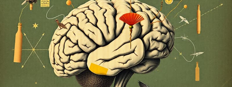
- The central nervous system (CNS) comprises the brain and spinal cord.
- The brain is the most complex structure known to humans.
- The brain develops from a single neural tube in the embryo.
- The anterior end of the neural tube forms three primary brain vesicles: prosencephalon (forebrain), mesencephalon (midbrain), and rhombencephalon (hindbrain).
- The posterior end of the neural tube develops into the spinal cord.
- The primary brain vesicles differentiate into secondary brain vesicles: the forebrain becomes the telencephalon (endbrain) and diencephalon (interbrain), the midbrain remains unchanged, and the hindbrain becomes the metencephalon (afterbrain) and myelencephalon (spinal brain).
- The telencephalon develops into cerebral hemispheres, forming the cerebrum.
- The midbrain and hindbrain segments together form the brainstem.
- The brain's rapid growth causes it to fold, a process called gyrification.
- The adult brain is typically divided into four main regions: cerebral hemispheres, diencephalon, brainstem, and cerebellum.
- The brain contains cavities called ventricles filled with cerebrospinal fluid and lined with ependymal cells.
- The CNS contains four types of neuroglia: astrocytes, microglial cells, oligodendrocytes, and ependymal cells.
Cerebral Hemispheres
- The cerebral hemispheres constitute the majority of the brain's mass.
- The surface of the cerebral hemispheres is characterized by ridges called gyri, separated by grooves called sulci, and deeper grooves called fissures.
- The longitudinal fissure separates the two hemispheres.
- Each hemisphere is divided into five lobes: frontal, parietal, temporal, occipital, and insula.
- Each hemisphere consists of three regions: cerebral cortex (gray matter), internal region of white matter, and basal nuclei (gray matter regions within the white matter).
- The cerebral cortex is the most recently evolved part of the brain and is responsible for conscious thought.
- The cerebral cortex contains six layers of interneurons, glia, and blood vessels.
- Specific regions of the cortex, called domains, are responsible for particular motor and sensory functions.
- These domains are classified as motor areas, sensory areas, and association areas.
- Higher mental functions, such as memory and language, are distributed across the cortex.
- Each hemisphere controls the opposite side of the body.
Motor Areas
- The primary motor cortex controls voluntary movement.
- Different parts of the body are represented in specific areas of the primary motor cortex.
- The motor homunculus is a representation of the human body with body parts scaled proportionally to the amount of neurons controlling them.
- The premotor cortex plans and sequences movements into complex tasks.
- Broca's area controls muscles involved in speech production.
- The frontal eye field controls voluntary eye movement.
Sensory Areas
- The primary somatosensory cortex receives sensory information from the skin and other areas.
- The somatosensory association cortex integrates sensory information to provide a comprehensive understanding of perceived objects.
- The primary visual cortex and visual association area receive and integrate visual information.
- The primary auditory cortex and auditory association area process auditory information.
- The olfactory cortex processes odors.
- The gustatory cortex perceives taste.
- The visceral sensory area provides conscious perception of visceral sensations.
- The vestibular cortex allows us to perceive balance and equilibrium.
Multimodal Association Areas
- Multimodal association areas communicate with multiple brain regions.
- These areas include the anterior and posterior association areas and the limbic association area.
Diencephalon
- The diencephalon is located at the center of the brain.
- It consists of the thalamus, hypothalamus, and epithalamus.
- The thalamus receives and relays information to the cerebral cortex.
- The hypothalamus controls the autonomic nervous system, regulates body temperature, hunger, thirst, sleep cycles, emotions, and the endocrine system.
- The hypothalamus houses the pituitary gland.
- The epithalamus contains the pineal gland and helps regulate sleep.
Brain Stem
- The brainstem consists of the midbrain, pons, and medulla oblongata.
- The medulla oblongata connects to the spinal cord.
Cerebellum
- The cerebellum consists of two hemispheres and regulates muscle contractions for smooth, coordinated movement.
Brain Protection
- The brain is protected by meninges: connective tissue membranes situated between the brain and the skull.
- The meninges consist of three layers: dura mater, arachnoid mater, and pia mater.
Spinal Cord
- The spinal cord extends from the base of the skull to just past the ribs.
- It is protected by the vertebral column, cerebrospinal fluid, and the meninges.
- Thirty-one pairs of spinal nerves attach to the spinal cord.
- The gray matter of the spinal cord forms a butterfly shape and is composed of multipolar neurons.
- The gray matter contains dorsal and ventral horns, which connect to skeletal muscles and other structures.
- The gray matter is surrounded by white matter containing nerve fibers that transmit information between the spinal cord and the brain.
- These fibers can be ascending, descending, or transverse based on their direction of travel.
- The white matter is involved in numerous pathways that will be further investigated later.
Studying That Suits You
Use AI to generate personalized quizzes and flashcards to suit your learning preferences.


