Podcast
Questions and Answers
Which type of glial cell is responsible for myelinating axons in the central nervous system?
Which type of glial cell is responsible for myelinating axons in the central nervous system?
- Astrocytes
- Ependymal cells
- Microglia
- Oligodendrocytes (correct)
What is the primary function of astrocytes in the central nervous system?
What is the primary function of astrocytes in the central nervous system?
- Myelination of axons
- Signaling between neurons
- Production of cerebrospinal fluid
- Physical support and repair (correct)
Which of the following statements about ependymal cells is true?
Which of the following statements about ependymal cells is true?
- They myelinate axons in the peripheral nervous system.
- They are responsible for providing structural support in the CNS.
- They are primarily involved in immune response in the CNS.
- They line the ventricles and the central canal of the spinal cord. (correct)
What is the main structural feature of oligodendrocytes that differs from other glial cells?
What is the main structural feature of oligodendrocytes that differs from other glial cells?
Which characteristic is used to identify dendrites and cell bodies in neuronal tissue?
Which characteristic is used to identify dendrites and cell bodies in neuronal tissue?
Flashcards
Neurons
Neurons
Permanent signal-transmitting cells in the nervous system, composed of dendrites, cell bodies, and axons.
Myelin
Myelin
Multilayer wrapping of electrical insulation around axons, increasing the speed of signal transmission via saltatory conduction.
Astrocytes
Astrocytes
Largest and most abundant glial cells in the CNS providing support, removing neurotransmitters, and part of the blood-brain barrier.
Oligodendrocytes
Oligodendrocytes
Signup and view all the flashcards
Ependymal cells
Ependymal cells
Signup and view all the flashcards
Study Notes
Cells of the Nervous System
-
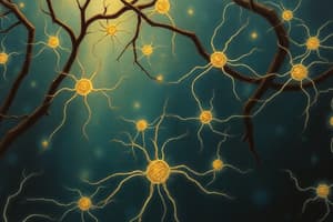
Neurons are signal-transmitting cells composed of dendrites (input), cell bodies, and axons (output).
-
Dendrites and cell bodies are visible with Nissl staining (stains RER), while axons do not have RER.
-
Neurofilament protein and synaptophysin are neuronal markers.
-
Astrocytes provide physical support, repair, remove excess neurotransmitters.
-
They also form a part of the blood-brain barrier and buffer glycogen.
-
GFAP proteins identify astrocytes
-
Myelinate CNS axons (including cranial nerves II)
-
Oligodendrocytes myelinate multiple axons in the CNS (around 30 axons are myelinated by a single oligodendrocyte).
-
They are identified histologically as "fried eggs."
-
Oligodendrocytes are prevalent in white matter
-
They are damaged in multiple sclerosis, leukodystrophies, and progressive multifocal leukoencephalopathy.
-
Ependymal cells line the ventricles and central canal of the spinal cord.
-
Ciliated and microvilli are used to circulate CSF and absorb it.
-
Specialized ependymal cells in the choroid plexus produce cerebrospinal fluid (CSF).
-
Microglia are phagocytic cells in the CNS, acting as scavengers.
-
Activated microglia release inflammatory mediators (e.g., nitric oxide, glutamate).
-
They are involved in HIV-associated dementia.
-
Satellite cells surround neuron cell bodies in ganglia.
-
Schwann cells myelinate axons in the PNS (cranial nerves III-XII).
-
Schwann cells myelinate single axons.
-
They are damaged in Guillain-Barré Syndrome
CNS Glial Cells
- CNS glial cells originate mostly from the neuroectoderm, except microglia, which come from the mesoderm.
- PNS glial cells originate from the neural crest ectoderm.
- Myelin is a multilayered sheath, electrically insulating axons.
- Increased insulation leads to faster conduction velocity ("saltatory conduction") at nodes of Ranvier.
Neuron Action Potential
- Resting potential: the membrane is more permeable to K+ than Na+.
- Depolarization: Na+ channels open, Na+ flows inward.
- Repolarization: Na+ channels close, and K+ channels open, K+ flows outward.
- Hyperpolarization: K+ channels close slowly, leading to slightly more negative potential than resting.
- Na+/K+ pump restores ion concentrations to original levels.
Sensory Receptors
- Free nerve endings: pain and temperature receptors found in all body tissues except cartilage and the eye lens.
- Meissner corpuscles: located in hairless skin, adapt quickly to light touch, low-frequency vibration, skin indentation.
- Pacinian corpuscles: located in deeper skin layers, ligaments, and joints; adapt quickly to deep pressure.
- Merkel discs: adapt slowly to pressure (deep static touch), shape, and edge.
- Ruffini corpuscles: adapt slowly; detect skin stretching, joint angle change.
Studying That Suits You
Use AI to generate personalized quizzes and flashcards to suit your learning preferences.


