Podcast
Questions and Answers
What is the primary function of lysosomes in the cell?
What is the primary function of lysosomes in the cell?
- Intracellular digestion (correct)
- Energy production
- Protein synthesis
- DNA replication
Which process is NOT associated with lysosomes?
Which process is NOT associated with lysosomes?
- Transport of oxygen (correct)
- Recycling of cell membrane
- Synthesis of carbohydrates
- Formation of lysosome
In which process do lysosomes play a significant role?
In which process do lysosomes play a significant role?
- Cellular respiration
- Apoptosis
- Intracellular digestion (correct)
- Cell division
What is formed as part of the overall functioning of lysosomes?
What is formed as part of the overall functioning of lysosomes?
Which of the following is a secondary function of lysosomes?
Which of the following is a secondary function of lysosomes?
What is the primary role of the cristae in cellular respiration?
What is the primary role of the cristae in cellular respiration?
What is the shape of the cristae in steroid-secreting cells?
What is the shape of the cristae in steroid-secreting cells?
Which of the following processes occurs on the cristae?
Which of the following processes occurs on the cristae?
Which enzymes are found within the cristae?
Which enzymes are found within the cristae?
What would happen if the enzymes located in the cristae were malfunctioning?
What would happen if the enzymes located in the cristae were malfunctioning?
What property of the tails in a membrane contributes to their positioning at the center?
What property of the tails in a membrane contributes to their positioning at the center?
What is the first step in the process when a ligand binds to its receptors on the surface of a cell?
What is the first step in the process when a ligand binds to its receptors on the surface of a cell?
What gives rise to the trilaminar appearance in membranes?
What gives rise to the trilaminar appearance in membranes?
What structure is formed as the invaginated coated pit continues to develop?
What structure is formed as the invaginated coated pit continues to develop?
Which characteristic is NOT associated with the membrane tails?
Which characteristic is NOT associated with the membrane tails?
What is the role of staining in observing membrane structure?
What is the role of staining in observing membrane structure?
After the formation of a coated vesicle, what is the next stage that occurs with the early endosome?
After the formation of a coated vesicle, what is the next stage that occurs with the early endosome?
What is the final destination of the early endosome as it progresses through the cytoplasm?
What is the final destination of the early endosome as it progresses through the cytoplasm?
Which statement about the membrane's structure is accurate?
Which statement about the membrane's structure is accurate?
Which of the following statements about receptor-ligand interactions is true?
Which of the following statements about receptor-ligand interactions is true?
What structures are formed when the two membranes fuse together?
What structures are formed when the two membranes fuse together?
Which cellular components does the nuclear pore connect?
Which cellular components does the nuclear pore connect?
What is the primary function of nuclear pores in a cell?
What is the primary function of nuclear pores in a cell?
Which of the following best describes the location of nuclear pores?
Which of the following best describes the location of nuclear pores?
Which statement about nuclear pores is incorrect?
Which statement about nuclear pores is incorrect?
Where are peripheral proteins primarily located in the cell membrane?
Where are peripheral proteins primarily located in the cell membrane?
What is a defining feature of integral proteins?
What is a defining feature of integral proteins?
What is the term for integral proteins that extend from one side of the membrane to the other?
What is the term for integral proteins that extend from one side of the membrane to the other?
Which of the following accurately describes peripheral proteins?
Which of the following accurately describes peripheral proteins?
Which statement about integral proteins is true?
Which statement about integral proteins is true?
Flashcards
Hydrophobic Tails
Hydrophobic Tails
The nonpolar tails of a phospholipid that repel water and are found in the interior of the cell membrane.
Cell Membrane Structure
Cell Membrane Structure
The cell membrane's trilaminar appearance is due to stain deposition on the head regions of phospholipids, not the tails.
Peripheral Proteins
Peripheral Proteins
Proteins located on the inner and outer surfaces of the cell membrane, mostly on the inner surface.
Integral Proteins
Integral Proteins
Signup and view all the flashcards
Transmembrane Proteins
Transmembrane Proteins
Signup and view all the flashcards
Ligand binding to receptors
Ligand binding to receptors
Signup and view all the flashcards
Coated pit formation
Coated pit formation
Signup and view all the flashcards
Coated vesicle formation
Coated vesicle formation
Signup and view all the flashcards
Early endosome movement
Early endosome movement
Signup and view all the flashcards
Late endosome formation
Late endosome formation
Signup and view all the flashcards
Cristae shape
Cristae shape
Signup and view all the flashcards
Cristae function
Cristae function
Signup and view all the flashcards
Nuclear Pore Formation
Nuclear Pore Formation
Signup and view all the flashcards
Lysosome function
Lysosome function
Signup and view all the flashcards
Cell Membrane Recycling
Cell Membrane Recycling
Signup and view all the flashcards
CHO Synthesis
CHO Synthesis
Signup and view all the flashcards
Study Notes
Cell Membrane
- The plasma membrane (cell membrane), plasma lemma, or limiting membrane is extremely thin (7.5 to 10 nm).
- It's not visible under Light Microscopy (LM) without special stains (e.g., silver).
- Electron Microscopy (EM) reveals a trilaminar appearance: two electron-dense layers separated by a translucent layer.
- The membrane is primarily composed of a phospholipid bilayer with glycolipids and cholesterol.
- Phospholipid molecules have a hydrophilic head and hydrophobic tails.
- The hydrophilic heads face the intracellular and extracellular environments.
- The hydrophobic tails position themselves within the membrane.
- The trilaminar appearance results from the staining of the hydrophilic heads on either side of the membrane.
- Proteins are embedded in the membrane.
- Proteins can be peripheral (located on the membrane surface) or integral (integrated within the bilayer), and some span both the inner and outer surfaces (transmembrane proteins).
- Carbohydrates are also present predominantly on the outer membrane surface, linked to lipids (glycolipids) or proteins (glycoproteins) forming a glycocalyx.
- The cell membrane acts as a barrier and regulates the passage of substances into and out of the cell, maintaining a constant intracellular environment.
- It also plays a role in cell signaling.
Vesicular Transport
- Endocytosis and exocytosis are two types of vesicular transport.
- Endocytosis involves the pinching off of the cell membrane to form vesicles that engulf materials from outside the cell.
- Pinocytosis ("cell drinking") is one form; it engulfs fluids.
- Phagocytosis ("cell eating") involves engulfing larger particles or cells.
- Receptor-mediated endocytosis is a specialized form where specific receptors bind foreign molecules, initiating the process.
- Exocytosis is the release of materials from intracellular vesicles to the extracellular environment.
- The process is affected by calcium levels in the cytoplasm; higher calcium leads to more exocytosis.
- Exocytosis pathways include constitutive (continuous) and regulated (stimulated).
Mitochondria
- Mitochondria have a distinctive shape, typically spherical or rod-like.
- They are not visible in light microscopy (LM) without special vital stains like Janus green B.
- Mitochondria are localized in cells according to their energy utilization, e.g., more prominent in active cells like heart muscle (cardiac cells) as well as in ion-transferring cells and spermatozoa.
- They have their own DNA and can replicate independently from the cell.
- Mitochondrial DNA and RNA are contained within the organelle.
- The interior of the mitochondria is partitioned by inner membranes; folds are called cristae.
- The inner membrane encloses the matrix, containing enzymes necessary for the citric acid/Krebs cycle, and the electron transport chain is present there.
- The process of oxidative phosphorylation occurs on the cristae, generating ATP (energy).
Endoplasmic Reticulum
- Endoplasmic reticulum (ER) is a network of channels and sacs formed by continuous membrane.
- It is classified as either rough (RER), having ribosomes attached to its surface, or smooth (SER), lacking ribosomes.
- RER appears as a basophilic area in the cytoplasm, while SER is not visible in light microscopy.
- RER is associated with protein synthesis and modification, targeting proteins for secretion or insertion into the cell membrane.
- SER is involved in steroid hormone synthesis, lipid metabolism, drug detoxification, calcium storage, and glycogen metabolism.
Golgi Apparatus
- The Golgi apparatus is composed of flattened sacs (cisternae).
- The Golgi apparatus is usually organized in stacks with dilated peripheries.
- The Golgi apparatus has two faces: the cis (receiving) face and the trans (shipping) face.
- The cis face is associated with small vesicles coming from the ER, carrying immature proteins.
- The trans face is associated with large secretory vesicles containing mature proteins.
- Functions include protein modification (such as glycosylation), concentration, packaging of proteins into secretory vesicles, lysosome formation, and membrane recycling.
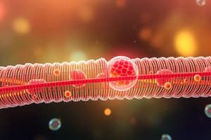
Lysosomes
- Lysosomes are sites for intracellular digestion.
- Their enzymes optimally function in acidic pH.
- The organelle can be seen in electron microscopy (EM).
- Lysosomes are types: primary and secondary, containing enzymes that degrade cellular substances.
Peroxisomes
- Peroxisomes are membrane-enclosed organelles.
- Function in removing hydrogen peroxide and breaking down fatty acids into smaller molecules.
- Peroxisomes utilize oxidative enzymes and generate hydrogen peroxide as a by-product.
Ribosomes
- Ribosomes are non-membrane bound organelles.
- Ribosomes are composed of rRNA and proteins.
- Two forms are free in the cytoplasm and bound to RER.
- Ribosomes function in protein synthesis by translating mRNA into amino acid chains.
Proteasomes
- Proteasomes are non-membrane bound.
- They are responsible for protein degradation.
- They target improperly folded or damaged proteins for destruction.
Cytoskeleton
- Cytoskeleton is vital for maintaining cell shape and facilitating internal movement.
- Composed of microtubules, intermediate filaments, and microfilaments, or actin filaments.
Centrioles
- Centrioles organize microtubules during cell division, playing a critical role in forming the mitotic spindle, determining the basis of cilia, and forming flagella.
- Centrioles are composed of microtubules arranged in a specific configuration.
Cilia
- Cilia are hair-like projections on the cell surface.
- Cilia are responsible for moving substances across the cell surface.
- Cilia have a characteristic structure of microtubules, organized in a 9+2 arrangement
Filaments
- Filaments are protein fibers supporting cell structure.
- Three types are thinner actin, intermediate filaments, and thick myosin filaments.
Inclusions
- Inclusions are stored materials within the cell.
- Includes stored food, pigments, and crystals. Lipofuscin is a type of aging pigment. Melanin is a natural skin pigment. Hemoglobin is an oxygen-carrying protein pigment found in red blood cells. Glycogen is a type of stored carbohydrate.
Nucleus
- The nucleus is the largest and most prominent organelle in the cell.
- It has a rounded or oval shape but can be irregular, and a central location within a cell body.
- The nucleus can be observed by light microscopy (LM) and contains chromatin, nucleolus, nuclear envelope, and nuclear matrix.
- Chromatin comprises DNA and associated proteins.
- The nucleolus is a region within the nucleus where ribosome components are synthesized.
- The nuclear envelope ensures the separation of the nucleus from the cytoplasm. It has a double membrane. Nuclear pores allow passage into and out of the nucleus.
- The nuclear matrix provides structural support to the nucleus.
Studying That Suits You
Use AI to generate personalized quizzes and flashcards to suit your learning preferences.