Podcast
Questions and Answers
What phase is the cell in during Interphase?
What phase is the cell in during Interphase?
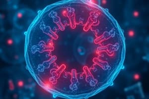
Interphase
What phase is the cell in during Prophase?
What phase is the cell in during Prophase?
Prophase
What phase is the cell in during Metaphase?
What phase is the cell in during Metaphase?
Metaphase
What phase is the cell in during Anaphase?
What phase is the cell in during Anaphase?
What phase is the cell in during Telophase?
What phase is the cell in during Telophase?
The basic unit of structure and function of life is the __________.
The basic unit of structure and function of life is the __________.
A cell that is growing and conducting usual activities is in _________________.
A cell that is growing and conducting usual activities is in _________________.
The nuclear envelope has dissolved and chromosomes are visible but have not migrated when a cell is in __________________.
The nuclear envelope has dissolved and chromosomes are visible but have not migrated when a cell is in __________________.
Chromosomes line up on the mitotic spindle in the middle of the cell during ___________________.
Chromosomes line up on the mitotic spindle in the middle of the cell during ___________________.
Chromosomes are migrating apart from the middle of the cell in ____________________.
Chromosomes are migrating apart from the middle of the cell in ____________________.
Chromosomes have finished moving apart and nuclear envelopes start to reform during __________________.
Chromosomes have finished moving apart and nuclear envelopes start to reform during __________________.
A structure located in the cell that houses chromatin; it divides during mitosis is the __________________.
A structure located in the cell that houses chromatin; it divides during mitosis is the __________________.
The double-layered membrane that surrounds the cell is the ______________________________.
The double-layered membrane that surrounds the cell is the ______________________________.
A double-layered membrane that encloses the nucleus is the __________________________.
A double-layered membrane that encloses the nucleus is the __________________________.
What plane cuts the body into right and left portions?
What plane cuts the body into right and left portions?
What plane cuts the body into equal right and left portions?
What plane cuts the body into equal right and left portions?
What plane cuts the body into anterior and posterior portions?
What plane cuts the body into anterior and posterior portions?
What plane cuts the body into superior and inferior portions?
What plane cuts the body into superior and inferior portions?
The knee is ____________________ to the foot.
The knee is ____________________ to the foot.
The shoulder is _____________________ to the sternum.
The shoulder is _____________________ to the sternum.
The abdomen is __________________ to the thorax.
The abdomen is __________________ to the thorax.
Name the type of tissue in the picture.
Name the type of tissue in the picture.
Name the type of tissue in the picture.
Name the type of tissue in the picture.
Name the type of tissue in the picture.
Name the type of tissue in the picture.
Name the type of tissue in the top part of this picture.
Name the type of tissue in the top part of this picture.
Name the type of tissue in the picture.
Name the type of tissue in the picture.
Name the type of tissue in the picture.
Name the type of tissue in the picture.
Name the type of tissue in the picture.
Name the type of tissue in the picture.
Name the type of tissue in the picture.
Name the type of tissue in the picture.
Name the type of tissue in the picture.
Name the type of tissue in the picture.
Name the circled structures.
Name the circled structures.
Name the large purple cells in the picture.
Name the large purple cells in the picture.
Name the type of fiber indicated by the arrow.
Name the type of fiber indicated by the arrow.
Name the type of tissue in the picture.
Name the type of tissue in the picture.
Name the circled structures.
Name the circled structures.
Name the structure indicated by the yellow circles.
Name the structure indicated by the yellow circles.
Name the type of cell indicated by the arrow.
Name the type of cell indicated by the arrow.
Name the structure indicated on the microscope.
Name the structure indicated on the microscope.
Name the structure indicated on the microscope.
Name the structure indicated on the microscope.
Name the structure indicated on the microscope.
Name the structure indicated on the microscope.
Name the structure indicated on the microscope.
Name the structure indicated on the microscope.
Name the structure indicated on the microscope.
Name the structure indicated on the microscope.
Name the structure indicated on the microscope.
Name the structure indicated on the microscope.
Name the structure indicated on the microscope.
Name the structure indicated on the microscope.
Name the structure indicated on the microscope.
Name the structure indicated on the microscope.
Name the structure indicated on the microscope.
Name the structure indicated on the microscope.
Multiplying the power of the objective lens by the power of the ocular lens will give the _____________ __________________ of an image viewed through a microscope.
Multiplying the power of the objective lens by the power of the ocular lens will give the _____________ __________________ of an image viewed through a microscope.
Flashcards are hidden until you start studying
Study Notes
Cell Cycle Phases
- Interphase: The cell is actively growing and performing normal functions; preparation for division.
- Prophase: The nuclear envelope disintegrates; chromosomes become visible but remain unassigned in position.
- Metaphase: Chromosomes align along the mitotic spindle at the cell's equatorial plane.
- Anaphase: Sister chromatids are pulled apart to opposite poles of the cell.
- Telophase: Chromosomes have separated and begin to de-condense; nuclear envelopes reform around each set.
Cellular Structures
- Cell: The fundamental structural and functional unit of life.
- Nucleus: Houses chromatin; undergoes division during mitosis.
- Plasma membrane: A double-layered membrane that encloses the cell, controlling entry and exit of substances.
- Nuclear envelope: A double-layered membrane that surrounds and protects the nucleus.
Anatomical Planes
- Sagittal plane: Divides the body into right and left portions.
- Midsagittal plane: Cuts the body into equal right and left halves along the midline.
- Frontal (coronal) plane: Divides the body into anterior (front) and posterior (back) sections.
- Transverse plane: Splits the body into superior (upper) and inferior (lower) sections.
Anatomical Directions
- Proximal: Indicates that a body part is closer to the point of attachment than another part (e.g., knee to foot).
- Lateral: Refers to a position further away from the midline of the body (e.g., shoulder to sternum).
- Inferior: Denotes a position lower than another structure (e.g., abdomen to thorax).
Tissue Types
- Simple columnar epithelium: A type of tissue with taller, column-like cells.
- Simple cuboidal epithelium: Composed of cube-shaped cells.
- Stratified squamous epithelium: Multiple layers of flat cells; provides protection.
- Pseudostratified columnar epithelium: Appears layered due to differing cell heights but is a single layer.
- Mesenchyme: A form of connective tissue found in the embryo.
- Hyaline cartilage: Provides support and flexibility; lacks visible fibers.
- Areolar tissue: A loose connective tissue that supports and binds other tissues.
- Adipose tissue: Stores fat; serves as insulation and energy reserve.
- Blood: Transports nutrients, waste, and gases throughout the body.
Cellular Components
- Erythrocytes: Red blood cells responsible for oxygen transport.
- Leukocytes: White blood cells that play roles in immune defense.
- Collagen: A key extracellular matrix protein providing strength to tissues.
- Osseous tissue: Also known as bone tissue; provides structure and support.
- Lacunae: Small cavities within tissue, often found in bone or cartilage.
- Lamellae: Concentric layers of bone matrix in osseous tissue.
- Chondrocyte: A cell found in cartilage responsible for maintaining cartilage matrix.
Microscope Components
- Ocular Lens: The eyepiece through which an object is viewed; typically provides 10x magnification.
- Objective Lens: Shorter lenses that provide varying levels of magnification (e.g., 4x, 10x, 40x).
- Stage: The platform that holds the slide being observed.
- Mechanical Stage: A device that allows precise movement of the slide for better viewing.
- Iris Diaphragm: Regulates the amount of light that reaches the specimen.
- Condenser: Focuses light onto the specimen to improve clarity.
- Rotating Nosepiece: Holds multiple objective lenses; allows for easy switching.
- Course Adjustment Knob: Used to make large adjustments to focus the image.
- Fine Adjustment Knob: Used for precise focus adjustments.
- Total Magnification: Calculated by multiplying the power of the ocular lens by the power of the objective lens.
Studying That Suits You
Use AI to generate personalized quizzes and flashcards to suit your learning preferences.