Podcast
Questions and Answers
Which organelle plays a crucial role in the production of lipids?
Which organelle plays a crucial role in the production of lipids?
- Golgi Apparatus
- Rough Endoplasmic Reticulum
- Mitochondria
- Smooth Endoplasmic Reticulum (correct)
What is the primary function of lysosomes?
What is the primary function of lysosomes?
- Degradation of macromolecules (correct)
- Modification of proteins for transport
- Protein synthesis
- Energy production
Which of the following is a type of cell injury caused by toxins?
Which of the following is a type of cell injury caused by toxins?
- Direct-acting toxins
- Latent toxins
- Reactive Oxygen Species (ROS)
- All of the above (correct)
Which of the following is NOT a function of the cytoskeleton?
Which of the following is NOT a function of the cytoskeleton?
What is a key characteristic of reversible cell injury?
What is a key characteristic of reversible cell injury?
Which of the following is NOT an irreversible sign of cell injury?
Which of the following is NOT an irreversible sign of cell injury?
What is the role of the plasma membrane in cell function?
What is the role of the plasma membrane in cell function?
Which cellular component is directly involved in the process of oxidative phosphorylation?
Which cellular component is directly involved in the process of oxidative phosphorylation?
What is the main difference between apoptosis and necrosis?
What is the main difference between apoptosis and necrosis?
Which cellular adaptation involves an increase in cell size?
Which cellular adaptation involves an increase in cell size?
Which of these organelles is NOT involved in the process of protein synthesis?
Which of these organelles is NOT involved in the process of protein synthesis?
Besides lysosomes, which other organelle is involved in protein degradation?
Besides lysosomes, which other organelle is involved in protein degradation?
Which of the following is an example of physiologic hyperplasia?
Which of the following is an example of physiologic hyperplasia?
Which cellular adaptation is characterized by a change in cell type?
Which cellular adaptation is characterized by a change in cell type?
In which scenario would atrophy be most likely to occur?
In which scenario would atrophy be most likely to occur?
What does the term 'Left Shift' refer to in the context of blood cell analysis?
What does the term 'Left Shift' refer to in the context of blood cell analysis?
What is the primary cause of leukopenia?
What is the primary cause of leukopenia?
Which of the following is NOT a characteristic of the inflammatory response?
Which of the following is NOT a characteristic of the inflammatory response?
What is the primary mechanism of action for NSAIDs in fever reduction?
What is the primary mechanism of action for NSAIDs in fever reduction?
Which of the following is a key characteristic of the scar formation repair mechanism?
Which of the following is a key characteristic of the scar formation repair mechanism?
What is the role of macrophages in tissue repair?
What is the role of macrophages in tissue repair?
What is the primary factor determining whether a tissue will regenerate or scar?
What is the primary factor determining whether a tissue will regenerate or scar?
Which of the following is NOT a factor that stimulates cell proliferation during tissue regeneration?
Which of the following is NOT a factor that stimulates cell proliferation during tissue regeneration?
Which of the following factors can directly impair tissue repair by interfering with the formation of granulation tissue?
Which of the following factors can directly impair tissue repair by interfering with the formation of granulation tissue?
Which of these is an example of an intrinsic factor that can impair tissue repair?
Which of these is an example of an intrinsic factor that can impair tissue repair?
Which of these is a condition that can lead to excessive production of extracellular matrix (ECM), potentially resulting in the formation of keloids?
Which of these is a condition that can lead to excessive production of extracellular matrix (ECM), potentially resulting in the formation of keloids?
Which of the following statements accurately describes the relationship between cellular aging and tissue repair?
Which of the following statements accurately describes the relationship between cellular aging and tissue repair?
Which of these pigments is an endogenous accumulation that can occur within cells?
Which of these pigments is an endogenous accumulation that can occur within cells?
Which of the following conditions is NOT directly linked to fatty liver changes?
Which of the following conditions is NOT directly linked to fatty liver changes?
What is the primary mechanism responsible for the accumulation of lipofuscin within cells?
What is the primary mechanism responsible for the accumulation of lipofuscin within cells?
Which of the following is an example of a condition associated with exogenous carbon accumulation in the lungs?
Which of the following is an example of a condition associated with exogenous carbon accumulation in the lungs?
What primarily induces vasodilation in acute inflammation?
What primarily induces vasodilation in acute inflammation?
What is one of the early manifestations of vasodilation during acute inflammation?
What is one of the early manifestations of vasodilation during acute inflammation?
What is the effect of endothelial cell retraction during an inflammatory response?
What is the effect of endothelial cell retraction during an inflammatory response?
What can result from endothelial injury during inflammation?
What can result from endothelial injury during inflammation?
Which of the following best describes the term 'vascular leakage' in the context of inflammation?
Which of the following best describes the term 'vascular leakage' in the context of inflammation?
What is the primary component involved in fat necrosis that results in saponification?
What is the primary component involved in fat necrosis that results in saponification?
Which type of necrosis is characterized by a 'moth eaten' appearance on microscopy?
Which type of necrosis is characterized by a 'moth eaten' appearance on microscopy?
In which type of necrosis do dead cells leave behind a creamy yellow liquid?
In which type of necrosis do dead cells leave behind a creamy yellow liquid?
What key cellular changes are associated with necrosis?
What key cellular changes are associated with necrosis?
What is the main characteristic of fibroid necrosis observable under microscopy?
What is the main characteristic of fibroid necrosis observable under microscopy?
Which type of necrosis is associated with the formation of granulomas?
Which type of necrosis is associated with the formation of granulomas?
What distinguishes apoptosis from necrosis?
What distinguishes apoptosis from necrosis?
Which biomarkers can indicate damage to cardiac muscle cells during necrosis?
Which biomarkers can indicate damage to cardiac muscle cells during necrosis?
Flashcards
Nucleus
Nucleus
The part of a cell that stores DNA and is responsible for mRNA processing.
Mitochondria
Mitochondria
Organelles that produce energy and are involved in apoptosis through oxidative phosphorylation.
Lysosomes
Lysosomes
Cell organelles that contain enzymes to digest macromolecules.
Ribosomes
Ribosomes
Signup and view all the flashcards
Stem Cells
Stem Cells
Signup and view all the flashcards
Cell Injury Types
Cell Injury Types
Signup and view all the flashcards
Acute Inflammation
Acute Inflammation
Signup and view all the flashcards
Apoptosis
Apoptosis
Signup and view all the flashcards
Toxic Cell Injury
Toxic Cell Injury
Signup and view all the flashcards
Reversible Cell Injury
Reversible Cell Injury
Signup and view all the flashcards
Necrosis
Necrosis
Signup and view all the flashcards
Hypertrophy
Hypertrophy
Signup and view all the flashcards
Hyperplasia
Hyperplasia
Signup and view all the flashcards
Atrophy
Atrophy
Signup and view all the flashcards
Metaplasia
Metaplasia
Signup and view all the flashcards
Vasodilation
Vasodilation
Signup and view all the flashcards
Histamine
Histamine
Signup and view all the flashcards
Increased permeability
Increased permeability
Signup and view all the flashcards
Exudate
Exudate
Signup and view all the flashcards
Endothelial cells retraction
Endothelial cells retraction
Signup and view all the flashcards
Pyogenic Infection
Pyogenic Infection
Signup and view all the flashcards
Caseous Necrosis
Caseous Necrosis
Signup and view all the flashcards
Fat Necrosis
Fat Necrosis
Signup and view all the flashcards
Fibrinoid Necrosis
Fibrinoid Necrosis
Signup and view all the flashcards
Coagulative Necrosis
Coagulative Necrosis
Signup and view all the flashcards
Liquefactive Necrosis
Liquefactive Necrosis
Signup and view all the flashcards
Biomarkers in Necrosis
Biomarkers in Necrosis
Signup and view all the flashcards
Left shift
Left shift
Signup and view all the flashcards
Neutrophilia
Neutrophilia
Signup and view all the flashcards
Lymphocytosis
Lymphocytosis
Signup and view all the flashcards
Eosinophilia
Eosinophilia
Signup and view all the flashcards
Leukopenia
Leukopenia
Signup and view all the flashcards
Inflammation effects
Inflammation effects
Signup and view all the flashcards
Tissue Repair Mechanisms
Tissue Repair Mechanisms
Signup and view all the flashcards
Macrophages in repair
Macrophages in repair
Signup and view all the flashcards
Factors that inhibit tissue repair
Factors that inhibit tissue repair
Signup and view all the flashcards
Cutaneous wound healing phases
Cutaneous wound healing phases
Signup and view all the flashcards
Primary union
Primary union
Signup and view all the flashcards
Secondary union
Secondary union
Signup and view all the flashcards
Cellular aging
Cellular aging
Signup and view all the flashcards
Intracellular accumulations
Intracellular accumulations
Signup and view all the flashcards
Lipofuscin
Lipofuscin
Signup and view all the flashcards
Calcification
Calcification
Signup and view all the flashcards
Study Notes
Pathogen 3.1: Cells and Inflammation
- This unit examines cells and the inflammatory process.
Objectives
- Analyze the components of a typical cell and their function.
- Analyze how cells coordinate and integrate their functions.
- Interpret the role of growth factors in cellular activity.
- Understand stem cells, and their roles in regenerative medicine.
- Illustrate how cells are injured (reversible and irreversible), including changes in cytoplasm and nucleus.
- Analyze common causes of cell injury.
- Contrast four types of cellular adaptations.
- Differentiate between metaplasia and dysplasia.
- Discuss cellular aging.
- Compare necrosis and apoptosis as cell death forms.
- Compare different forms of necrosis and provide examples.
- Analyze cellular and events of inflammation.
- Define vascular changes in acute inflammation.
- Debate the role of leukocytes in an inflammatory response, including margination, diapedesis, emigration, exudation, chemotaxis, phagocytosis, and microbicidal substances.
- Analyze the complement system, cytokines, and clotting system in inflammation.
- Compare and contrast acute and chronic inflammation.
- Describe the formation of granulomas and analyze pathologic terms related to inflammation (serous, fibrinous, purulent, abscess, ulcer, wound, scar, keloid).
- Investigate the typical local and systemic symptoms of inflammation.
- Analyze the pathogenesis of fever.
The Cell as a Unit of Health and Disease
- Cells are the basic units of health and disease.
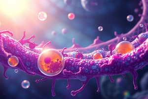
Essential Components of Animal Cells
- Nucleus: DNA storage, transcription, mRNA processing
- Ribosomes: Translation
- Mitochondria: Energy production, apoptosis
- Endoplasmic Reticulum (ER): Rough - protein synthesis, Smooth - lipid synthesis, calcium storage
- Golgi Apparatus: Modifies proteins for transport
- Lysosomes: Digestive enzymes for macromolecules
- Proteasomes: Degrade proteins
- Cytoskeleton: Cell shape, polarity, organization
- Plasma membrane: Regulated movement of solutes, cell-to-cell interaction
Lysosomes/Proteasomes
- Lysosomes contain hydrolases to break down substances.
- Proteasomes dismantle denatured or misfolded proteins.
- Lysosomes form in three ways - pinocytosis, receptor-mediated endocytosis, and autophagy.
Plasma Membrane
- Permeable: Very small particles, non-polar particles (water, O2, CO2, ethanol, steroids, vit D)
- Impermeable: Polar particles, large particles (proteins, glucose, ions)
- Ways to get through:
- Passive transport (channels, carriers)
- Receptor-mediated
- Active transport (pumps - ATPase)
- Endocytosis (caveolae, receptor-mediated, phagocytosis)
- Exocytosis
ER/Golgi
- Rough ER synthesizes transmembrane proteins and lipids; chaperones ensure proper folding.
- Smooth ER synthesizes steroids, catabolizes lipid-soluble molecules, and stores calcium.
- Golgi apparatus packages proteins for organelles or plasma membrane; modifies N-linked oligosaccharides.
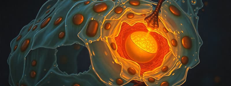
Mitochondria
- Oxidative phosphorylation
- Cell death (apoptosis)
Receptors
- Extracellular and intracellular receptors
- Ligands bind to receptors, initiating intracellular function or DNA interaction
- Effects: transcription
Kinase Activity
- Kinase is a molecule that phosphorylates other molecules, initiating cellular activity
- Receptor tyrosine kinases (RTKs)
- G-protein coupled receptors
- Steroids
Transcription Factors
- DNA-binding domains permit specific binding to short DNA sequences (promotor region or enhancers).
- Some elements interact with genes.
Growth Factors
- Promote cell entry into the cell cycle and replication.
- Enhance cellular component synthesis (nucleic acids, proteins, lipids, carbohydrates).
- Prevent apoptosis.
- Involved in regeneration and repair.
Stem Cells
- Totipotent stem cells give rise to all differentiated tissues.
- Adult stem cells replace damaged cells of the same type.
- Regenerative medicine uses stem cells to heal patients.
Cell Injury
- Underlying causes: Hypoxia/ischemia, infection, autoimmune diseases, toxins, immunologic reactions, physical agents, genetic abnormality, nutritional imbalance, aging
- Reversible injury: Intracellular swelling, mitochondrial/ER disturbance, plasma membrane blebbing, chromatin clumping
- Irreversible injury: mitochondrial failure, loss of membrane structure and function, DNA damage
Toxic Cell Injury
- Direct-acting toxins combine with cellular components, disrupting their function (mercury).
- Latent toxins are converted into reactive metabolites that damage target cells.
- Reactive Oxygen Species (ROS) are highly reactive and damage cellular components.
Reversible Injury Signs
- Intracellular swelling
- Mitochondrial and ER disturbance
- Plasma membrane blebbing
- Clumping of chromatin in nucleus
- Myelin figures (phospholipids of damaged membrane)
Cell Injury: When Badness Happens
- Normal cell (homeostasis) → Injurious stimulus → Reversible injury (if mild or transient) → Irreversible injury (severe, progressive) → Necrosis (or apoptosis)
- Irreversible signs: Inability to restore mitochondrial function, Loss of plasma membrane structure or function, Loss of DNA structural integrity
Apoptosis vs. Necrosis
- Apoptosis: Controlled programmed cell death. Organized disassembly, membrane bound, no inflammation.
- Necrosis: Unprogrammed violent, eventual lysis, swelling, inflammation, usually widespread.
- Apoptosis vs. necrosis overview.
Adapting to Cellular Injury
- Types of Adaptation: Hypertrophy, hyperplasia, atrophy, metaplasia.
Hypertrophy
- Size of cells increases. (due to increased demands, in relation to muscle for example)
- Pathological when concerning heart muscle
Hyperplasia
- Increased cell numbers. (physiological - puberty, pregnancy; pathological - if growth control is lost)
- Hyperplasia alone does not cause cancer
Atrophy
- Decrease in cell size (due to loss of workload, loss of innervation, diminished blood supply, nutritional difficulties etc).
- Aging is a factor.
Metaplasia
- Change in cell type. (smokers' bronchi, Barrett's esophagus)
- Distinguish between metaplasia and dysplasia
Metaplasia vs Dysplasia
- Dysplasia is disorderly proliferation of cells
- Polarity and uniformity are lost
- Dysplasia does not indicate cancer, though it is on the pathway to becoming cancerous
Types of Necrosis
- Coagulative
- Liquefactive
- Caseous
- Fat necrosi
- Fibrinoid
- Key features included: Increased eosinophilia.Nuclear changes (pyknosis, karyorrhexis, karyolysis)Fate of cells in types of necrosis: Coagulative, Liquefactive, Caseous, Fat, Fibrinoid.Biomarkers in necrosis: Identification of affected organs using biomarkers like creatine kinase, contractile protein troponin, alkaline phosphatase.
Apoptosis
- Controlled dismantling of the cell. "Edible" fragments break off.
- This process occurs without inflammation because it's done in a controlled manner.
- Types: Physiologic (embryonic development, immune system) and Pathologic (severe or irreversible damage, cancer emergence)
- Pathways: Intrinsic (Mitochondrial) and Extrinsic (Death Receptor)
Intrinsic Pathway
- The intrinsic pathway is the most common cause of physiological and pathological apoptosis.
- If the intrinsic pathway is not stopped, mitochondria release cytochrome C.
- This initiates the formation of caspase cascades.
- Key features like the role of BCL-2, BAD protein, and P53 are mentioned.
Extrinsic Pathway
- For apoptosis, the presence of antigens recognizes if a cell needs to be destroyed.
- Two main types - TNF-alpha and Fas ligand.
- These receptors initiate a caspase cascade.
- This is mainly for self-reactive T cells (in the thymus)
- Intrinsic/Extrinsic pathway
Pathways leading to cell demise
- Hypoxia, ischemia - Cellular injury -> Necrosis
- Multiple injurious stimuli - cellular Damage to proteins, lipids, nucleic acids -> Apoptosis
- Mutations, cell stress, infections - Accumulation of misfolded proteins - -> Apoptosis
- Radiation, other insults - DNA damage -> Apoptosis
- Infections, immunologic disorders, inflammation - Toxic molecules -> Necrosis/Apoptosis
Causes of Inflammation
- Infections, Bacteria, Viruses, Fungal, Parasitic, Microbial toxins, Tissue Necrosis, Foreign bodies, Immune reactions (hypersensitivity).
Two Big Things Happen in inflammation
- More fluid is sent to the area.
- Cells in the body actively "go to war".
External Manifestations of Inflammation
- Rubor - redness, Calor - heat, Tumor - swelling, Dolor - Pain, loss of function
Inflammation: Recognition
- Microbes and necrotic cells are recognized by foreign invaders from outside of the cell
- Internal stimuli within the cell, as cellular distress
- Circulating proteins that indicate damage has occurred
Inflammation: Stages
- Recognition of threat
- Vascular tissue response to insult
- Leukocytes and plasma proteins recruitment
- Destruction of offending substance
- Termination of sequence.
- Repair of injured tissue.
Recognition: Foreign Invaders
- Phagocytes (capture foreign invaders) & dendritic cells
- Detection of infectious invaders, which then trigger secretion of specific proteins.
- Cytokines for inflammation, anti-viral cytokines (interferons), cytokines & membrane proteins for lymphocyte activation.
Recognition of Internal Cellular Damage
- Signals inside the cytoplasm indicate issues like uric acid build-up, reduced ATP, reduced intracellular potassium and DNA in the cytoplasm.
- DAMPs (Damage-associated molecular patterns) are activated.
- Multiprotein cytosolic complex (inflammasome) recruits leukocytes, inducing inflammation
Recognition of Circulating Proteins
- Complement system reacts against microbes, produces inflammation mediators, destroys infectious organisms, activates further inflammatory cells, and opsonizes targeted agents.
Inflammation: Vascular Changes
- Two big steps: increased blood flow (vasodilation) and increased vessel permeability. Exudate (high protein, cellular debris) by contrast transudate (low content, less cells).
Changes in Vessels: Vasodilation
- Histamine is released from mast cells causing vasodilation and increased blood flow, one of the first signs of acute inflammation
- Vasodilation leads to increased permeability and exudative fluid build-up.
- Blood flow slows, and vessels become engorged.
Changes in Vessels: Permeability
- Histamine, Bradykinin, Leukotrienes cause endothelial cells to retract and open inter-endothelial spaces creating leakage.
- endothelial injury occurs due to tissue damage - microbes, burns.
- Continues until vessels are thrombosed or repaired.
Lymph Involvement
- Lymphatic vessels become involved, draining away extravascular fluid and its contents.
- Lymphangitis and secondary inflammation can occur, which sometimes lead to lymph node enlargement (lymphadenopathy).
Acute vs Chronic Inflammation
- Acute: Minutes to days, exudation of fluid, plasma proteins, edema, neutrophils, and macrophages
- Chronic: Longer duration, more tissue destruction, macrophages, blood vessel proliferation, fibrosis.
Three outcomes of acute inflammation
- Complete resolution
- Healing by connective tissue replacement or scarring/fibrosis
- Chronic inflammation
Chronic Inflammation
- Prolonged, lasting weeks to months.
- Inflammation, tissue injury and repair occur simultaneously and in varying combinations.
Inflammation: Destruction, Termination and Repair
- The body's inflammatory response may destroy the source of inflammation.
- If the offending agent isn't destroyed, the inflammation becomes chronic.
- Mechanisms for repair and tissue healing may then occur.
Inflammation Recap
- Inflammation is a favorable host response to foreign invaders.
- Vascular reactions and cellular responses are key parts of this process.
- The major steps include recognition, recruitment of leukocytes, and removal of the offending agent.
- The outcome can be acute or chronic inflammation, depending on the body's ability to destroy the source of damage.
Mediators of Inflammation
- Multifunctional mediators.
- Effects: Vasodilation, vasoconstriction, altered permeability, activation of inflammatory cells, chemotaxis, cytotoxicity, tissue degradation, pain, and fever.
- Two types: Cellular and plasma-derived.
- Major cells are macrophages, dendritic cells, and mast cells.
- Plasma-derived mediators are compliment proteins
Mediators: (Cell-derived)
- Histamine and serotonin
- Prostaglandins and leukotrienes
- Cytokines (TNF, IL-1, IL-6)
- Chemokines
- Platelet-activating factor
Mediators: (Plasma-derived)
- Complement system
- Kinins
Mediators: Suppression
- Lipoxins (suppress inflammation, and inhibit leukocyte recruitment)
- Pharmacologic interventions
- Cyclooxygenase inhibitors (COX inhibitors) NSAIDS (COX-2 of particular interest)
- Lipoxygenase Inhibitors
- Corticosteroids (reduce transcription of genes)
- Leukotriene receptor antagonists
Morphological Patterns of Acute Inflammation
- Serous inflammation (cell-poor fluid)
- Fibrinous inflammation (fibrinogen passes through vessels)
- Purulent inflammation (exudate, pus)
- Ulcers (defect in an organ or tissue surface)
- Granulomatous inflammation (collections of activated macrophages (epithelioid cells & giant cells))
Effects of Inflammation
- Systemic effects like fever (high temperature (>38°C), sometimes <36°C), Elevated temperature, pyrogens (chemicals that trigger fever), leukocytosis, left shift (immature neutrophils).
- Additional effects: chills, anorexia, somnolence, malaise, septic shock.
Fever Pathogenesis
- Treatment with NSAIDs (turn down prostaglandins), Tylenol (unknown mechanism but possibly targets prostaglandin production).
Tissue Repair Mechanisms
- Tissue repair involves either regeneration or scarring.
- Different tissue exhibit different capacities to regenerate or heal.
Tissue Repair Factors that Inhibit Repair
- Infection,
- diabetes
- Nutritional status
- Glucocorticoids
- Mechanical factors
- Poor perfusion
- Foreign bodies
- Type and extent of injury
- Site of injury; different tissues/organs heal differently
Cutaneous Wound Healing
- Main phases: inflammation, granulation tissue formation, and ECM remodeling.
- Primary union (first intention): more rapid healing.
- Secondary union (secondary intention): more extensive scarring.
Cellular Aging Processes
- Accumulations of errors in DNA (ROS, mutations)
- Decreased cellular replication
- Cells have a limited capacity for replication.
- Build up of various substances
- Steatosis (triglycerides)
- Cholesterol
- Proteins
- Glycogen Pigments (exogenous & endogenous)
Fatty Liver Changes
- Related to alcohol abuse, diabetes with obesity NASH (non-alcoholic steatohepatitis)
Atherosclerosis/Cholesterol
- Accumulation of cholesterol in vessels impacting blood flow
Exogenous Carbon
- Anthracosis (coal miner's lung): accumulation of carbon.
Lipofuscin
- Yellow-brown pigment (wear and tear pigment)
- Free-radical peroxidation of membrane lipids
Calcification
- Aortic valve.
Summary of Presented Information
- This detailed unit covers essential biological processes, including cells, inflammation, apoptosis, necrosis, and tissue repair.
Studying That Suits You
Use AI to generate personalized quizzes and flashcards to suit your learning preferences.