Podcast
Questions and Answers
What is the result of a severe desmoglein 3 deficiency?
What is the result of a severe desmoglein 3 deficiency?
- Formation of desmosomes
- Formation of hemidesmosomes
- Blistering of the skin with leakage of body fluids (correct)
- Increased cell-cell adhesion
Which type of intermediate filaments is found in epithelial cells?
Which type of intermediate filaments is found in epithelial cells?
- Desmin filaments
- Laminin filaments
- Keratin filaments (correct)
- Actin filaments
What is the function of anchoring junctions?
What is the function of anchoring junctions?
- To regulate cell division
- To regulate cell signaling pathways
- To attach cells to each other or to the matrix (correct)
- To provide mechanical support to cells
Which type of junctions is involved in cell-cell adhesion?
Which type of junctions is involved in cell-cell adhesion?
What is the function of hemidesmosomes?
What is the function of hemidesmosomes?
Which protein is involved in the attachment of intermediate filaments to hemidesmosomes?
Which protein is involved in the attachment of intermediate filaments to hemidesmosomes?
What is the consequence of autoantibodies attacking type XVII collagen in bullous pemphigoid?
What is the consequence of autoantibodies attacking type XVII collagen in bullous pemphigoid?
What is the consequence of mutations in plectin?
What is the consequence of mutations in plectin?
What is the function of desmoglein and desmocollin?
What is the function of desmoglein and desmocollin?
What is the result of desmosome disruption in skin autoimmune diseases like Pemphigus?
What is the result of desmosome disruption in skin autoimmune diseases like Pemphigus?
What type of proteins are desmoglein and desmocollin?
What type of proteins are desmoglein and desmocollin?
Where are desmosomes most abundant?
Where are desmosomes most abundant?
What is the role of desmoplakin, plakoglobin, and plakophilin?
What is the role of desmoplakin, plakoglobin, and plakophilin?
What is the purpose of desmosomes?
What is the purpose of desmosomes?
What is the result of desmoglein and desmocollin binding to anchor proteins?
What is the result of desmoglein and desmocollin binding to anchor proteins?
Where are desmosomes found?
Where are desmosomes found?
What is the main function of anchoring junctions?
What is the main function of anchoring junctions?
What is the role of integrins in focal adhesions?
What is the role of integrins in focal adhesions?
What is the consequence of mutations in plectin?
What is the consequence of mutations in plectin?
What is the function of hemidesmosomes?
What is the function of hemidesmosomes?
What is the role of the cytoplasmic domain of integrins in focal adhesions?
What is the role of the cytoplasmic domain of integrins in focal adhesions?
What type of collagen is attacked by autoantibodies in bullous pemphigoid?
What type of collagen is attacked by autoantibodies in bullous pemphigoid?
What is the main function of desmoglein and desmocollin?
What is the main function of desmoglein and desmocollin?
What is the characteristic of hemidesmosomes?
What is the characteristic of hemidesmosomes?
What is the primary function of microvilli?
What is the primary function of microvilli?
What are the proteins that cross-link actin filaments in microvilli?
What are the proteins that cross-link actin filaments in microvilli?
What is the characteristic of glycocalix around microvilli?
What is the characteristic of glycocalix around microvilli?
What is the function of cilia in epithelial cells?
What is the function of cilia in epithelial cells?
Where are cilia typically found in the human body?
Where are cilia typically found in the human body?
What is the structure of cilia composed of?
What is the structure of cilia composed of?
What is the composition of a triplet microtubule in a basal body?
What is the composition of a triplet microtubule in a basal body?
What is the structure that extends from the basal body into the apical cytoplasm?
What is the structure that extends from the basal body into the apical cytoplasm?
What is the function of basal bodies in the organization of cilia and flagella?
What is the function of basal bodies in the organization of cilia and flagella?
What is the difference between a basal body and a centriole?
What is the difference between a basal body and a centriole?
What is the significance of ciliary movement during development?
What is the significance of ciliary movement during development?
What is the characteristic of cilia in patients with Kartagener's Syndrome?
What is the characteristic of cilia in patients with Kartagener's Syndrome?
What is the role of nodal cilia in development?
What is the role of nodal cilia in development?
What is the main function of the flagellum in mammalian sperm?
What is the main function of the flagellum in mammalian sperm?
What is the characteristic of microtubules?
What is the characteristic of microtubules?
What is the significance of the helical beating of cilia at the node?
What is the significance of the helical beating of cilia at the node?
What is the main difference between flagella and cilia?
What is the main difference between flagella and cilia?
What is the function of sterocilia?
What is the function of sterocilia?
What is the composition of protofilaments in microtubules?
What is the composition of protofilaments in microtubules?
What is the function of the basal body in a ciliated cell?
What is the function of the basal body in a ciliated cell?
What is the structure of the axoneme in flagella and cilia?
What is the structure of the axoneme in flagella and cilia?
What is the characteristic of centrioles?
What is the characteristic of centrioles?
What type of anchoring junctions are responsible for binding cells to the extracellular matrix?
What type of anchoring junctions are responsible for binding cells to the extracellular matrix?
What is the function of the cytoplasmic domain of integrins in focal adhesions?
What is the function of the cytoplasmic domain of integrins in focal adhesions?
Which protein is involved in the attachment of intermediate filaments to hemidesmosomes?
Which protein is involved in the attachment of intermediate filaments to hemidesmosomes?
What is the consequence of mutations in plectin?
What is the consequence of mutations in plectin?
Which type of collagen is attacked by autoantibodies in bullous pemphigoid?
Which type of collagen is attacked by autoantibodies in bullous pemphigoid?
What is the characteristic of hemidesmosomes?
What is the characteristic of hemidesmosomes?
What is the role of integrins in focal adhesions?
What is the role of integrins in focal adhesions?
What type of anchoring junctions are involved in cell-cell adhesion?
What type of anchoring junctions are involved in cell-cell adhesion?
What is the main role of cytoplasmic plaque proteins in desmosomes?
What is the main role of cytoplasmic plaque proteins in desmosomes?
Which of the following is NOT a cytoplasmic plaque protein?
Which of the following is NOT a cytoplasmic plaque protein?
What is the main function of desmoplakin in desmosomes?
What is the main function of desmoplakin in desmosomes?
Which of the following cytoplasmic plaque proteins is involved in the attachment of intermediate filaments to desmosomes?
Which of the following cytoplasmic plaque proteins is involved in the attachment of intermediate filaments to desmosomes?
What is the consequence of disrupting the cytoplasmic plaque proteins in desmosomes?
What is the consequence of disrupting the cytoplasmic plaque proteins in desmosomes?
Which of the following proteins is involved in the attachment of cadherins to intermediate filaments?
Which of the following proteins is involved in the attachment of cadherins to intermediate filaments?
What is the role of plakophilin in desmosomes?
What is the role of plakophilin in desmosomes?
What is the function of fibrin in blood clots?
What is the function of fibrin in blood clots?
Which of the following is a characteristic of desmosomes?
Which of the following is a characteristic of desmosomes?
What is the enzyme that cleaves fibrinogen to form fibrin?
What is the enzyme that cleaves fibrinogen to form fibrin?
What is the function of von Willebrand factor in thrombus formation?
What is the function of von Willebrand factor in thrombus formation?
What is the site of storage of von Willebrand factor in endothelial cells?
What is the site of storage of von Willebrand factor in endothelial cells?
What is the enzyme that cleaves fibrin to disperse blood clots?
What is the enzyme that cleaves fibrin to disperse blood clots?
What is the precursor molecule of plasmin?
What is the precursor molecule of plasmin?
What is the function of fibrin in promoting angiogenesis?
What is the function of fibrin in promoting angiogenesis?
What is the role of tissue plasminogen activator in fibrinolysis?
What is the role of tissue plasminogen activator in fibrinolysis?
What is the purpose of the plaque proteins in desmosomes?
What is the purpose of the plaque proteins in desmosomes?
Which type of junctions are desmosomes?
Which type of junctions are desmosomes?
What is the role of desmoglein and desmocollin in desmosomes?
What is the role of desmoglein and desmocollin in desmosomes?
In which tissues are desmosomes most abundant?
In which tissues are desmosomes most abundant?
What is the result of desmosome disruption in skin autoimmune diseases like Pemphigus?
What is the result of desmosome disruption in skin autoimmune diseases like Pemphigus?
What is the role of desmoplakin in desmosomes?
What is the role of desmoplakin in desmosomes?
What type of filaments are attached to desmosomes?
What type of filaments are attached to desmosomes?
What is the function of desmosomes in tissues?
What is the function of desmosomes in tissues?
What type of proteins do Integrins bind to in the basal lamina?
What type of proteins do Integrins bind to in the basal lamina?
What is the characteristic feature of Hemidesmosomes?
What is the characteristic feature of Hemidesmosomes?
Which type of intermediate filaments is attached to Hemidesmosomes?
Which type of intermediate filaments is attached to Hemidesmosomes?
What is the consequence of autoantibodies attacking Type XVII Collagen in Bullous Pemphigoid?
What is the consequence of autoantibodies attacking Type XVII Collagen in Bullous Pemphigoid?
Which protein is involved in the attachment of intermediate filaments to Hemidesmosomes?
Which protein is involved in the attachment of intermediate filaments to Hemidesmosomes?
What is the function of Hemidesmosomes?
What is the function of Hemidesmosomes?
What is the structure that connects an epithelial cell to the basal lamina?
What is the structure that connects an epithelial cell to the basal lamina?
What is the consequence of mutations in Plectin?
What is the consequence of mutations in Plectin?
What is the function of desmosomes in cells?
What is the function of desmosomes in cells?
What type of filaments are anchored to desmosomes?
What type of filaments are anchored to desmosomes?
What is the structure of a desmosome?
What is the structure of a desmosome?
What is the role of desmosomes in vertebrate development?
What is the role of desmosomes in vertebrate development?
What is the function of adherens junctions?
What is the function of adherens junctions?
What is the role of α-catenin in adherens junctions?
What is the role of α-catenin in adherens junctions?
What is the structure of an adherens junction?
What is the structure of an adherens junction?
What is the function of vinculin in adherens junctions?
What is the function of vinculin in adherens junctions?
What is the effect of autoantibodies attacking type XVII collagen in bullous pemphigoid?
What is the effect of autoantibodies attacking type XVII collagen in bullous pemphigoid?
What is the result of a severe desmoglein 3 deficiency in the skin?
What is the result of a severe desmoglein 3 deficiency in the skin?
What is the consequence of mutations in plectin?
What is the consequence of mutations in plectin?
Which type of junction is responsible for attaching epithelial cells to the basal lamina?
Which type of junction is responsible for attaching epithelial cells to the basal lamina?
Which type of anchoring junctions are responsible for binding cells to the extracellular matrix in skin?
Which type of anchoring junctions are responsible for binding cells to the extracellular matrix in skin?
What is the function of integrins in hemidesmosomes?
What is the function of integrins in hemidesmosomes?
What is the role of integrins in focal adhesions in skin?
What is the role of integrins in focal adhesions in skin?
What is the function of hemidesmosomes in skin?
What is the function of hemidesmosomes in skin?
What is the consequence of autoantibodies attacking type XVII collagen in bullous pemphigoid?
What is the consequence of autoantibodies attacking type XVII collagen in bullous pemphigoid?
Which protein is involved in the attachment of intermediate filaments to hemidesmosomes in skin?
Which protein is involved in the attachment of intermediate filaments to hemidesmosomes in skin?
What is the result of mutations in plectin?
What is the result of mutations in plectin?
What is the characteristic of hemidesmosomes?
What is the characteristic of hemidesmosomes?
What is the characteristic of hemidesmosomes in skin?
What is the characteristic of hemidesmosomes in skin?
Which skin disease is caused by autoantibodies attacking type XVII collagen in the basal lamina?
Which skin disease is caused by autoantibodies attacking type XVII collagen in the basal lamina?
What type of intermediate filaments are involved in hemidesmosomes?
What type of intermediate filaments are involved in hemidesmosomes?
What is the function of plectin in hemidesmosomes?
What is the function of plectin in hemidesmosomes?
What is the result of desmosome disruption in skin autoimmune diseases like Pemphigus?
What is the result of desmosome disruption in skin autoimmune diseases like Pemphigus?
Which protein is responsible for the mechanical binding of cells in desmosomes?
Which protein is responsible for the mechanical binding of cells in desmosomes?
What is the characteristic of desmosomes in skin autoimmune diseases like Pemphigus?
What is the characteristic of desmosomes in skin autoimmune diseases like Pemphigus?
What is the consequence of autoantibodies binding to desmosomal cadherins in Pemphigus?
What is the consequence of autoantibodies binding to desmosomal cadherins in Pemphigus?
What is the role of plaque proteins in desmosomes?
What is the role of plaque proteins in desmosomes?
What is the characteristic of desmosomes in tissues that are exposed to mechanical stress?
What is the characteristic of desmosomes in tissues that are exposed to mechanical stress?
What is the function of desmoplakin in desmosomes?
What is the function of desmoplakin in desmosomes?
What is the consequence of desmosome disruption in skin autoimmune diseases?
What is the consequence of desmosome disruption in skin autoimmune diseases?
Study Notes
Anchoring Junctions
- Anchoring junctions can be subclassified according to the cytoskeletal element that is involved.
- There are two types of anchoring junctions:
- Actin filament attachment sites
- Intermediate filament attachment sites
Actin Filament Attachment Sites
- Cell-cell junction (ADHERENS JUNCTIONS)
- Cell-matrix junctions (FOCAL ADHESIONS)
Intermediate Filament Attachment Sites
- Cell-cell junctions (DESMOSOMES)
- Cell-matrix junctions (HEMIDESMOSOMES)
Desmosomes
- Strongest points of cell adhesion that provide mechanical binding
- Most abundant in tissues that are exposed to mechanical stress (epidermis of the skin, heart muscle)
- Cell-to-cell binding depends on cadherin family of proteins called desmoglein and desmocollin
- Desmosomes contain plaque-shaped structures on the cytoplasmic face of the junction, which provide attachment sites for intermediate filaments
- Plaque proteins are plakoglobins, desmoplakins, and plakophilins
- Two types of cadherins (desmoglein and desmocollin) link adjacent cells together, and cytoplasmic plaque (desmoplakin, plakoglobin, and plakophilin) links the cadherins
- Desmosomes give tissues mechanical strength
- Desmosomes are found in many tissues, especially abundant in skin (epidermis), heart muscle, and the neck of the uterus
Importance of Desmosome Junctions
- Demonstrated by some of the skin autoimmune diseases, such as PEMPHIGUS
- Affected individuals make antibodies against their own desmosomal cadherins, which bind to and disrupt the desmosomes that hold their skin epithelial cells together
Hemidesmosomes
- Resemble desmosomes morphologically
- Cell uses hemidesmosomes to attach to the basal lamina
- Cell-to-matrix adhesion proteins, such as INTEGRINS, are involved in hemidesmosomes
- Hemidesmosomes have only a single dense plaque on the cytoplasmic surface of the hemidesmosome (hemi=half) that anchors loops of intermediate filaments
- Schematic model of hemidesmosome connecting an epithelial cell to the basal lamina
- Extracellular domains of the integrins bind to laminin protein in the basal lamina, and an intracellular domain binds to an anchor protein (plectin) that binds to keratin intermediate filaments
Hemidesmosome-Related Diseases
- In a blistering skin disease called bullous pemphigoid, autoantibodies attack type XVII collagen
- Mutations in plectin cause skin blisters associated with late-onset muscular dystrophy
Focal Adhesions
- Bind the cells to the extracellular matrix
- Cell-to-matrix adhesion proteins, such as INTEGRINS, are responsible for the binding to the matrix
- Cytoplasmic domain of the integrin binds indirectly to actin filaments
- Integrin's extracellular domains bind to components of the extracellular matrix, while the cytoplasmic tail of the β subunit binds indirectly to actin
Microvillus Structure
- A bundle of parallel actin filaments extend 0.5μ down into the apical cytoplasm and enter into the terminal web
- Actin filaments are cross-linked into closely packed bundles by actin-bundling proteins, villin, and fimbrin, forming the core of a microvillus
- Actin filaments are attached to the plasma membrane by lateral arms consisting of myosin I and calmodulin
- Glycocalix is thicker around the microvilli, forming a striated border, and is PAS+
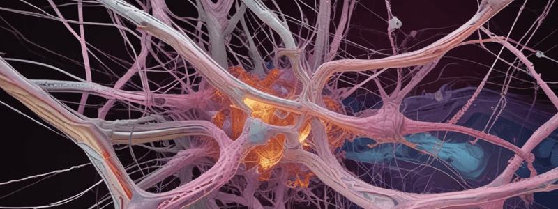
Cilia and Flagella
- Cilia are eyelash or hair-like processes from the cell surface, motile, and longer than microvilli (5-10μ long, 0.2μ in diameter)
- Under the electron microscope, they have a complex internal structure composed of microtubules
- 250 or more cilia are found in each cell, arranged in parallel rows, and function to move fluid over the surface of the cell
- Found in epithelial cells of the upper respiratory tract, uterine tubes (oviducts), and efferent ducts (Ductus efferentes)
Basal Body Structure
- At the base of a cilium, a central pair of single microtubules terminates
- Each of the peripheral doublets is continuous with a triplet microtubule of the basal body
- Three microtubules are fused together and form a triplet microtubule
- Basal body resembles a centriole but contains some accessory structures such as basal foot and rootlet
Cilia Function
- Ciliary movement is important during development
- Dynein arms are absent from cilia in patients with Kartagener's Syndrome
- Nodal Cilia cannot move during development, and internal organs cannot be located at their normal positions
- Helical beating of cilia at the node, and the origins of left-right asymmetry
Flagella Structure and Function
- Flagellum propels sperm, is longer than cilia (100-200 μ), and has the same internal structure as cilia (Axoneme 9+2)
- Has a different type of movement (undulating wave type of movement) and is less in number (one or two in a single cell)
- Mammalian spermium contains 9 additional dense fibers around the axoneme (9+9+2), which serves a protective function
Microtubule Structure
- Microtubules are hollow tube-like or pipe-like structures
- The wall of the microtubule is composed of 13 protofilaments
- Protofilaments are composed of Tubulin subunits (dimer), which consist of Tubulin α and Tubulin β
Gap Junctions
- Site of firm adhesion of cells
- Allow free interchange of substances
- Important role in regulation of intrauterine development and differentiation
- Coordinate function among groups of cells
Anchoring Junctions
- Two types: Hemidesmosomes and Focal Adhesions
Hemidesmosomes
- Have a single dense plaque on the cytoplasmic surface
- Anchor loops of intermediate filaments
- Integrins bind to laminin protein in the basal lamina
- Intracellular domain binds to an anchor protein (plectin) that binds to keratin intermediate filaments
- Found in epithelial cells, attach to basal lamina
- Mutations in plectin cause skin blisters
- Autoantibodies attack type XVII collagen in bullous pemphigoid, a blistering skin disease
Focal Adhesions
- Bind cells to the extracellular matrix
- Cell-matrix adhesion proteins; integrins responsible for binding to the matrix
- Cytoplasmic domain of the integrin binds indirectly to actin filaments
- Integrin's extracellular domains bind to components of extracellular matrix
- Cytoplasmic tail of the β subunit binds indirectly to actin
Adhesion Proteins
- Fibrin: a major ECM component of blood clots
- Forms an elastic network to which cells and other ECM components bind
- Polymerization of fibrin to form the network occurs when its precursor molecule fibrinogen is cleaved by the enzyme thrombin
- Fibrin has binding interactions with various extracellular components, including fibronectin, heparin, growth factors, and cytokines
- Fibrinolysis is mediated by plasmin, which cleaves fibrin
Von Willebrand Factor
- Plays a key role in the major response of platelets to vascular injury by mediating the initiation and progression of thrombus formation
- Enables cell adhesion to develop by forming a bridge between collagen in the vessel wall and blood platelets
Desmosomes
- Strongest points of cell adhesion that provide mechanical binding
- Most abundant in tissues that are exposed to mechanical stress (epidermis of the skin, heart muscle)
- Cell-to-cell binding depends on cadherin family of proteins called desmoglein and desmocollin
- Contain plaque-shaped structures on the cytoplasmic face of the junction which provide attachment sites for intermediate filaments
- Plaque proteins are plakoglobins, desmoplakins, and plakophilins
- Found in many tissues, especially abundant in skin, heart muscle, and the neck of the uterus
- Importance of desmosome junctions is demonstrated by some skin autoimmune diseases, such as pemphigus, where affected individuals make antibodies against their own desmosomal cadherins
Desmosomes
- Strongest points of cell adhesion that provide mechanical binding
- Most abundant in tissues exposed to mechanical stress (epidermis of the skin, heart muscle)
- Contain plaque-shaped structures on the cytoplasmic face of the junction, providing attachment sites for intermediate filaments
- Plaque proteins include plakoglobins, desmoplakins, and plakophilins
- Two types of cadherins (desmoglein and desmocollin) link adjacent cells together
- Cytoplasmic plaque (desmoplakin, plakoglobin, and plakophilin) links the cadherins to intermediate filaments
Importance of Desmosomes
- Desmosomal cadherins are targeted in autoimmune diseases such as Pemphigus, leading to blistering of the skin
- Affected individuals make antibodies against their own desmosomal cadherins, disrupting desmosomes and causing skin epithelial cells to separate
Intermediate Filaments
- Types of intermediate filaments include keratin filaments in epithelial cells and desmin filaments in heart and muscle cells
- Intermediate filaments can be subclassified according to the cytoskeletal element involved
Anchoring Junctions
- Subclassified into two types:
- Intermediate filament attachment sites
- Cell-cell junctions (desmosomes)
- Cell-matrix junctions (hemidesmosomes)
- Actin filament attachment sites
- Cell-cell junctions (adherens junctions)
- Cell-matrix junctions (focal adhesions)
- Intermediate filament attachment sites
Hemidesmosomes
- Resemble desmosomes morphologically
- Cell uses hemidesmosomes to attach to the basal lamina
- Contain a single dense plaque on the cytoplasmic surface that anchors loops of intermediate filaments
- Schematic model shows an epithelial cell attached to the basal lamina via hemidesmosomes
- Autoantibodies attack type XVII collagen in bullous pemphigoid, a blistering skin disease
- Mutations in plectin cause skin blisters associated with late-onset muscular dystrophy
Adherens Junctions
- Actin filament bundles are attached by intracellular anchor proteins to cadherins
- Cadherins are transmembrane proteins that bind to those of adjacent cells, tying actin filament bundles together
- Adherens junctions in the form of adhesion belts between epithelial cells in the small intestine
- Role in early development: controlled contraction of actin filament bundles causes epithelial cells to narrow and form a tube in vertebrate development
Anchoring Junctions
- Anchoring junctions can be subclassified according to the cytoskeletal element involved
- Two types of anchoring junctions:
- Actin filament attachment sites
- Cell-cell junctions (ADHERENS JUNCTIONS)
- Cell-matrix junctions (FOCAL ADHESIONS)
- Intermediate filament attachment sites
- Cell-cell junctions (DESMOSOMES)
- Cell-matrix junctions (HEMIDESMOSOMES)
- Actin filament attachment sites
Desmosomes
- Desmosomes are strongest points of cell adhesion that provide mechanical binding
- Most abundant in tissues that are exposed to mechanical stress (Epidermis of the skin, heart muscle)
- Cell to cell binding depends on cadherin family of proteins called desmoglein and desmocollin
- Desmosomes contain plaque-shaped structures on the cytoplasmic face of the junction which provide attachment sites for intermediate filaments
- Plaque proteins: plakoglobins, desmoplakins, plakophilins
Hemidesmosomes
- Hemidesmosomes resemble desmosomes morphologically
- Hemidesmosomes have only a single dense plaque on the cytoplasmic surface of the hemidesmosome (hemi=half) that anchors loops of intermediate filaments
- Hemidesmosomes connect epithelial cells to the basal lamina
- Integrin (α6β4) and type XVII collagen (also called BPAG2) attach to the basal lamina
Importance of Desmosomes and Hemidesmosomes
- In a blistering skin disease called bullous pemphigoid, autoantibodies attack type XVII collagen
- Mutations in plectin cause skin blisters associated with late-onset muscular dystrophy
- Cataract, heart malformations, and skin blistering are examples of diseases related to desmosome and hemidesmosome dysfunction
Studying That Suits You
Use AI to generate personalized quizzes and flashcards to suit your learning preferences.
Related Documents
Description
This quiz covers the basics of intermediate filaments and anchoring junctions in cells, including types of filaments and their locations in different cell types.


