Podcast
Questions and Answers
What is the primary characteristic of the matrix in cartilage?
What is the primary characteristic of the matrix in cartilage?
- It is rigid and brittle.
- It is entirely solid and non-flexible.
- It is rubbery and flexible. (correct)
- It is fluid-like and unstable.
Which of the following cell types are found in cartilage?
Which of the following cell types are found in cartilage?
- Adipocytes and macrophages.
- Osteoblasts and osteocytes.
- Fibroblasts and mast cells.
- Chondroblasts and chondrocytes. (correct)
What is the role of the perichondrium in cartilage?
What is the role of the perichondrium in cartilage?
- It is responsible for the formation of hyaline cartilage.
- It acts as a rigid outer layer.
- It provides nerve supply to the cartilage.
- It serves as a nutrient source via diffusion. (correct)
What are the three types of cartilage based on?
What are the three types of cartilage based on?
How does cartilage provide support to soft tissues?
How does cartilage provide support to soft tissues?
What is the primary function of chondrocytes in cartilage?
What is the primary function of chondrocytes in cartilage?
What type of cartilage is found in the intervertebral disc?
What type of cartilage is found in the intervertebral disc?
Which structure primarily provides strength and limited mobility at joints?
Which structure primarily provides strength and limited mobility at joints?
What occurs when there is herniation of the nucleus pulposus?
What occurs when there is herniation of the nucleus pulposus?
Which of the following is a characteristic of cartilage?
Which of the following is a characteristic of cartilage?
What occurs at the primary ossification center?
What occurs at the primary ossification center?
In which part of a long bone does the secondary ossification center develop?
In which part of a long bone does the secondary ossification center develop?
Which zone of the epiphyseal cartilage involves the accumulation of glycogen and alkaline phosphatase enzyme?
Which zone of the epiphyseal cartilage involves the accumulation of glycogen and alkaline phosphatase enzyme?
What is the function of osteoblasts during the ossification process?
What is the function of osteoblasts during the ossification process?
What occurs in the zone of invasion of the epiphyseal cartilage?
What occurs in the zone of invasion of the epiphyseal cartilage?
What is the appearance of chondrocytes during tissue preparation?
What is the appearance of chondrocytes during tissue preparation?
What is formed by clusters of dividing chondrocytes?
What is formed by clusters of dividing chondrocytes?
What is the primary function of protein-forming cells in cartilage?
What is the primary function of protein-forming cells in cartilage?
What type of growth does cartilage undergo when young chondrocytes divide?
What type of growth does cartilage undergo when young chondrocytes divide?
What is a characteristic of elastic cartilage?
What is a characteristic of elastic cartilage?
What changes occur in old chondrocytes?
What changes occur in old chondrocytes?
What type of collagen is primarily formed in cartilage?
What type of collagen is primarily formed in cartilage?
What distinguishes tough cartilage from hyaline cartilage?
What distinguishes tough cartilage from hyaline cartilage?
What initiates the acute inflammatory response during the healing process of a fracture?
What initiates the acute inflammatory response during the healing process of a fracture?
Which method allows for the demonstration of bone cells and soft tissue?
Which method allows for the demonstration of bone cells and soft tissue?
What is characteristic of the grinding method used on bones?
What is characteristic of the grinding method used on bones?
Which type of bone is classified as compact bone?
Which type of bone is classified as compact bone?
What is found under the periosteum of a long bone?
What is found under the periosteum of a long bone?
What are Haversian systems also known as?
What are Haversian systems also known as?
Which of the following statement regarding Volkmann's canal is correct?
Which of the following statement regarding Volkmann's canal is correct?
Which anatomical classification refers to bones shaped like cubes?
Which anatomical classification refers to bones shaped like cubes?
What is the tubular system formed with T-tubules called in cardiac muscle at the level of Z-line?
What is the tubular system formed with T-tubules called in cardiac muscle at the level of Z-line?
Where are glycogen granules mainly concentrated in cardiac muscle cells?
Where are glycogen granules mainly concentrated in cardiac muscle cells?
Which hormone is contained in atrial granules within the heart?
Which hormone is contained in atrial granules within the heart?
What do desmosomes and adherent junctions do in cardiac muscle cells?
What do desmosomes and adherent junctions do in cardiac muscle cells?
What shape do the discs of cardiac muscle cells resemble?
What shape do the discs of cardiac muscle cells resemble?
What is located in the right ventricle and contains Purkinje muscle fibers?
What is located in the right ventricle and contains Purkinje muscle fibers?
What is a characteristic feature of Purkinje muscle fibers compared to ordinary cardiac muscle fibers?
What is a characteristic feature of Purkinje muscle fibers compared to ordinary cardiac muscle fibers?
What prevents gap junctions in cardiac muscle cells from being damaged during contractions?
What prevents gap junctions in cardiac muscle cells from being damaged during contractions?
Flashcards
What is cartilage?
What is cartilage?
Cartilage is a specialized type of connective tissue with a rubbery matrix, allowing it to withstand mechanical stress.
How does cartilage receive nutrients?
How does cartilage receive nutrients?
Cartilage is avascular, meaning it lacks blood vessels. It receives oxygen and nutrients from the surrounding connective tissue (perichondrium) or synovial fluid in joints.
What cells make up cartilage?
What cells make up cartilage?
Cartilage is made up of chondroblasts and chondrocytes, cells responsible for producing the matrix.
What is hyaline cartilage?
What is hyaline cartilage?
Signup and view all the flashcards
What is the perichondrium?
What is the perichondrium?
Signup and view all the flashcards
Chondrocytes
Chondrocytes
Signup and view all the flashcards
Chondroblasts
Chondroblasts
Signup and view all the flashcards
Interstitial Growth
Interstitial Growth
Signup and view all the flashcards
Appositional Growth
Appositional Growth
Signup and view all the flashcards
Elastic Cartilage
Elastic Cartilage
Signup and view all the flashcards
Hyaline Cartilage
Hyaline Cartilage
Signup and view all the flashcards
Fibrocartilage
Fibrocartilage
Signup and view all the flashcards
Perichondrium
Perichondrium
Signup and view all the flashcards
What are chondrocytes?
What are chondrocytes?
Signup and view all the flashcards
What is elastic cartilage?
What is elastic cartilage?
Signup and view all the flashcards
What is the annulus fibrosus?
What is the annulus fibrosus?
Signup and view all the flashcards
What is the nucleus pulposus?
What is the nucleus pulposus?
Signup and view all the flashcards
Endochondral ossification
Endochondral ossification
Signup and view all the flashcards
Primary Ossification Center
Primary Ossification Center
Signup and view all the flashcards
Secondary Ossification Center
Secondary Ossification Center
Signup and view all the flashcards
Periosteal bone collar
Periosteal bone collar
Signup and view all the flashcards
Zone of Calcification
Zone of Calcification
Signup and view all the flashcards
Intercalated Discs
Intercalated Discs
Signup and view all the flashcards
Gap Junctions (in Intercalated Discs)
Gap Junctions (in Intercalated Discs)
Signup and view all the flashcards
Transverse Component of Intercalated Discs
Transverse Component of Intercalated Discs
Signup and view all the flashcards
Lateral Component of Intercalated Discs
Lateral Component of Intercalated Discs
Signup and view all the flashcards
Desmosomes & Adherent Junctions
Desmosomes & Adherent Junctions
Signup and view all the flashcards
Purkinje Fibers
Purkinje Fibers
Signup and view all the flashcards
Moderator Band
Moderator Band
Signup and view all the flashcards
Purkinje Fibers' Sarcoplasm
Purkinje Fibers' Sarcoplasm
Signup and view all the flashcards
Decalcification: How does it work?
Decalcification: How does it work?
Signup and view all the flashcards
Grinding method: What does it show?
Grinding method: What does it show?
Signup and view all the flashcards
Periosteum: What is it?
Periosteum: What is it?
Signup and view all the flashcards
External circumferential lamellae: What are they?
External circumferential lamellae: What are they?
Signup and view all the flashcards
Haversian system (osteon): What is it?
Haversian system (osteon): What is it?
Signup and view all the flashcards
Volkmann's canal: What is it?
Volkmann's canal: What is it?
Signup and view all the flashcards
Lacunae: What do they contain?
Lacunae: What do they contain?
Signup and view all the flashcards
Canaliculi: What are they?
Canaliculi: What are they?
Signup and view all the flashcards
Study Notes
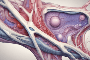
Cartilage
- Specialized connective tissue with a rubbery matrix, designed to withstand mechanical stress.
- Composed of chondroblasts (cells that produce matrix) and chondrocytes (cells within matrix).
- Matrix includes ground substance (abundant, firm, and rubbery), and collagen and elastic fibers.
- Avascular; relies on diffusion of oxygen and nutrients from surrounding connective tissues (perichondrium) or synovial fluid in joints.
- Lacks blood vessels and nerves.
- Three types: hyaline, elastic, and fibrocartilage, distinguished by the type and amount of fibers in the matrix.
Hyaline Cartilage
- Most common type, appearing translucent and glassy.
- Found in fetal skeletons, articular surfaces of bones, costal cartilage, and respiratory passages (nose, larynx, trachea, and bronchi).
- Matrix is mostly collagen type II.
- Chondrocytes are arranged in lacunae and are typically in small groups.
- Surrounded by perichondrium (a layer of dense connective tissue).
- Supports soft tissues with flexibility, helps with weight-bearing and shock absorption, and facilitates easy movement at joints.
Perichondrium
-
Capsule-like covering around hyaline cartilage (except at articular surfaces).
-
Consists of two layers: outer fibrous (white fibrous connective tissue) containing fibroblasts that secrete collagen type I, as well as blood vessels and nerves; inner chondrogenic (cellular) with chondroblasts, crucial for cartilage nutrition (by diffusion).
-
Essential for cartilage nutrition and growth, and also for attachment of muscles.
-
Collagen fibers: Type II, not visible by light microscopy (LM) due to their thinness and similar refractive index to the ground substance. Visible after digestion of the matrix by enzymes.
-
Ground substance: produced by chondrocytes and chondroblasts, rubbery, homogeneous, and transparent; characterized by its high content of proteoglycans, glycoproteins, and water; stained metachromatically (e.g., with toluidine blue).
Cartilage Cells
- Chondroblasts: young cells that actively produce cartilage matrix, flattened or oval with a basophilic cytoplasm and pale nucleus; located on the surface of cartilage; give rise to chondrocytes.
- Chondrocytes: mature cells maintained within spaces (lacunae) located within the matrix, rounded cells; typically seen within groups, with pale cytoplasm and a darker-staining nucleus; become trapped as the matrix hardens around the cells.
Cartilage Function
- Supports soft tissues with flexibility
- Keeps airway patent (open)
- Tissue attachment & weight bearing, shock absorbing
- Smooth surface for easy movement of bones
- Development and growth of bones
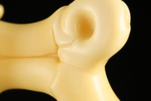
Bone
- Highly vascularized, hard connective tissue composed of a calcified matrix.
- Provides support, protection (e.g., brain, bone marrow), and mineral storage (mainly calcium).
- Matrix: organic (collagen fibers, ground substance) and inorganic (calcium salts).
- Covers bone surface: periosteum (outer) and endosteum (inner).
- Three types of cells (in order): osteogenic, osteoblasts, and osteocytes, and osteoclasts.
Bone Composition
- Organic Parts (35%): Type I collagen fibers in thick bundles and ground substance (glycosaminoglycans, glycoproteins).
- Inorganic Parts (65%): Calcium salts (mainly calcium phosphate and carbonate) deposited within and on surface of collagen bundles. These give the bone its hardness.
Bone Coverings
- Periosteum: Tough, fibrous outer layer of dense irregular connective tissue; contains blood vessels and nerves; covers outer surface of bone, except at articular surfaces. Essential for bone nutrition, growth, and repair, and attachment of tendons and ligaments.
- Endosteum: Delicate inner layer of connective tissue; lining the bone's inner surface, including the medullary cavity and trabeculae; contains osteogenic cells that aid in bone growth by adding from inside (internal growth), in bone repair, and nutrition of the bone surface
Bone Covering Function
- Periosteum:
- Protection of bone
- Attachment for muscles, tendons, ligaments.
- Bone nutrition (through blood vessels)
- Growth and repair
- Endosteum:
- Protection of bone surface
- Bone growth from the inside (internal growth)
- Essential part of bone remodeling
Bone Cells
- Osteogenic cells: Undifferentiated mesenchymal stem cells from the periosteum or endosteum. Give rise to osteoblasts.
- Osteoblasts: Immature bone-forming cells; found on bone surfaces. Secrete a matrix composed mainly of collagen and proteins, which later calcifies to make bone.
- Osteocytes: Mature osteoblasts that are encased in the bone matrix; maintain bone tissue. Connect to each other through canaliculi - allowing them to communicate via processes in the hard matrix.
- Osteoclasts: Bone-resorbing cells; participate in remodeling and regulate calcium homeostasis. Large, multinucleated cells that breakdown bone tissue by decalcification.
Bone Classifications
-
Anatomical: Long, short, flat, irregular
-
Histological: Compact, spongy
-
Compact Bone: Dense, outer layer of bone; composed of osteons (Haversian systems), cylindrical units of calcified bone lamellae surrounding a central canal with nerves and blood vessels. Interstitial lamellae fill gaps between osteons. Outer circumferential and inner circumferential lamellae surround the entire outer/inner surface of compact bone respectively.
-
Spongy Bone: Porous inner layer of bone, found in the interior of flat bones, and within epiphyses of long bones; consists of trabeculae, interlocking bony struts, filled with bone marrow; Provides strength and lightness.
-
Volkmann's canals: Canals that traverse compact bone and connect Haversian canals with bone marrow and periosteum.
-
Perforating fibers of Sharpey: Collagen fibers that attach tendons and ligaments to the bone matrix.
Muscle Tissue
- Muscle tissue specialized for contraction, facilitating movement.
- Divided into three types - skeletal, cardiac, and smooth - each possessing distinct structural and functional characteristics.
Skeletal Muscle Tissue
- Attaches to bones, enabling voluntary movement.
- Composed of elongated muscle cells called fibers.
- Striated appearance due to orderly arrangement of contractile proteins.
- Multinucleated, with nuclei located peripherally.
- Requires signal from the nervous system to contract.
Cardiac Muscle Tissue
- Found exclusively in the heart.
- Branched, striated appearance.
- Intercalated discs (specialized junctions) connect adjacent cells, facilitating rapid transmission of signals for coordinated contraction.
- Involuntary movement.
- Single nucleus located centrally.
Smooth Muscle Tissue
- Located in walls of internal organs and blood vessels.
- Non-striated appearance, with a spindle-shaped cell form.
- Single nucleus in the cell's center.
- Involuntary contraction, controlled by autonomic nervous system and hormones.
Skin
- Protective covering of the body.
- Two main layers - epidermis, dermis and hypodermis
- Hypodermis - important for insulation.
- The function of the skin is protection.
- Skin repairs through various cells.
- Also a site for many receptors and cell communication
Epidermis
- Outermost layer of skin, mostly keratinized stratified squamous epithelium.
- Avascular, nutrition by diffusion from dermis.
- Consists of five layers — Stratum basale, Stratum spinosum, Stratum granulosum, Stratum lucidum, and Stratum corneum.
Dermis
- Deep layer of skin, consisting of connective tissue (CT), rich in collagen and elastin fibers.
- Responsible for skin's strength, flexibility, and elasticity.
- Contains blood vessels, nerves, and specialized sensory receptors.
- Two sublayers - papillary layer and reticular layer.
Hypodermis
- Subcutaneous tissue, not a part of skin.
- Layer below dermis, highly vascular, mostly adipose CT, providing insulation and cushioning.
- Connects skin to underlying tissues.
Skin Appendages
- (Hair Follicles):
- Epithelial tube extending into the dermis or hypodermis.
- Contains hair shaft (outer, part of the hair shaft).
- Muscle bundles which attached to hair follicle.
- (Sebaceous glands):
- Simple alveolar glands associated with hair follicles; secrete sebum (oily substance) that keeps hair and skin soft and waterproof.
- (Sweat glands):
- Produce sweat (water, salts, urea); responsible for thermoregulation and excretion. Two types — eccrine (most abundant) and apocrine (associated with hair follicles; larger secretion volume).
Studying That Suits You
Use AI to generate personalized quizzes and flashcards to suit your learning preferences.