Podcast
Questions and Answers
What is the name of the anatomical region where the heart is located?
What is the name of the anatomical region where the heart is located?
- Abdominal cavity
- Cranial cavity
- Pelvic cavity
- Thoracic cavity (correct)
What is the name of the author of the document that discusses the cardiovascular system?
What is the name of the author of the document that discusses the cardiovascular system?
- Marcela Bezdickova (correct)
- Week 1
- The Heart
- Cardiovascular System
What is the name of the document as indicated by the title?
What is the name of the document as indicated by the title?
- Week 1
- Cardiovascular System (correct)
- Mediastinum
- The Heart
What is the format of the document?
What is the format of the document?
What is the date the document was created?
What is the date the document was created?
Flashcards
Cardiovascular System
Cardiovascular System
The system in the body that comprises the heart and blood vessels, responsible for circulating blood.
Heart
Heart
A muscular organ that pumps blood throughout the body, part of the cardiovascular system.
Mediastinum
Mediastinum
The central compartment of the thoracic cavity that contains the heart, trachea, and other structures.
Blood Vessels
Blood Vessels
Signup and view all the flashcards
Circulation
Circulation
Signup and view all the flashcards
Study Notes
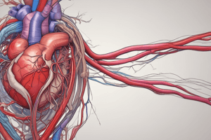
Cardiovascular System: The Heart
-
Location and Size:
- Located in the middle mediastinum, within the thoracic cavity, between the lungs.
- Roughly 12 cm long, 8 cm wide, and 6 cm thick.
- Weighs approximately 250-300 grams (Females 250g, Males 300g).
- About two-thirds of the heart is located in the mid-portion of the body cavity
-
Mediastinum:
- The central part of the thoracic cavity.
- Extends from the sternum to the thoracic vertebrae, first rib to diaphragm, and between the lungs.
- Contains the heart, great vessels, phrenic and cardiac nerves, thoracic duct, lymph nodes, esophagus, and the trachea.
-
Functions of the Heart:
- Pumps blood: Self-conducting system for contraction (heartbeat). A functional circulation system with two pumps (ventricles), in a series arrangement, including systemic and pulmonary circulation.
- Regulates blood supply and blood pressure: Adjusts heart rate and force of contraction to meet the body's changing needs (exercise, temperature changes, digestion).
- Routes blood: Separates pulmonary and systemic circulations. One-way flow is ensured by valves.
-
Sections of Heart:
- Right border: Right atrium
- Left border: Left ventricle and left atrium
- Superior border: Both atria, ascending aorta, and pulmonary trunk
- Inferior border: Right ventricle and apical part of left ventricle
- Anterior surface: Right ventricle and parts of left ventricle and right atrium
- Inferior surface: Both ventricles, mainly left ventricle
- Base: Primarily left atrium
- Left pulmonary surface: Left ventricle and parts of left atrium
- Apex: Tip of left ventricle (located at the 5th left intercostal space at the midclavicular line)
-
Surface of Heart
- Right pulmonary surface: Right atrium
- Base is also called posterior surface
- Inferior surface is also called diaphragmaic surface
External and Internal Structures of the Heart
-
External:
- Superior vena cava (SVC): Carries deoxygenated blood to the right atrium.
- Right pulmonary veins: Carry oxygenated blood from the lungs to the left atrium.
- Left pulmonary veins: Carry oxygenated blood from the lungs to the left atrium..
- Pulmonary trunk and its branches
- Aorta arch
- Inferior vena cava (IVC) : Carries deoxygenated blood to the right atrium.
- Pulmonary trunk
- Aorta arch
-
Internal: Right atrium, right ventricle, left atrium, Left Ventricle
-
Heart Valves:
- Atrioventricular (AV) Valves:
- Tricuspid valve: Between the right atrium and ventricle
- Mitral (bicuspid) valve: Between the left atrium and ventricle
- Semilunar Valves:
- Pulmonary valve: Between the right ventricle and pulmonary artery
- Aortic valve: Between the left ventricle and the aorta
- Atrioventricular (AV) Valves:
Pericardium
-
**Membranous Structure:**Encloses the heart and the great vessels.
- Fibrous layer: The most superficial fibrous connective tissue layer that prevents heart overfilling. -Continuous with the outermost layer (adventitia) of the great vessels.
- Serous layer: Double-layered membrane with a parietal layer (fused to the fibrous pericardium) & visceral layer (part of the epicardium)- reduce heart friction during beating.
- Pericardial cavity: Space between the parietal and visceral layers, containing about 20 ml of pericardial fluid.
-
Clinical Relevance:
- Pericarditis: Inflammation of the pericardium (various causes). Clinical symptoms include chest pain, pressure, mild discomfort, cough, palpitations, and lightheadedness.
- Pericardial effusion: Buildup of excess fluid in the pericardial cavity.
-
Heart Wall Layers:
- Epicardium: Outermost layer (visceral layer of the pericardium).
- Myocardium: Thickest, major muscle layer regulating heart contractions/relaxations.
- Endocardium: Innermost layer (smooth endothelial lining).
Coronary Arteries and Venous Drainage
-
Coronary Arteries: Supply oxygenated blood to the heart muscle.
- Right Coronary Artery (RCA): Supplies blood primarily to the right structures of the heart
- Left Coronary Artery (LCA): Supplies the left structures of the heart and about 2/3rds of the septum
-
Venous Drainage: Cardiac veins collect deoxygenated blood and drain into the coronary sinus, which then enters the right atrium.
Ischemic Cardiac Diseases
- Inadequate oxygen supply to the heart muscle: Obstruction of coronary arteries causing myocardial ischemia (lack of blood supply).
- Heart attack: Most frequent symptom of ischemia - often due to artery blockage.
- Clinical presentations: Differ based on location of artery blockage (e.g., inferior, lateral, anterior).
- Coronary artery disease: Accumulation of atherosclerotic plaque in coronary arteries leading to reduced blood flow.
Mediastinum Regions
- Superior mediastinum: Upper chest, above the heart, contains great vessels, trachea, esophagus, nerves, and thymus.
- Middle mediastinum: Center of the chest; Contains the heart and major blood vessels (aorta, pulmonary artery, etc.)
- Anterior mediastinum: In front of the middle mediastinum and contains mostly connective tissue..
- Posterior mediastinum: Below the middle mediastinum contains large blood vessels, esophagus, and veins, vagus nerve.
Aortic Arch Branches
- Brachiocephalic trunk: Branches into the right common carotid artery and right subclavian artery. Supplies blood to the right side of the head and neck and right upper limb
- Left common carotid artery: Supplies blood to the left side of the head and neck.
- Left subclavian artery: Supplies blood to the left upper limb.
- Descending aorta: Further branches supply the intercostal spaces and other tissues around the thorax and abdomen.
Carotid Arteries and Internal Jugular Veins
- Common carotid artery: Main blood supply to the head and neck; pulse palpitation is possible here close to the heart.
- Internal carotid artery: Supplies the brain
- External carotid artery: Supplies the face and neck.
- Internal jugular vein: Drains blood from the face, brain, and neck, and joins the subclavian vein to form the brachiocephalic veins. (both left and right brachiocephalic veins combine at the superior vena cava that empties into the right atrium)
Studying That Suits You
Use AI to generate personalized quizzes and flashcards to suit your learning preferences.


