Podcast
Questions and Answers
What is the primary function of the cardiovascular system?
What is the primary function of the cardiovascular system?
- Bringing oxygen and nutrients while removing waste products (correct)
- Removing only oxygen from the body
- Only carrying hormones between the body's cells
- Transporting only oxygen to cells
What is the anatomical position of the heart in most people?
What is the anatomical position of the heart in most people?
- At the lower right of the abdomen
- Vertically aligned in the center of the chest
- Directly behind the sternum without any angle
- Obliquely with the right side below and in front of the left (correct)
Which layer surrounds the heart and provides protection?
Which layer surrounds the heart and provides protection?
- Endocardium
- Pericardium (correct)
- Epicardium
- Myocardium
How many chambers does the heart have?
How many chambers does the heart have?
What is the pointed end of the heart referred to as?
What is the pointed end of the heart referred to as?
What are the two main types of valves found in the heart?
What are the two main types of valves found in the heart?
What is the fibrous layer of the pericardium composed of?
What is the fibrous layer of the pericardium composed of?
Which layer of the serous pericardium adheres to the heart's surface?
Which layer of the serous pericardium adheres to the heart's surface?
What is the primary function of pericardial fluid?
What is the primary function of pericardial fluid?
Which layer of the heart wall is primarily responsible for contraction?
Which layer of the heart wall is primarily responsible for contraction?
What separates the right and left ventricles?
What separates the right and left ventricles?
Which chamber receives deoxygenated blood from the body?
Which chamber receives deoxygenated blood from the body?
What type of valve is the mitral valve?
What type of valve is the mitral valve?
Which chamber of the heart has the thickest walls?
Which chamber of the heart has the thickest walls?
What is the function of the semilunar valves?
What is the function of the semilunar valves?
Which blood vessels return oxygenated blood to the heart?
Which blood vessels return oxygenated blood to the heart?
What is the primary function of the aortic valve?
What is the primary function of the aortic valve?
What is the characteristic feature of the tricuspid valve?
What is the characteristic feature of the tricuspid valve?
How many times per minute does the SA node typically generate an impulse?
How many times per minute does the SA node typically generate an impulse?
Which of the following accurately describes the role of the AV node?
Which of the following accurately describes the role of the AV node?
What are the unique characteristics of pacemaker cells?
What are the unique characteristics of pacemaker cells?
Where is the sinoatrial (SA) node located?
Where is the sinoatrial (SA) node located?
What happens if the SA node fails to fire?
What happens if the SA node fails to fire?
What is the function of the Purkinje fibers?
What is the function of the Purkinje fibers?
Flashcards are hidden until you start studying
Study Notes
Cardiovascular System
- The circulatory system consists of the heart, blood vessels, and lymphatics.
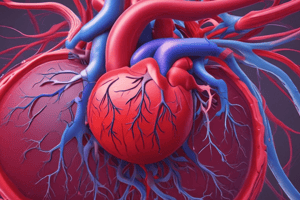
- The heart is made of two separate pumps: the right side pumps blood to the lungs for oxygenation, and the left side pumps oxygenated blood to the rest of the body.
- The heart is located in the mediastinum (cavity between the lungs) beneath the sternum between the 2nd and 6th ribs.
- The heart rests obliquely with its right side below and almost in front of the left. The broad part or top of the heart is at its upper right, and its pointed end (apex) is at its lower left. The apex is where the heart sounds are loudest.
Heart Structure
- The heart is surrounded by a sac called the pericardium.
- The heart wall is composed of three layers: myocardium, endocardium, and epicardium.
- The heart contains four chambers (two atria and two ventricles) and four valves (two atrioventricular and two semilunar valves).
Pericardium
- The pericardium is a fibroserous sac surrounding the heart and the roots of the great vessels.
- The fibrous pericardium is composed of tough, white fibrous tissue that fits loosely around the heart, protecting it.
- The serous pericardium is the thin smooth inner portion, containing two layers: the parietal layer (lines the inside of the fibrous pericardium) and the visceral layer (adheres to the surface of the heart).
- The pericardial space, located between the fibrous and serous pericardium, contains pericardial fluid to lubricate the surfaces and allow easy movement.
The Wall
- The epicardium is the outer layer, and the visceral layer of the serous pericardium. It is made up of squamous epithelial cells overlying connective tissue.
- The myocardium is the middle layer, forming most of the heart wall. Composed of striated muscle fibers, it causes the heart to contract.
- The endocardium is the inner layer of the heart. It consists of endothelial tissue with small blood vessels and bundles of smooth muscle.
Inside the Heart
- Two Atria:
- The atria are the upper chambers separated by the interatrial septum.
- The right atrium receives blood from the superior and inferior venae cavae.
- The left atrium, which is smaller but has thicker walls, forms the uppermost part of the heart's left border. It receives blood from the two pulmonary veins.
- Two Ventricles:
- The ventricles are the lower chambers separated by the interventricular septum.
- They receive blood from the atria.
- The ventricles are larger and have thicker walls than the atria due to their highly developed musculature.
- The right ventricle pumps blood to the lungs.
- The left ventricle, larger than the right, pumps blood through all other vessels of the body.
The Valves
- The heart contains four valves: two AV valves and two semilunar valves.
- The valves allow forward flow of blood through the heart and prevent backflow.
- The AV valves separate the atria from the ventricles.
- The right AV valve, called the tricuspid valve, prevents backflow from the right ventricle into the right atrium.
- The left AV valve, called the mitral valve, prevents backflow from the left ventricle into the left atrium.
- The semilunar valves prevent backflow from the arteries into the ventricles.
- The pulmonic valve prevents backflow from the pulmonary artery into the right ventricle.
- The aortic valve prevents backflow from the aorta into the left ventricle.
Tricuspid and Mitral Valves
- The tricuspid valve has three triangular cusps, or leaflets.
- The mitral valve (bicuspid valve) contains two cusps, a large anterior and a smaller posterior.
- Chordae tendineae attach the cusps of the AV valves to papillary muscles in the ventricles.
Semilunar Valves
- The semilunar valves have three cusps shaped like half-moons.
Conduction System
- The conduction system of the heart contains pacemaker cells with three unique characteristics:
- Automaticity: ability to generate an electrical impulse automatically.
- Conductivity: ability to pass the impulse to the next cell.
- Contractility: ability to shorten the fibers in the heart when receiving the impulse.
Heart Pacemaker
- The sinoatrial (SA) node, located on the endocardial surface of the right atrium near the superior vena cava, is the heart's normal pacemaker, generating an impulse between 60 and 100 times per minute.
- The firing of the SA node spreads an impulse throughout the atria, resulting in atrial contraction.
AV Node
- The atrioventricular (AV) node, situated low in the septal wall of the right atrium, slows impulse conduction between the atria and ventricles, allowing time for the contracting atria to fill the ventricles with blood before ventricular contraction.
Bundle of His and Purkinje Fibers
- From the AV node, the impulse travels to the bundle of His (modified muscle fibers), branching off to the right and left bundles.
- The impulse then travels to the Purkinje fibers, the distal portions of the left and right bundle branches.
- The Purkinje fibers fan across the surface of the ventricles from the endocardium to the myocardium.
- As the impulse spreads, it brings the signal to the blood-filled ventricles to contract.
Conduction System Safety Mechanisms
- If the SA node fails to fire, the AV node will generate an impulse between 40 and 60 times per minute.
Studying That Suits You
Use AI to generate personalized quizzes and flashcards to suit your learning preferences.


