Podcast
Questions and Answers
What is the primary function of the heart within the cardiovascular system?
What is the primary function of the heart within the cardiovascular system?
- Serve as the main site for nutrient absorption
- Filter waste products from blood
- Act as a muscular pumping device (correct)
- Regulate blood pressure
What is the primary purpose of capillaries in the cardiovascular system?
What is the primary purpose of capillaries in the cardiovascular system?
- Return blood to the heart
- Increase the pressure of circulating blood
- Conduct blood through tissues and facilitate exchange of materials (correct)
- Carry blood away from the heart
What does hemodynamics refer to?
What does hemodynamics refer to?
- The study of blood cell formation
- The regulation of blood pH levels
- The mechanisms that influence blood circulation (correct)
- The process of nutrient absorption in blood
How does the cardiovascular system respond to the activity levels of different cells?
How does the cardiovascular system respond to the activity levels of different cells?
What are the major components of the cardiovascular system?
What are the major components of the cardiovascular system?
What two things are required to maintain effective blood circulation?
What two things are required to maintain effective blood circulation?
Which of the following statements about veins is correct?
Which of the following statements about veins is correct?
What happens to blood volume distribution when certain tissues become more active?
What happens to blood volume distribution when certain tissues become more active?
Which statement best describes arteries in the cardiovascular system?
Which statement best describes arteries in the cardiovascular system?
What role do capillaries play in the cardiovascular system?
What role do capillaries play in the cardiovascular system?
What mechanism is essential for distributing blood to more active tissues?
What mechanism is essential for distributing blood to more active tissues?
Which component of the cardiovascular system is primarily responsible for pumping blood?
Which component of the cardiovascular system is primarily responsible for pumping blood?
What is the main purpose of hemodynamics in the body?
What is the main purpose of hemodynamics in the body?
What type of blood do arteries primarily carry?
What type of blood do arteries primarily carry?
What occurs when cells become more active in terms of blood circulation?
What occurs when cells become more active in terms of blood circulation?
How does the cardiovascular system maintain homeostasis?
How does the cardiovascular system maintain homeostasis?
What defines the closed system of blood vessels in the cardiovascular system?
What defines the closed system of blood vessels in the cardiovascular system?
What happens to blood volume during increased activity levels of specific tissues?
What happens to blood volume during increased activity levels of specific tissues?
What are trabeculae carinae?
What are trabeculae carinae?
The atria are called 'pumping chambers' of the heart.
The atria are called 'pumping chambers' of the heart.
What is the function of heart valves?
What is the function of heart valves?
Which chamber of the heart is primarily responsible for pumping blood to the entire body?
Which chamber of the heart is primarily responsible for pumping blood to the entire body?
What is the septum in the heart?
What is the septum in the heart?
The left ventricle has a thinner myocardial wall compared to the right ventricle.
The left ventricle has a thinner myocardial wall compared to the right ventricle.
What type of valves guard the opening between the atria and ventricles?
What type of valves guard the opening between the atria and ventricles?
What is the first step of the cardiac cycle?
What is the first step of the cardiac cycle?
What does isovolumic mean?
What does isovolumic mean?
Where is the Sinoatrial (SA) node located?
Where is the Sinoatrial (SA) node located?
Which heart sound is caused by the contraction of the ventricles?
Which heart sound is caused by the contraction of the ventricles?
Which side of the heart moves blood through the pulmonary circulation?
Which side of the heart moves blood through the pulmonary circulation?
What causes the second heart sound 'dupp'?
What causes the second heart sound 'dupp'?
The cardiac cycle illustrates changes in pressure gradients in the left atrium, left ventricle, and ______.
The cardiac cycle illustrates changes in pressure gradients in the left atrium, left ventricle, and ______.
What occurs during passive ventricular filling?
What occurs during passive ventricular filling?
Heart murmurs are normal heart sounds.
Heart murmurs are normal heart sounds.
What is the determinant of arterial blood pressure?
What is the determinant of arterial blood pressure?
Which phase of the cardiac cycle occurs after atrial systole?
Which phase of the cardiac cycle occurs after atrial systole?
What does the atrioventricular (AV) node do?
What does the atrioventricular (AV) node do?
What is the function of the atrioventricular bundle (bundle of His)?
What is the function of the atrioventricular bundle (bundle of His)?
Pacemaker fibers are contractile cardiac muscles.
Pacemaker fibers are contractile cardiac muscles.
What does the P wave in an electrocardiogram (ECG) represent?
What does the P wave in an electrocardiogram (ECG) represent?
What does the T wave reflect in an ECG?
What does the T wave reflect in an ECG?
The ________ is a single heartbeat or pumping cycle.
The ________ is a single heartbeat or pumping cycle.
In heart failure, the residual volume left in the ventricles may greatly exceed the amount ejected during systole.
In heart failure, the residual volume left in the ventricles may greatly exceed the amount ejected during systole.
What does the QRS complex represent in an ECG?
What does the QRS complex represent in an ECG?
What can the U wave in an ECG indicate?
What can the U wave in an ECG indicate?
What is vasodilation?
What is vasodilation?
What is the control center for the vasomotor control mechanism?
What is the control center for the vasomotor control mechanism?
A sudden increase in arterial blood pressure stimulates the aortic and carotid baroreceptors.
A sudden increase in arterial blood pressure stimulates the aortic and carotid baroreceptors.
Which factors influence venous return? (Select all that apply)
Which factors influence venous return? (Select all that apply)
When a person is lying flat, the pull of gravity is equal _____ and _____ the heart.
When a person is lying flat, the pull of gravity is equal _____ and _____ the heart.
What role do venous pumps play during venous return?
What role do venous pumps play during venous return?
What effect does changes in total blood volume have on venous return?
What effect does changes in total blood volume have on venous return?
What system is triggered when blood pressure in the kidney is low?
What system is triggered when blood pressure in the kidney is low?
What are the basic components of the cardiovascular system?
What are the basic components of the cardiovascular system?
What are the structures of the heart that serve to pump blood?
What are the structures of the heart that serve to pump blood?
What are the deflection waves of a normal ECG and why are they significant?
What are the deflection waves of a normal ECG and why are they significant?
What are the basic steps of the cardiac cycle?
What are the basic steps of the cardiac cycle?
What is the primary principle of circulation?
What is the primary principle of circulation?
What factors affect stroke volume and heart rate?
What factors affect stroke volume and heart rate?
What is the significance of peripheral resistance in the cardiovascular system?
What is the significance of peripheral resistance in the cardiovascular system?
What is the significance of the vasomotor control mechanism?
What is the significance of the vasomotor control mechanism?
How do the respiratory pump and skeletal muscle pump assist the venous return of blood to the heart?
How do the respiratory pump and skeletal muscle pump assist the venous return of blood to the heart?
What are the hormonal mechanisms that regulate blood volume?
What are the hormonal mechanisms that regulate blood volume?
What is the significance of the pulse mechanism?
What is the significance of the pulse mechanism?
What are major pulse points in the body?
What are major pulse points in the body?
What is hemodyamics?
What is hemodyamics?
What are the three layers that make up the heart wall?
What are the three layers that make up the heart wall?
What is the pericardium?
What is the pericardium?
What are the chambers of the heart?
What are the chambers of the heart?
What are the valves of the heart responsible for?
What are the valves of the heart responsible for?
What factors determine arterial pressure?
What factors determine arterial pressure?
What happens if cardiac output increases?
What happens if cardiac output increases?
Peripheral resistance affects the blood leaving the arteries.
Peripheral resistance affects the blood leaving the arteries.
What is the Starling Law of the Heart?
What is the Starling Law of the Heart?
What increases the strength of myocardial contraction?
What increases the strength of myocardial contraction?
What is an Ejection Fraction (EF)?
What is an Ejection Fraction (EF)?
A higher afterload results in a higher stroke volume.
A higher afterload results in a higher stroke volume.
What initiates each heartbeat?
What initiates each heartbeat?
What influences heart rate?
What influences heart rate?
What is peripheral resistance?
What is peripheral resistance?
What is a factor that can increase blood viscosity?
What is a factor that can increase blood viscosity?
Flashcards are hidden until you start studying
Study Notes
Cardiovascular System Overview
- Comprises the heart and a closed network of blood vessels.
- Heart functions as a muscular pump, circulating blood throughout the body.
- Blood circulates continuously, passing through various organ systems multiple times.
Components of Blood Vessels
- Arteries: Transport blood away from the heart, maintaining high pressure.
- Capillaries: Facilitate the exchange of materials (nutrients, oxygen, waste) between blood and tissues.
- Veins: Return blood toward the heart, typically under lower pressure.
Homeostasis and Blood Flow
- Continuous and controlled blood movement is essential for homeostasis.
- Various regulatory mechanisms ensure integration and proper function of the cardiovascular system.
Hemodynamics
- Refers to the mechanisms that influence blood circulation and flow.
- Essential for delivering necessary materials to cells and removing waste products.
- Different organs require varying blood volumes per minute based on their activity levels.
Blood Volume and Activity Levels
- Active cells (e.g., muscle during exercise) require increased blood supply compared to less active cells.
- Blood distribution adjusts dynamically; more blood is directed to active tissues while diverting from less active ones.
Key Functions
- Maintain continuous blood circulation.
- Regulate the volume and distribution of blood flow to accommodate metabolic demands of tissues.
Learning Outcomes
- Basic components of the cardiovascular system include the heart, arteries, capillaries, and veins.
- The heart's structures, including chambers and valves, facilitate blood pumping throughout the body.
- Normal ECG deflection waves indicate electrical activity and heart function; they are crucial for diagnosing cardiac issues.
- The cardiac cycle encompasses stages of the heart's contraction and relaxation, essential for blood flow.
- High arterial blood pressure is vital for effective circulation and organ perfusion, indicating cardiovascular health.
- Factors affecting stroke volume and heart rate include physical activity, hormonal influences, and autonomic nervous system regulation.
- Peripheral resistance significantly impacts blood flow and pressure; it is affected by vessel diameter and blood viscosity.
- Vasomotor control mechanisms adjust blood vessel diameter, influencing blood distribution and pressure.
- The respiratory pump and skeletal muscle pump enhance venous return to the heart, aiding circulation.
- Hormonal mechanisms regulate blood volume to maintain homeostasis and fluid balance.
- The pulse mechanism signifies the heart's pumping action, with implications for overall cardiovascular health.
- Major pulse points include the radial artery, carotid artery, femoral artery, and others for monitoring heart rate.
Function of the Heart and Blood Vessels
- The cardiovascular system, or circulatory system, circulates blood to meet bodily needs.
- The heart acts as a muscular pump, driving blood through a closed system of vessels.
- Arteries carry blood away from the heart, capillaries facilitate material exchange, and veins return blood to the heart.
- Homeostasis relies on the continuous, controlled movement of blood.
Hemodynamics
- Hemodynamics is the study of blood circulation and its mechanisms.
- Circulatory function sustains cellular metabolism by transporting oxygen and nutrients while removing waste.
- The volume of blood flow varies among organs according to their activity levels.
- Active cells necessitate increased blood supply to meet metabolic demands.
- Blood circulation ensures replenishment of oxygen and nutrients while maintaining waste elimination.
- Control mechanisms regulate circulation, ensuring flow maintenance and volume distribution according to organ needs.
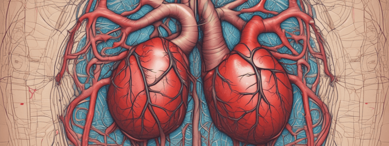
Overview of the Heart
- The heart comprises four chambers, categorizing it as a four-chambered organ.
- Located in the mediastinum, the heart's apex points leftward, resting on the diaphragm.
- The heart attains adult shape and size from puberty to about 25 years.
- The pericardium is a multilayered sac surrounding the heart, consisting of a fibrous layer and a serous layer.
- The fibrous pericardium offers protective support, while the serous pericardium has parietal and visceral sections, with pericardial fluid reducing friction.
- The heart wall is composed of three distinct layers:
- Epicardium (Outer Layer): Visceral layer of the serous pericardium.
- Myocardium (Middle Layer): Thick, contractile muscle responsible for heart contractions.
- Endocardium (Inner Layer): Lining of the heart’s interior, made of specialized endothelium, crucial for smooth blood flow.
- The heart valves ensure unidirectional blood flow, vital for effective pumping action.### Heart Chambers
- The heart consists of four chambers: two atria (upper chambers) and two ventricles (lower chambers).
- Atria:
- Each atrium is divided into left and right by the interatrial septum.
- Functions as receiving chambers, collecting blood from veins.
- Atria contract and relax alternately to receive blood and pump it to ventricles.
- Thin myocardium due to low pressure requirements in blood movement.
- Ventricles:
- Each ventricle is divided into left and right by the interventricular septum.
- Acts as pumping chambers, receiving blood from atria and ejecting it into arteries.
- Thicker myocardium due to the need for stronger contractions to pump blood farther.
Blood Circulation
- The left pump (left side of the heart) drives systematic circulation, distributing blood throughout the body.
- Excludes blood flow to the lungs.
- The right pump (right side of the heart) facilitates pulmonary circulation, sending blood to the lungs for oxygenation.
Heart Valves
- Heart valves ensure unidirectional blood flow.
- There are four valves crucial for heart function:
- Atrioventricular (AV) Valves (two):
- Located between atria and ventricles, preventing backflow.
- Have cusps or pointed flaps.
- Semilunar Valves (two):
- Located at the exits of ventricles, ensuring blood flows into arteries (pulmonary and aorta).
- Atrioventricular (AV) Valves (two):
Cardiac Contraction
- Cardiac muscle contracts autorhythmically, maintaining a natural rhythm.
- Action potentials coordinate cardiac muscle contractions across the myocardium for effective pumping.
- Rate adjustment of cardiac contraction occurs through autonomic nerve signals.
Electrical Conduction System
- The heart's electrical conduction system comprises four main structures:
- Sinoatrial (SA) Node:
- The primary pacemaker, initiating contraction impulses.
- Located at the right atrial epicardium near the superior vena cava.
- Action potentials travel swiftly to both atria.
- Atrioventricular (AV) Node:
- Receives impulses from the SA node, slowing them for complete atrial contraction and ventricular filling.
- Atrioventricular Bundle (Bundle of His):
- Conducts impulses from the AV node to the ventricles via right and left bundle branches.
- Purkinje Fibers:
- Ensure rapid conduction of impulses throughout ventricular muscles, facilitating synchronized contractions.
- Sinoatrial (SA) Node:
Summary of Functions
- The overall structure and function of the heart involves cooperation between different chambers and valves to ensure efficient blood circulation and response to physiological demands.### Cardiac Conduction System
- Involves specialized cardiac muscle fibers, different from ordinary muscle, enabling rapid conduction of signals.
- Pacemaker fibers initiate electrical signals critical for heart rhythm.
- During pacemaker activity, K+ ions leak out, while Na+ and Ca++ ions flow in, causing depolarization.
- This generates an intrinsic rhythm characterized by continuous action potentials.
- Conduction fibers are specifically adapted to rapidly transmit action potentials throughout the heart's syncytium.
Electrocardiogram (ECG)
- Records electrical activity of the heart, reflecting impulses that precede contractions.
- ECG is created by attaching electrodes to the limbs and chest to record voltage changes.
- Healthy ECG includes P wave (atrial depolarization), QRS complex (ventricular depolarization), and T wave (ventricular repolarization).
- Typical duration of QRS complex ranges from 0.07 to 0.11 seconds.
ECG Wave Details
- P wave: Represents atrial depolarization; corresponds to electrical impulses from the SA node.
- QRS complex: Indicates ventricular depolarization; masks atrial repolarization.
- T wave: Reflects ventricular repolarization; inversion suggests myocardial damage.
- U wave: May appear following the T wave, associated with late repolarization of Purkinje fibers; abnormalities could indicate hypokalemia or digoxin toxicity.
Cardiac Cycle
- Describes one complete heartbeat, including both contraction (systole) and relaxation (diastole) phases.
- The cycle illustrates pressure gradients within the heart's chambers.
- In heart failure, residual volume in ventricles may significantly exceed ejected volume during systole.
Phases of the Cardiac Cycle
- Atrial Systole: Initiated by P wave; atria contract, pushing blood into ventricles, keeping AV valves open.
- Isovolumic Ventricular Contraction: Begins with R wave; intraventricular pressure rises, closing AV valves and producing the first heart sound.
- Ejection Phase: Occurs when SL valves open; blood is pumped from ventricles into the pulmonary artery and aorta, marked by increased pressure.
Functional Aspects
- Diastasis refers to reduced ventricular filling.
- The cardiac cycle integrates the changes in pressure within the left atrium, left ventricle, and aorta, essential for effective heart function.
Studying That Suits You
Use AI to generate personalized quizzes and flashcards to suit your learning preferences.


