Podcast
Questions and Answers
If blood flow ceases, which of the following immediate consequences would occur in the body?
If blood flow ceases, which of the following immediate consequences would occur in the body?
- A rapid increase in nutrient and oxygen levels.
- Exhaustion of nutrient and oxygen supplies, leading to waste accumulation. (correct)
- An immediate drop in body temperature due to lack of circulation.
- Immediate activation of the immune response.
Consider a scenario where a person's heart rate consistently averages 50 beats per minute. How would this condition be classified, and what potential issue could arise from it?
Consider a scenario where a person's heart rate consistently averages 50 beats per minute. How would this condition be classified, and what potential issue could arise from it?
- Bradycardia; potential for insufficient oxygen delivery to tissues. (correct)
- Arrhythmia; potential for normal blood pressure.
- Normal sinus rhythm; no issues expected.
- Tachycardia; potential for increased cardiac output.
Which of the following statements correctly describes the systemic and pulmonary circuits?
Which of the following statements correctly describes the systemic and pulmonary circuits?
- The pulmonary circuit carries blood to the lungs, while the systemic circuit carries blood to the rest of the body. (correct)
- The pulmonary circuit carries oxygenated blood to the body, while the systemic circuit carries deoxygenated blood to the lungs.
- Both circuits operate in parallel, supplying blood to all organs simultaneously.
- Both circuits carry the same type of blood, differing only in pressure.
What is the primary functional difference between arteries and veins in the cardiovascular system?
What is the primary functional difference between arteries and veins in the cardiovascular system?
What is the physiological significance of the pericardial fluid found within the pericardial cavity?
What is the physiological significance of the pericardial fluid found within the pericardial cavity?
Why is the myocardium composed of primarily cardiac muscle tissue?
Why is the myocardium composed of primarily cardiac muscle tissue?
How do intercalated discs contribute to the coordinated function of cardiac muscle cells?
How do intercalated discs contribute to the coordinated function of cardiac muscle cells?
What is the specific role of desmosomes within the structure of intercalated discs?
What is the specific role of desmosomes within the structure of intercalated discs?
What is the functional significance of the cardiac skeleton?
What is the functional significance of the cardiac skeleton?
An individual has damage to left atrium. What would be expected?
An individual has damage to left atrium. What would be expected?
Which cardiac surface primarily consists of the right and left ventricles?
Which cardiac surface primarily consists of the right and left ventricles?
Which groove separates the atria from the ventricles?
Which groove separates the atria from the ventricles?
What structural characteristic distinguishes ventricles from atria?
What structural characteristic distinguishes ventricles from atria?
Clinical intervention is required for someone with a damaged interatrial septum. What direct physiological consequence could arise?
Clinical intervention is required for someone with a damaged interatrial septum. What direct physiological consequence could arise?
The right atrium receives deoxygenated blood from which three sources?
The right atrium receives deoxygenated blood from which three sources?
What is the function of the fossa ovalis, and where is it located?
What is the function of the fossa ovalis, and where is it located?
What is the primary function of the right atrioventricular valve (tricuspid valve)?
What is the primary function of the right atrioventricular valve (tricuspid valve)?
What happens if the chordae tendineae are damaged or ruptured?
What happens if the chordae tendineae are damaged or ruptured?
What is the primary function of the moderator band in the right ventricle, and where is it located?
What is the primary function of the moderator band in the right ventricle, and where is it located?
The left atrioventricular valve prevents backflow of blood from the left ventricle to the left atrium. What is another name for this valve?
The left atrioventricular valve prevents backflow of blood from the left ventricle to the left atrium. What is another name for this valve?
Which heart chamber has the thickest wall, and why?
Which heart chamber has the thickest wall, and why?
What is the destination of blood that passes through the aortic valve?
What is the destination of blood that passes through the aortic valve?
How many valves are in the human heart?
How many valves are in the human heart?
What are the four components of both atrioventricular valves?
What are the four components of both atrioventricular valves?
What happens to the atrioventricular valves during ventricular contraction, and why is this important?
What happens to the atrioventricular valves during ventricular contraction, and why is this important?
During the cardiac cycle, what occurs when pressure in the atria exceeds pressure in the ventricles?
During the cardiac cycle, what occurs when pressure in the atria exceeds pressure in the ventricles?
If there is a blockage of blood flow in the right coronary artery what is likely to happen?
If there is a blockage of blood flow in the right coronary artery what is likely to happen?
What would be the impact of a blocked circumflex branch on the left coronary artery?
What would be the impact of a blocked circumflex branch on the left coronary artery?
Which cardiac vein runs alongside the posterior interventricular artery?
Which cardiac vein runs alongside the posterior interventricular artery?
Where does venous blood from the coronary circulation ultimately drain?
Where does venous blood from the coronary circulation ultimately drain?
What is the main difference between systole and diastole?
What is the main difference between systole and diastole?
What event characterizes atrial systole?
What event characterizes atrial systole?
Which of the following is the correct sequence of the cardiac conducting system?
Which of the following is the correct sequence of the cardiac conducting system?
What is the role of nodal cells in the cardiac cycle?
What is the role of nodal cells in the cardiac cycle?
What would happen if the atrioventricular node failed to slow the electrical impulse from the sinoatrial node?
What would happen if the atrioventricular node failed to slow the electrical impulse from the sinoatrial node?
Which of the following best describes the role of the bundle branches in the cardiac cycle?
Which of the following best describes the role of the bundle branches in the cardiac cycle?
How does the autonomic nervous system influence heart rate?
How does the autonomic nervous system influence heart rate?
How would a decrease in parasympathetic activity affect heart rate?
How would a decrease in parasympathetic activity affect heart rate?
If the heart pumps approximately 2.9 gallons of blood per minute, and can vary between 5 and 30 liters per minute, what is the most likely reason for this wide variation in pumping capacity?
If the heart pumps approximately 2.9 gallons of blood per minute, and can vary between 5 and 30 liters per minute, what is the most likely reason for this wide variation in pumping capacity?
Imagine a scenario where a person experiences trauma that directly damages the fibrous pericardium. What immediate risk is most concerning?
Imagine a scenario where a person experiences trauma that directly damages the fibrous pericardium. What immediate risk is most concerning?
A patient is diagnosed with a condition affecting the pericardial cavity. Which of the following would be the most likely consequence of reduced pericardial fluid?
A patient is diagnosed with a condition affecting the pericardial cavity. Which of the following would be the most likely consequence of reduced pericardial fluid?
A histological analysis of cardiac tissue reveals an abundance of mitochondria and myoglobin. What does this suggest about the metabolic requirements of cardiac muscle cells?
A histological analysis of cardiac tissue reveals an abundance of mitochondria and myoglobin. What does this suggest about the metabolic requirements of cardiac muscle cells?
If a toxin interferes with the function of desmosomes in cardiac muscle, disrupting their ability to bind adjacent cells, what is the most likely direct consequence?
If a toxin interferes with the function of desmosomes in cardiac muscle, disrupting their ability to bind adjacent cells, what is the most likely direct consequence?
A cardiologist observes that a patient's cardiac cells are not effectively connected by gap junctions. What physiological consequence is most likely to arise from this observation?
A cardiologist observes that a patient's cardiac cells are not effectively connected by gap junctions. What physiological consequence is most likely to arise from this observation?
After a heart attack, scar tissue forms within the cardiac skeleton. What is the most concerning long-term effect of this change in tissue composition?
After a heart attack, scar tissue forms within the cardiac skeleton. What is the most concerning long-term effect of this change in tissue composition?
If the base of the heart is considered the superior border, which chamber primarily contributes to forming the heart's right border?
If the base of the heart is considered the superior border, which chamber primarily contributes to forming the heart's right border?
What would happen if there was damage to the anterior surface of the heart, what structures would likely be affected?
What would happen if there was damage to the anterior surface of the heart, what structures would likely be affected?
What is the functional significance of the coronary sulcus in the heart's external anatomy?
What is the functional significance of the coronary sulcus in the heart's external anatomy?
During an autopsy, it's noted that the interatrial septum is unusually thin. What condition might this have contributed to during the individual's life?
During an autopsy, it's noted that the interatrial septum is unusually thin. What condition might this have contributed to during the individual's life?
A patient's echocardiogram reveals the presence of pectinate muscles. Which heart chamber are these structures located in?
A patient's echocardiogram reveals the presence of pectinate muscles. Which heart chamber are these structures located in?
In a developing fetus, the foramen ovale allows blood to bypass the lungs. What specific structure does this opening become in a mature adult heart?
In a developing fetus, the foramen ovale allows blood to bypass the lungs. What specific structure does this opening become in a mature adult heart?
If a patient's tricuspid valve is stenotic (narrowed), what direct effect will this have on blood flow?
If a patient's tricuspid valve is stenotic (narrowed), what direct effect will this have on blood flow?
What is the most likely consequence of damage to or absence of the moderator band in the right ventricle?
What is the most likely consequence of damage to or absence of the moderator band in the right ventricle?
A patient presents with mitral valve prolapse. What is the alternative name for the mitral valve that could help clarify its function?
A patient presents with mitral valve prolapse. What is the alternative name for the mitral valve that could help clarify its function?
A patient's echocardiogram shows significant hypertrophy (enlargement) of the left ventricular wall compared to the right. How does this relate to the function of the left ventricle?
A patient's echocardiogram shows significant hypertrophy (enlargement) of the left ventricular wall compared to the right. How does this relate to the function of the left ventricle?
After passing through the aortic valve, where does blood flow next?
After passing through the aortic valve, where does blood flow next?
During ventricular diastole, the AV valves open. What mechanical action directly causes the AV valves to open during this phase of the cardiac cycle?
During ventricular diastole, the AV valves open. What mechanical action directly causes the AV valves to open during this phase of the cardiac cycle?
If the circumflex branch of the left coronary artery becomes blocked, depriving the heart tissue of oxygen, which area may experience the most damage?
If the circumflex branch of the left coronary artery becomes blocked, depriving the heart tissue of oxygen, which area may experience the most damage?
Which of the following accurately describes the path venous blood takes in coronary circulation?
Which of the following accurately describes the path venous blood takes in coronary circulation?
During atrial systole, what crucial event occurs that directly contributes to ventricular filling?
During atrial systole, what crucial event occurs that directly contributes to ventricular filling?
What role do internodal pathways play in the cardiac cycle?
What role do internodal pathways play in the cardiac cycle?
In the sequence of cardiac conduction, what follows after the AV node slows the electrical impulse?
In the sequence of cardiac conduction, what follows after the AV node slows the electrical impulse?
What is the primary role of Purkinje fibers in the cardiac cycle?
What is the primary role of Purkinje fibers in the cardiac cycle?
If the sympathetic nervous system is activated, releasing norepinephrine, what are the expected effects on the heart?
If the sympathetic nervous system is activated, releasing norepinephrine, what are the expected effects on the heart?
Stimulation of the cardioinhibitory center results in which of the following?
Stimulation of the cardioinhibitory center results in which of the following?
What is the most likely outcome of increased vagal tone on the heart?
What is the most likely outcome of increased vagal tone on the heart?
What is the primary function of the chordae tendineae?
What is the primary function of the chordae tendineae?
What would have to happen for the semilunar valves to open?
What would have to happen for the semilunar valves to open?
If you were trying to listen (auscultate) to the heart, and you needed the heart to beat at a slower-than-normal heart rate, what would you look for?
If you were trying to listen (auscultate) to the heart, and you needed the heart to beat at a slower-than-normal heart rate, what would you look for?
Where would you find the mitral valve?
Where would you find the mitral valve?
Where will blood go after it leaves the left ventricle?
Where will blood go after it leaves the left ventricle?
Flashcards
What is the heart's primary function?
What is the heart's primary function?
Keeps the blood in motion, ensuring nutrient and oxygen supply to tissues and waste removal.
How many times does the heart beat per day?
How many times does the heart beat per day?
Averages around 100,000 times, or about 70 beats per minute.
What is the approximate size of the heart?
What is the approximate size of the heart?
The heart is roughly the size of a clenched fist.
How many chambers does the heart have?
How many chambers does the heart have?
Signup and view all the flashcards
Into which two circuits does the heart pump blood?
Into which two circuits does the heart pump blood?
Signup and view all the flashcards
What is an artery?
What is an artery?
Signup and view all the flashcards
What is a vein?
What is a vein?
Signup and view all the flashcards
What are capillaries?
What are capillaries?
Signup and view all the flashcards
What is the pericardium?
What is the pericardium?
Signup and view all the flashcards
What is the fibrous pericardium?
What is the fibrous pericardium?
Signup and view all the flashcards
What is the serous pericardium?
What is the serous pericardium?
Signup and view all the flashcards
What is the visceral layer of the pericardium?
What is the visceral layer of the pericardium?
Signup and view all the flashcards
What is the parietal layer of the pericardium?
What is the parietal layer of the pericardium?
Signup and view all the flashcards
What is the pericardial cavity?
What is the pericardial cavity?
Signup and view all the flashcards
What is pericardial fluid?
What is pericardial fluid?
Signup and view all the flashcards
What is the epicardium?
What is the epicardium?
Signup and view all the flashcards
What is the myocardium?
What is the myocardium?
Signup and view all the flashcards
What is the endocardium?
What is the endocardium?
Signup and view all the flashcards
Is cardiac muscle tissue highly vascularized?
Is cardiac muscle tissue highly vascularized?
Signup and view all the flashcards
What are intercalated discs?
What are intercalated discs?
Signup and view all the flashcards
What are intercalated discs in cardiac muscle?
What are intercalated discs in cardiac muscle?
Signup and view all the flashcards
What is functional syncytium?
What is functional syncytium?
Signup and view all the flashcards
What is the function of the cardiac skeleton?
What is the function of the cardiac skeleton?
Signup and view all the flashcards
What is the base of the heart?
What is the base of the heart?
Signup and view all the flashcards
What is the apex of the heart?
What is the apex of the heart?
Signup and view all the flashcards
What forms the right border of the heart?
What forms the right border of the heart?
Signup and view all the flashcards
What does the interatrial groove separate?
What does the interatrial groove separate?
Signup and view all the flashcards
What is the coronary sulcus?
What is the coronary sulcus?
Signup and view all the flashcards
What is the anterior interventricular sulcus?
What is the anterior interventricular sulcus?
Signup and view all the flashcards
What is the posterior interventricular sulcus?
What is the posterior interventricular sulcus?
Signup and view all the flashcards
Describe the left and right atria.
Describe the left and right atria.
Signup and view all the flashcards
Describe the left and right ventricles.
Describe the left and right ventricles.
Signup and view all the flashcards
What is the interatrial septum?
What is the interatrial septum?
Signup and view all the flashcards
What is the interventricular septum?
What is the interventricular septum?
Signup and view all the flashcards
What is the function of the right atrium?
What is the function of the right atrium?
Signup and view all the flashcards
What is the function of the right ventricle?
What is the function of the right ventricle?
Signup and view all the flashcards
What is the function of chordae tendineae and papillary muscles in the right ventricle?
What is the function of chordae tendineae and papillary muscles in the right ventricle?
Signup and view all the flashcards
What is the function of the left atrium?
What is the function of the left atrium?
Signup and view all the flashcards
What is the function of the left ventricle?
What is the function of the left ventricle?
Signup and view all the flashcards
What are the heart valves?
What are the heart valves?
Signup and view all the flashcards
During AV valve operation, what do the Papillary muscles do?
During AV valve operation, what do the Papillary muscles do?
Signup and view all the flashcards
What is regurgitation?
What is regurgitation?
Signup and view all the flashcards
What is the role of coronary arteries?
What is the role of coronary arteries?
Signup and view all the flashcards
What are the major branches of the right coronary artery?
What are the major branches of the right coronary artery?
Signup and view all the flashcards
What are the major branches of the left coronary artery?
What are the major branches of the left coronary artery?
Signup and view all the flashcards
What is the role of coronary veins?
What is the role of coronary veins?
Signup and view all the flashcards
What does the cardiac cycle consist of?
What does the cardiac cycle consist of?
Signup and view all the flashcards
What is diastole?
What is diastole?
Signup and view all the flashcards
What is systole?
What is systole?
Signup and view all the flashcards
What are the two kinds of conducting cells?
What are the two kinds of conducting cells?
Signup and view all the flashcards
What is the sinoatrial (SA) node?
What is the sinoatrial (SA) node?
Signup and view all the flashcards
What is the atrioventricular (AV) node?
What is the atrioventricular (AV) node?
Signup and view all the flashcards
What do the bundle branches do?
What do the bundle branches do?
Signup and view all the flashcards
What is the difference between Tachycardia and Bradycardia?
What is the difference between Tachycardia and Bradycardia?
Signup and view all the flashcards
What are nodal and conducting cells?
What are nodal and conducting cells?
Signup and view all the flashcards
What is the role of the SA node and the autonomic nervous system?
What is the role of the SA node and the autonomic nervous system?
Signup and view all the flashcards
What do nerves of the autonomic nervous system innervate in the heart?
What do nerves of the autonomic nervous system innervate in the heart?
Signup and view all the flashcards
What does the cardioacceleratory center do?
What does the cardioacceleratory center do?
Signup and view all the flashcards
What does the cardioinhibitory center do?
What does the cardioinhibitory center do?
Signup and view all the flashcards
Study Notes
Introduction
- The blood is kept in motion by the heart
- Nutrient and oxygen supplies run out and waste products build up if blood ceases moving
- Approximately 100,000 heartbeats occur daily, which is about 70 beats per minute
- The heart pumps approximately 1.5 million gallons of blood annually
- That equates to about 2.9 gallons each minute with fluctuations ranging from 5-30 liters per minute
Overview of the Cardiovascular System
- Roughly the size of a clenched fist, the heart
- Two atria and two ventricles form the heart's four chambers
- Blood is pumped by the heart across two circuits: pulmonary and systemic
Overview of Pulmonary and Systemic Circuits
- Arteries carry blood away from the heart
- Veins carry blood towards the heart
- Capillaries link arteries and veins together
The Pericardium
- The heart is surrounded by the pericardium which consists of two parts
- The heart is surrounded by an outer fibrous pericardium
- An inner serous pericardium of two layers:
- An inner visceral layer, is also called epicardium and is attached to the heart's surface
- An outer parietal layer that is adjacent to the fibrous pericardium
- The pericardial cavity is a space between the two serous layers that contains pericardial fluid
- Pericardial fluid lubricates to reduce friction
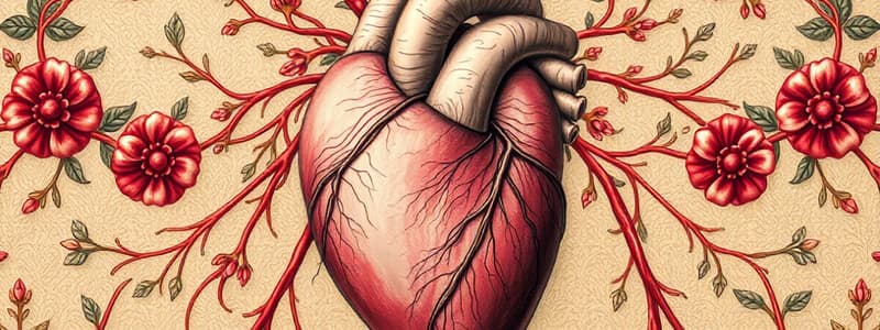
Heart Wall Structure
- The heart's wall consists of three layers: -The epicardium, an external surface, consists of visceral pericardium -The myocardium consists of cardiac tissue including cardiac muscle cells, connective tissue, blood vessels and nerves
- The endocardium is an internal endothelial surface
Cardiac Muscle Tissue
- Cardiac Muscle Tissue has a striated appearance
- Relies on aerobic respiration for fuel
- Contains numerous mitochondria and myoglobin
- Cardiac muscle tissue relies on an extensive circulatory supply
- Cardiac muscle cells contracts involuntarily
- Cells are linked by intercalated discs
Cardiac Muscle Tissue: Intercalated Discs
- Cardiac cells have specialized junctions from cell to cell
- Plasma membranes of adjacent cells are bound by desmosomes
- The myofibrils of neighboring cells are connected through intercalated discs
- Cardiac muscle cells linked via gap junctions
- Ions pass directly from cell to cell giving a direct electrical connection
- As one unit, the muscle cells are able to form a functional syncytium and contract
Cardiac Skeleton
- Each cardiac cell is wrapped is an elastic sheath
- Each muscle layer is wrapped in a sheet of fibrous material
- These fibrous sheets isolate the superficial layer from the deep layer muscles
- The base of the pulmonary trunk, ascending aorta and valves are encircled by these fibrous sheets
Function of the Cardiac Skeleton
- Stabilizes the position of cardiac cells, including the heart valves
- Provides support for blood vessels and nerves in the myocardium
- Helps distribute the forces of contraction to prevent overexpansion of the heart
- Provides elasticity so the recoils after contraction
- Isolates the atrial cells from the ventricular cells
Heart Orientation and Anatomy
- The heart is slightly to the left of the midsagittal plane
- Located in the mediastinum
- The base sits at the superior border
- The apex lays at the inferior portion
- Only the right atrium forms the right border, while the right ventricle forms the inferior border
- The heart is slightly rotated towards the left
- The right atrium, right ventricle and left ventricle form the anterior surface
- The left atrium and part of the right atrium make up the posterior surface
- The right and left ventricles form the diaphragmatic surface
Heart Chambers
- Sulci (grooves) on the external surface distinguish the four chambers
- The left and right atria are separated by an interatrial groove
- The atria and ventricles are separated by the coronary sulcus
- The left and right ventricles are separated by an anterior interventricular sulcus and a posterior interventricular sulcus
Left and Right Atria
- Positioned superior to the coronary sulcus, the left and right atria both have thin walls
- Each has an expandable auricle anteriorly
Left and Right Ventricles
- Positioned inferior to the coronary sulcus, the left and right ventricles both have walls that are thicker than that of the atria
- The left ventricular wall is thicker than the right ventricular wall
Heart: Internal Anatomy
- The frontal section displays the left and right atria separated by an interatrial septum, and left/right ventricles separated by an interventricular septum
- Folds of endocardium forming both atrioventricular valves exist between the atria and ventricles
The Right Atrium
- Receives deoxygenated venous blood by way of the superior vena cava, the inferior vena cava and the coronary sinus
- The coronary sinus enters at the posterior side
- Contains pectinate muscles on the anterior wall and auricle
- Contains the fossa ovalis a remanent of the foramen ovale, located on the interatrial septum and allowed fetal blood to bypass the lungs
The Right Ventricle
- Receives deoxygenated blood from the right atrium
- Blood flows into the right ventricle by way of the right atrioventricular valve, a tricuspid valve
- Blood leaves to the pulmonary trunk, flowing through the pulmonary valve to the right and left set of pulmonary arteries
Right Ventricle Valves
- The right AV valve connects to the papillary muscles by chordae tendineae
- There are three fibrous flaps (cusps) and papillary muscles
- Each cusp connects by chordae tendineae to distinct papillary muscles
- Papillary muscles and chordae tendineae prevent valve inversion during ventricular contraction
Right Ventricle Internal Surface
- The internal surface of the right ventricle consists of
- Muscular ridges called trabeculae carneae
- A moderator band exists only in the right ventricle
- The moderator band is a muscular band extending from the interventricular septum to the ventricular wall
- Prevents overexpansion of the thin-walled right ventricle
The Left Atrium
- Receives oxygenated blood from the lungs by way of the left and right pulmonary veins
- Pectinate muscles restricted to the auricle
- Blood passes by way of the left atrioventricular valve
- This is a bicuspid valve or left AV valve, also known as the mitral valve
The Left Ventricle
- Has a thicker wall for strong contractions that propels blood through the entire systemic circuit
- The right ventricle has a thinner wall as it only has to pump to the pulmonary circuit
- No moderator band
- Does have prominent trabeculae carneae
- Chordae tendineae connect the left AV valve to two cusps, and two papillary muscles
Blood Flow from the Left Ventricle
- Blood exits the left ventricle by flowing through the aortic valve, called the aortic semilunar valve
- Blood enters into the ascending aorta then travels to the aortic arch
- After the aortic arch, it travels down the descending aorta and into body parts (systemic)
Ventricle Structural Differences
- Right Ventricle
- Thinner wall that carries out weaker contractions. it contains a moderator band
- Left Ventricle
- Thicker wall that is the source for 6-7x more powerful contractions compared to the right ventricle
Valves of The Heart
- There are four valves
- Includes two atrioventricular (AV) valves: tricuspid and bicuspid
- Features two semilunar valves that contains aortic and pulmonary valves
Heart Valves
- Each atrioventricular (AV) valve has four parts:
- Ring of connective tissue which connects it to heart tissue while being a part of the heart's fibrous skeleton
- Contains cusps that connect to the papillary muscles by the chordae tendineae
- Papillary muscles contract in a manner as to prevent the inversion of AV valves during contraction.
Atrioventricular Valves During Cardiac Cycle
- Papillary muscles relax, opening the AV valves due to pressure in the atria
- Blood flows from atria to ventricle in this state. During this time is prevents backflow when the ventricles contract and increases pressure as the semilunar valves open
- Closure of valves prevents regurgitation of blood back into the atria which forces blood through open semilunar valves
Coronary Blood Vessels
- Originate at the base of the ascending aorta
- Supplies tissue by way of coronary circulation
- Contains these major arteries:
- Right Coronary Artery (RCA)
- Atrail Branches including a right marginal and posterior interventricular branch
- Left Coronary Artery(LCA)
- A curcumflex branch with left marginal and anterior interventricular branches
- Right Coronary Artery (RCA)
Right Coronary Artery
- The right auricle and the pulmonary trunk pass between each other
- Major branches are artial, right marginal, posterior interventricular and the conducting system branches
Major Branches of the Left Coronary Artery
- Major branches include the anterior interventricular which Branches to the posterior interventricular, which then leads to anatomoses
- Circumflex branch
- branches to the left marginal branch
- also has branches to the posterior left ventricle
Coronary Veins
- Coronary veins ultimately drain the venous blood to the right atrium
- This includes the main coronary veins
- Great cardiac vein that delivers blood to the coronary sinus
- Middle cardiac vein which also delivers blood to the coronary artery(vein)
- The coronary sinus drains directly into the posterior of the right atrium.
The Main Coronary Veins Include
- Posterior vein of the left ventricle parallels the posterior left ventricular branch,
- A small cardiac vein that parallels the right coronary artery
- An anterior cardiac vein with branches from the right ventricle cardiac cells
Coordination of Cardiac Contraction
- The cardiac cycle has alternating periods of contraction and relaxation
- The contraction is called systole that also is referring to the blood flowing into the ventricles (Atrial Systole & Ventricular Systole)
- Ventricular Systole also ejects blood into the arteries and the ascending aorta
- Relaxation is diastole as the chambers fill with blood
Cardiac Cycle Coordination
- Conducting cells coordinate cardiac contractions of cardiac cycle
- Nodal Cells includes sinoatrial and atrioventricular nodes which establish the rate with automatic membrane depolarization
- Conducting cells then Distributes stimuli to the myocardium
The Sinoatrial(SA) Node
- The SA node is located in the posterior wall of the right atrium close to where the superior vena cava is
- It is called cardiac pacemaker
- the pacemaker cells generates about 80-100 action potential per minute and can cause either of the following
- Bradycardia- slower than normal or Tachycardia that refers to a faster than normal heart rate
- The Atrioventricular (AV) node sits within the floor of the right atrium
Summary of Cardiac Conduction
- Impulse travels along internodal pathways from the SA node to the AV node, resulting in the atrial contraction
- The AV node slows the impulse
- The impulse goes from AV Node to the AV bundle
Cardiac Cycle: AV Bundle
- The AV bundle conducts the impulse moving along the interventricular septum before dividing to form the right and left bundle branches
- serves the right and left ventricle
- The Purkinje fibers receive the signals by the bundle branches
- those fibers connect to cardiac muscle cells where Ventricular Contraction follows
Autonomic Control of Heart Rate
- The heart rate can be altered by the SA Node settings
- Heart Rate is modified by impulses from the autonomic nervous system
- Nerves in the ANS innervate to these locations
- SA node, AV node, Cardiac cells, Smooth muscles in blood vessels
- The nodal tissue is affected by presence of various chemicals from the ANS
- Norepinephrine from the sympathetic division increases heart rate + force of the contraction.
- Acetylcholine decreases the heart rate and the forces of contraction from the parasympathetic
Cardiac Centers
- Heart rate gets modified by cardiac centers in the medulla oblongata by:
- Cardioacceleratory center
- Stimulates the sympathetic neurons that then increases heart rate increases
- Cardioinhibitory center
- which activates parasympathetic neurons
- the Vagus nerve plays a role and the Heart rated decreases
- which activates parasympathetic neurons
Studying That Suits You
Use AI to generate personalized quizzes and flashcards to suit your learning preferences.


