Podcast
Questions and Answers
What is the main reason why the wall of the left ventricle is thicker than the wall of the right ventricle?
What is the main reason why the wall of the left ventricle is thicker than the wall of the right ventricle?
- It pumps a smaller volume of blood
- It has a different type of valve
- It pumps blood to the lungs
- It generates a greater pressure than the right ventricle (correct)
What is the approximate pressure increase in the left ventricle when it contracts?
What is the approximate pressure increase in the left ventricle when it contracts?
- 20 mm Hg
- 80 mm Hg
- 120 mm Hg (correct)
- 60 mm Hg
What is the name of the valve located between the left atrium and left ventricle?
What is the name of the valve located between the left atrium and left ventricle?
- Semilunar valve
- Bicuspid valve
- Mitral valve (correct)
- Tricuspid valve
What happens to the atrioventricular valves when the ventricles relax?
What happens to the atrioventricular valves when the ventricles relax?
What is the function of the atrioventricular valves?
What is the function of the atrioventricular valves?
What type of valve is located in the aorta and pulmonary trunk?
What type of valve is located in the aorta and pulmonary trunk?
What happens to the semilunar cusps when the ventricles relax?
What happens to the semilunar cusps when the ventricles relax?
What is the function of the semilunar valves?
What is the function of the semilunar valves?
What percentage of oxygen does blood flowing through the coronary arteries of the heart typically give up?
What percentage of oxygen does blood flowing through the coronary arteries of the heart typically give up?
Where do the two coronary arteries originate from?
Where do the two coronary arteries originate from?
What is the most common cause of angina?
What is the most common cause of angina?
What is the primary reason why cardiac muscle is very dependent on an increased rate of blood flow during exercise?
What is the primary reason why cardiac muscle is very dependent on an increased rate of blood flow during exercise?
What is the typical percentage of oxygen released to skeletal muscle during rest?
What is the typical percentage of oxygen released to skeletal muscle during rest?
What is a common symptom of angina?
What is a common symptom of angina?
Where does the left coronary artery originate from?
Where does the left coronary artery originate from?
What is the effect of exercise on the percentage of oxygen released to skeletal muscle?
What is the effect of exercise on the percentage of oxygen released to skeletal muscle?
What is the reason for the self-excitation of the sinus nodal fibers?
What is the reason for the self-excitation of the sinus nodal fibers?
What happens to the sodium-calcium channels after 100-150 milliseconds of opening?
What happens to the sodium-calcium channels after 100-150 milliseconds of opening?
What is the effect of the increased potassium channels opening?
What is the effect of the increased potassium channels opening?
What is the purpose of hyperpolarization in the sinus nodal fibers?
What is the purpose of hyperpolarization in the sinus nodal fibers?
What happens to the potassium channels after the action potential is over?
What happens to the potassium channels after the action potential is over?
What is the potential at which the 'resting' potential reaches the threshold level for discharge?
What is the potential at which the 'resting' potential reaches the threshold level for discharge?
What is the result of the inward-leaking sodium and calcium ions overbalancing the outward flux of potassium ions?
What is the result of the inward-leaking sodium and calcium ions overbalancing the outward flux of potassium ions?
What level does the membrane potential drop to during hyperpolarization?
What level does the membrane potential drop to during hyperpolarization?
What is the purpose of electrodes placed on the surface of the body?
What is the purpose of electrodes placed on the surface of the body?
Why does the hyperpolarization state not last indefinitely?
Why does the hyperpolarization state not last indefinitely?
What does the P wave in an ECG represent?
What does the P wave in an ECG represent?
What is the minimum potential required for the sodium-calcium channels to become activated?
What is the minimum potential required for the sodium-calcium channels to become activated?
What is the process that continues indefinitely throughout a person's life?
What is the process that continues indefinitely throughout a person's life?
What is the record of the electrical events in the heart?
What is the record of the electrical events in the heart?
What is the characteristic of the heart that enables highly coordinated contractions?
What is the characteristic of the heart that enables highly coordinated contractions?
In which phase of the action potential in cardiac muscle do Ca++ channels begin to open?
In which phase of the action potential in cardiac muscle do Ca++ channels begin to open?
What is the function of the atrioventricular (AV) node in the conduction system of the heart?
What is the function of the atrioventricular (AV) node in the conduction system of the heart?
What is the result of the slow rate of action potential conduction in the AV node?
What is the result of the slow rate of action potential conduction in the AV node?
What is the name of the bundle of specialized cardiac muscle that transmits action potentials from the AV node to the ventricles?
What is the name of the bundle of specialized cardiac muscle that transmits action potentials from the AV node to the ventricles?
What is the location of the atrioventricular (AV) node in the heart?
What is the location of the atrioventricular (AV) node in the heart?
What is the function of the sinoatrial (SA) node in the conduction system of the heart?
What is the function of the sinoatrial (SA) node in the conduction system of the heart?
What is the result of the K+ channels closing at the end of the repolarization phase in skeletal muscle?
What is the result of the K+ channels closing at the end of the repolarization phase in skeletal muscle?
Flashcards are hidden until you start studying
Study Notes
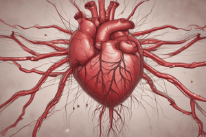
Heart Structure and Function
- The left ventricle wall is thicker than the right ventricle wall due to generating higher pressure (approx. 120 mm Hg) to pump blood to the entire body, compared to the right ventricle (approx. 1/5 of the left ventricle pressure) which pumps blood to the lungs.
- Atrioventricular valves (tricuspid and bicuspid/mitral) separate the atria from the ventricles, allowing blood to flow from atria to ventricles but preventing backflow.
- Semilunar valves (aortic and pulmonary) are located at the base of the aorta and pulmonary trunk, respectively, and prevent backflow from the aorta and pulmonary trunk into the ventricles.
Blood Supply to the Heart
- Coronary arteries, originating from the aorta, supply blood to the heart wall, with the left coronary artery supplying the anterior wall and most of the left ventricle, and the right coronary artery supplying most of the right ventricle.
- In a resting person, 70% of oxygen is released from blood flowing through the coronary arteries, compared to 25% from blood flowing through skeletal muscle arteries.
Coronary Artery Disease (CAD)
- Angina pectoris is the most common clinical manifestation of CAD, resulting from an imbalance between myocardial oxygen supply and demand, often due to atherosclerotic coronary artery obstruction.
- Clinical features of angina include chest pain or crushing sensation, radiation to the neck, jaw, arms, and back, as well as shortness of breath and dizziness.
Electrical Activity of the Heart
- Action potential in cardiac muscle has three phases: depolarization, repolarization, and final repolarization, involving the opening and closing of Na+, K+, and Ca++ channels.
- The conduction system of the heart consists of the sinoatrial (SA) node, atrioventricular (AV) node, atrioventricular bundle, and Purkinje fibers, which coordinate the contraction of atria and ventricles.
Electrocardiogram (ECG)
- An ECG records the electrical activity of the heart, measured through electrodes on the body surface, and consists of a P wave, QRS complex, and T wave.
- The P wave represents depolarization of the atrial myocardium, preceding atrial contraction.
Studying That Suits You
Use AI to generate personalized quizzes and flashcards to suit your learning preferences.