Podcast
Questions and Answers
What structure does 'arbor vitae' refer to?
What structure does 'arbor vitae' refer to?
- Cerebellum (correct)
- Corpus Callosum
- Pons
- Medulla Oblongata
What structure does 'cerebellum' refer to?
What structure does 'cerebellum' refer to?
- Temporal Lobe
- Optic Nerve
- Frontal Lobe
- Balance and coordination center (correct)
What structure does 'corpus callosum' refer to?
What structure does 'corpus callosum' refer to?
- Sensory processing
- Motor control
- Connects left and right brain hemispheres (correct)
- Speech production
What structure does 'midbrain' refer to?
What structure does 'midbrain' refer to?
What structure does 'pons' refer to?
What structure does 'pons' refer to?
What structure does 'hypothalamus' refer to?
What structure does 'hypothalamus' refer to?
What structure does 'thalamus' refer to?
What structure does 'thalamus' refer to?
What structure does 'cerebral aqueduct' refer to?
What structure does 'cerebral aqueduct' refer to?
What structure does 'fourth ventricle' refer to?
What structure does 'fourth ventricle' refer to?
What structure does 'third ventricle' refer to?
What structure does 'third ventricle' refer to?
What structure does 'medulla oblongata' refer to?
What structure does 'medulla oblongata' refer to?
What lobe does the arrow point to in 'frontal lobe'?
What lobe does the arrow point to in 'frontal lobe'?
What lobe does the arrow point to in 'occipital lobe'?
What lobe does the arrow point to in 'occipital lobe'?
What lobe does the arrow point to in 'parietal lobe'?
What lobe does the arrow point to in 'parietal lobe'?
What lobe does the arrow point to in 'temporal lobe'?
What lobe does the arrow point to in 'temporal lobe'?
What connects the 'pituitary gland' to the hypothalamus?
What connects the 'pituitary gland' to the hypothalamus?
Identify the structure: __________ (shown by the arrow; often referred to as the primary sensory cortex)
Identify the structure: __________ (shown by the arrow; often referred to as the primary sensory cortex)
Identify the structure: __________ (shown by the arrow; often referred to as the primary motor cortex)
Identify the structure: __________ (shown by the arrow; often referred to as the primary motor cortex)
Identify the structure: __________ (shown by the arrow; involved in emotion and memory)
Identify the structure: __________ (shown by the arrow; involved in emotion and memory)
What is the function of the 'insula'?
What is the function of the 'insula'?
What is the function of the 'pineal gland'?
What is the function of the 'pineal gland'?
What structure does 'Olfactory Nerve' refer to?
What structure does 'Olfactory Nerve' refer to?
What structure does 'Optic Nerve' refer to?
What structure does 'Optic Nerve' refer to?
What structure does 'Oculomotor nerve' refer to?
What structure does 'Oculomotor nerve' refer to?
What structure does 'Trigeminal' refer to?
What structure does 'Trigeminal' refer to?
What structure does 'medulla oblongata olive' refer to?
What structure does 'medulla oblongata olive' refer to?
What structure does 'medulla oblongata pyramid' refer to?
What structure does 'medulla oblongata pyramid' refer to?
Flashcards are hidden until you start studying
Study Notes
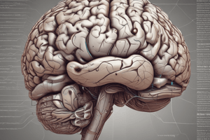
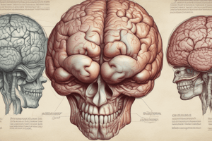
Brain Model Labels
- Arbor vitae: Refers to the tree-like structure of white matter found in the cerebellum, essential for coordination and balance.
- Cerebellum: Part of the hindbrain responsible for regulating motor control, posture, and movement coordination.
- Corpus callosum: Thick band of nerve fibers connecting the left and right cerebral hemispheres, facilitating interhemispheric communication.
- Midbrain: Located between the forebrain and hindbrain, playing a crucial role in vision, hearing, motor control, sleep/wake, arousal, and temperature regulation.
- Pons: Regulates breathing and relays signals between the cerebellum and cerebrum, involved in sleep and dreaming.
- Hypothalamus: Controls body temperature, hunger, thirst, and circadian rhythms; links the nervous system to the endocrine system via the pituitary gland.
- Thalamus: Acts as the brain's relay station, processing sensory information and transmitting it to appropriate cortical areas.
- Cerebral aqueduct: Connects the third and fourth ventricles, facilitating the flow of cerebrospinal fluid.
- Fourth ventricle: Cavities within the brain filled with cerebrospinal fluid, located between the brainstem and cerebellum.
- Third ventricle: A narrow cavity located between the two halves of the thalamus, containing cerebrospinal fluid.
- Medulla oblongata: Controls autonomic functions such as breathing, heart rate, and blood pressure; connects the brain to the spinal cord.
- Frontal lobe: Responsible for decision-making, problem-solving, emotional regulation, and motor function.
- Occipital lobe: The center for visual processing; interprets and makes sense of visual information from the eyes.
- Parietal lobe: Processes sensory information regarding the location of parts of the body, comprehension of objects, and spatial reasoning.
- Temporal lobe: Involved in processing auditory information, language comprehension, and memory storage.
- Pituitary gland & infundibulum: The pituitary gland regulates endocrine functions and is connected to the hypothalamus by the infundibulum, controlling hormone release.
- Central Sulcus: A prominent groove in the brain delineating the frontal and parietal lobes, critical for motor and sensory cortex separation.
- Lateral Sulcus: Separates the temporal lobe from the frontal and parietal lobes, playing a role in auditory processing.
- Longitudinal fissure: Divides the brain into left and right hemispheres.
- Transverse fissure: Separates the cerebellum from the cerebrum.
- Sulci: Grooves or indentations on the brain's surface, increasing the brain's surface area and allowing for more neurons.
- Gyri: The raised ridges or folds of brain tissue lying between the sulci, contributing to cortical surface area.
- Postcentral gyrus (primary sensory cortex): Receives tactile information, allowing for the processing of touch sensations from various parts of the body.
- Precentral gyrus (primary motor cortex): Directly involved in the planning and execution of voluntary movements.
- Insula: Plays a role in perception, motor control, self-awareness, and emotional regulation.
- Mammillary body: Part of the hypothalamus involved in recollective memory, often associated with the limbic system.
- Epithalamus: Houses the pineal gland which regulates circadian rhythms through melatonin secretion.
- Pineal gland: Endocrine gland that produces melatonin, regulating sleep-wake cycles.
- Subarachnoid space: The area between the arachnoid mater and pia mater, filled with cerebrospinal fluid providing cushioning for the brain.
- Dura Mater: The tough outer layer of the meninges, providing protection to the brain and spinal cord.
- Arachnoid Villi: Protrusions through the dura mater that allow cerebrospinal fluid to be absorbed into the bloodstream.
- Choroid plexus: A network of cells that produce cerebrospinal fluid within the ventricles of the brain.
- Lateral ventricles: Paired structures in the brain filled with cerebrospinal fluid.
- Olfactory Nerve: Responsible for the sense of smell, transmitting sensory information from the nasal cavity to the brain.
- Optic nerve: Transmits visual information from the retina to the brain.
- Oculomotor nerve: Controls most eye movements, as well as constriction of the pupil and maintaining an open eyelid.
- Trochlear nerve: Responsible for the movement of the superior oblique muscle of the eye, playing a key role in eye positioning.
- Trigeminal nerve: Responsible for sensation in the face and motor functions such as biting and chewing.
- Somatosensory Cortex: Area of the brain responsible for processing sensory input from the body, including touch, temperature, and pain.
- Primary Motor Cortex: Controls voluntary movements by sending signals to the muscles.
- Visual Cortex: Processes visual stimuli, interpreting information from the eyes for perception of the surrounding environment.
- Meninges: Three protective membranes covering the brain and spinal cord, including dura mater, arachnoid mater, and pia mater.
- Medulla oblongata olive: An area in the medulla involved in motor learning and coordination.
- Medulla oblongata pyramid: Contains descending motor pathways that relay messages to the spinal cord.
- Olfactory nerve: Responsible for transmitting information about smell.
- Optic Nerve: Critical for vision by transmitting signals from the eye to the brain.
- Trochlear: Assists with eye movement, particularly in controlling the muscle that moves the eye downward.
- Oculomotor: Involved in regulating eye movement and lens shape for focusing.
Studying That Suits You
Use AI to generate personalized quizzes and flashcards to suit your learning preferences.