Podcast
Questions and Answers
Damage to the arcuate fasciculus would most likely result in impairment of which function?
Damage to the arcuate fasciculus would most likely result in impairment of which function?
- Recognizing faces.
- Repeating spoken language. (correct)
- Understanding spoken words.
- Producing grammatically correct sentences.
A patient presents with an inability to recognize familiar faces, but can recognize familiar objects. Where is the most likely location of the brain lesion?
A patient presents with an inability to recognize familiar faces, but can recognize familiar objects. Where is the most likely location of the brain lesion?
- Prefrontal cortex.
- Visual association cortex. (correct)
- Parietal lobe.
- Primary motor cortex.
A stroke affecting the precentral gyrus in the left hemisphere would most likely result in what?
A stroke affecting the precentral gyrus in the left hemisphere would most likely result in what?
- Loss of sensation on the right side of the body.
- Visual deficits in the left visual field.
- Muscle weakness on the right side of the body. (correct)
- Impaired language comprehension.
What would most likely be the result of damage to the posterior parietal cortex?
What would most likely be the result of damage to the posterior parietal cortex?
Which of the following represents the most accurate path of the corticospinal tract?
Which of the following represents the most accurate path of the corticospinal tract?
The pulvinar nucleus and superior colliculus are involved in which phenomenon related to vision?
The pulvinar nucleus and superior colliculus are involved in which phenomenon related to vision?
A patient has damage to their primary somatosensory cortex. What is the MOST likely deficit they will experience?
A patient has damage to their primary somatosensory cortex. What is the MOST likely deficit they will experience?
If a person has damage to the motor cortex area corresponding to their hand, but retains function in the arm, what could be expected?
If a person has damage to the motor cortex area corresponding to their hand, but retains function in the arm, what could be expected?
Which white matter tract facilitates communication between the frontal lobe and occipital lobe within the same hemisphere?
Which white matter tract facilitates communication between the frontal lobe and occipital lobe within the same hemisphere?
A patient struggles with producing speech. They understand what is being said to them, but their own speech is halting and grammatically incorrect. Which area is most likely affected?
A patient struggles with producing speech. They understand what is being said to them, but their own speech is halting and grammatically incorrect. Which area is most likely affected?
In the dorsal/posterior column pathway, where does the second-order neuron synapse with the third-order neuron?
In the dorsal/posterior column pathway, where does the second-order neuron synapse with the third-order neuron?
What would be the MOST likely symptom of damage to the corpus callosum?
What would be the MOST likely symptom of damage to the corpus callosum?
In a representative somatosensory pathway, which neuron is responsible for crossing the midline to the opposite side of the brain?
In a representative somatosensory pathway, which neuron is responsible for crossing the midline to the opposite side of the brain?
What is the primary function of the precentral gyrus?
What is the primary function of the precentral gyrus?
Which of the following best explains the somatotopic organization of the primary motor cortex?
Which of the following best explains the somatotopic organization of the primary motor cortex?
What is a key characteristic of the neurons in the corticospinal tract?
What is a key characteristic of the neurons in the corticospinal tract?
A patient can understand speech but speaks in 'word salad'. Which area of the brain has been affected?
A patient can understand speech but speaks in 'word salad'. Which area of the brain has been affected?
You touch something hot and quickly pull your hand away. Which white matter tracts are primarily responsible for rapidly communicating information between sensory and motor areas to facilitate this response?
You touch something hot and quickly pull your hand away. Which white matter tracts are primarily responsible for rapidly communicating information between sensory and motor areas to facilitate this response?
A patient has an injury to the medulla oblongata that interrupts neuron #2 of their dorsal column pathway. What sensory perception would be affected?
A patient has an injury to the medulla oblongata that interrupts neuron #2 of their dorsal column pathway. What sensory perception would be affected?
A researcher discovers a previously unknown white matter tract in the brain. Axons within the tract connect regions of the frontal lobe in the left hemisphere with corresponding regions in the right hemisphere. This tract would MOST accurately be classified as what?
A researcher discovers a previously unknown white matter tract in the brain. Axons within the tract connect regions of the frontal lobe in the left hemisphere with corresponding regions in the right hemisphere. This tract would MOST accurately be classified as what?
Following a car accident, a patient reports being unable to feel pain or temperature in their left leg. However, they can still perceive fine touch and vibration in the same leg. Based on this information, where is the most likely location of the lesion?
Following a car accident, a patient reports being unable to feel pain or temperature in their left leg. However, they can still perceive fine touch and vibration in the same leg. Based on this information, where is the most likely location of the lesion?
A patient has difficulty understanding the emotional content of speech (e.g., recognizing sarcasm or anger in someone's tone). What area of the brain is most likely affected?
A patient has difficulty understanding the emotional content of speech (e.g., recognizing sarcasm or anger in someone's tone). What area of the brain is most likely affected?
A patient with damage to the motor cortex has difficulty moving their fingers individually. They can still make a fist, grasp objects, and move their arm without difficulty. What best explains this?
A patient with damage to the motor cortex has difficulty moving their fingers individually. They can still make a fist, grasp objects, and move their arm without difficulty. What best explains this?
What would be the effect of a lesion in the thalamus that specifically interrupts the somatosensory pathways from the body?
What would be the effect of a lesion in the thalamus that specifically interrupts the somatosensory pathways from the body?
A patient who has sustained damage to their primary visual cortex reports that they cannot consciously see anything, but they are still able to navigate around obstacles in a room. This is an example of:
A patient who has sustained damage to their primary visual cortex reports that they cannot consciously see anything, but they are still able to navigate around obstacles in a room. This is an example of:
If a person has a lesion that severs the corpus callosum completely, which of the following is most likely to occur?
If a person has a lesion that severs the corpus callosum completely, which of the following is most likely to occur?
If a patient has damage to the primary somatosensory cortex related to their right hand, they would likely have trouble doing what?
If a patient has damage to the primary somatosensory cortex related to their right hand, they would likely have trouble doing what?
In the spinothalamic tract, which neuron's axon crosses the midline immediately after synapsing?
In the spinothalamic tract, which neuron's axon crosses the midline immediately after synapsing?
Why does damage to the primary motor cortex often result in more pronounced deficits in fine motor control compared to gross motor movements?
Why does damage to the primary motor cortex often result in more pronounced deficits in fine motor control compared to gross motor movements?
A patient exhibits the inability to understand written or spoken language. Which area of the brain is most likely damaged?
A patient exhibits the inability to understand written or spoken language. Which area of the brain is most likely damaged?
An individual experiences a traumatic brain injury and can no longer recognize everyday objects by sight alone. What condition does this person most likely have?
An individual experiences a traumatic brain injury and can no longer recognize everyday objects by sight alone. What condition does this person most likely have?
What is the primary function of commissural fibers in the brain?
What is the primary function of commissural fibers in the brain?
Which of the following is the most accurate description of the sensory information processed by the dorsal/posterior column pathway?
Which of the following is the most accurate description of the sensory information processed by the dorsal/posterior column pathway?
If damage occurs to the ventral aspect of the brainstem where the corticospinal tract descends. Which side of the body would have paralysis?
If damage occurs to the ventral aspect of the brainstem where the corticospinal tract descends. Which side of the body would have paralysis?
What is the best description of projection tracts?
What is the best description of projection tracts?
How many neurons are between a sensory receptor (in the body) and somatosensory neuron in the postcentral gyrus?
How many neurons are between a sensory receptor (in the body) and somatosensory neuron in the postcentral gyrus?
What type of sensations does the spinothalamic tract percieve?
What type of sensations does the spinothalamic tract percieve?
Flashcards
What is a Sulcus?
What is a Sulcus?
The 'furrow' or 'valley' on the surface of the brain.
What is a Gyrus?
What is a Gyrus?
The 'hill' on the surface of the brain.
What is the central sulcus?
What is the central sulcus?
Separates the frontal and parietal lobes.
What is the parieto-occipital sulcus?
What is the parieto-occipital sulcus?
Signup and view all the flashcards
What is the lateral sulcus?
What is the lateral sulcus?
Signup and view all the flashcards
What is the transverse fissure?
What is the transverse fissure?
Signup and view all the flashcards
What is the frontal lobe's function?
What is the frontal lobe's function?
Signup and view all the flashcards
What is the parietal lobe's function?
What is the parietal lobe's function?
Signup and view all the flashcards
What is the occipital lobe's function?
What is the occipital lobe's function?
Signup and view all the flashcards
What is the temporal lobe's function?
What is the temporal lobe's function?
Signup and view all the flashcards
What makes up the diencephalon?
What makes up the diencephalon?
Signup and view all the flashcards
What is the role of the thalamus?
What is the role of the thalamus?
Signup and view all the flashcards
What is the role of the hypothalamus?
What is the role of the hypothalamus?
Signup and view all the flashcards
What are the parts of the brainstem?
What are the parts of the brainstem?
Signup and view all the flashcards
What is the role of the brainstem?
What is the role of the brainstem?
Signup and view all the flashcards
What is the cerebellum's function?
What is the cerebellum's function?
Signup and view all the flashcards
What are commissural tracts?
What are commissural tracts?
Signup and view all the flashcards
What is the corpus callosum?
What is the corpus callosum?
Signup and view all the flashcards
What are projection tracts?
What are projection tracts?
Signup and view all the flashcards
What is the corticospinal tract?
What is the corticospinal tract?
Signup and view all the flashcards
What are association tracts?
What are association tracts?
Signup and view all the flashcards
What is the primary motor cortex?
What is the primary motor cortex?
Signup and view all the flashcards
What is the precentral gyrus?
What is the precentral gyrus?
Signup and view all the flashcards
What is the somatosensory cortex?
What is the somatosensory cortex?
Signup and view all the flashcards
What is the postcentral gyrus?
What is the postcentral gyrus?
Signup and view all the flashcards
What is the homunculus?
What is the homunculus?
Signup and view all the flashcards
What kind of neurons are in the corticospinal tract?
What kind of neurons are in the corticospinal tract?
Signup and view all the flashcards
What is efferent information?
What is efferent information?
Signup and view all the flashcards
What is the upper motor neuron?
What is the upper motor neuron?
Signup and view all the flashcards
What is the lower motor neuron?
What is the lower motor neuron?
Signup and view all the flashcards
What neurons are in the Dorsal/Posterior Column Pathway?
What neurons are in the Dorsal/Posterior Column Pathway?
Signup and view all the flashcards
What neurons are in the Spinothalamic Tract?
What neurons are in the Spinothalamic Tract?
Signup and view all the flashcards
What is Agnosia?
What is Agnosia?
Signup and view all the flashcards
What is Aphasia?
What is Aphasia?
Signup and view all the flashcards
What is Apperceptive Agnosia?
What is Apperceptive Agnosia?
Signup and view all the flashcards
What is Associative Agnosia?
What is Associative Agnosia?
Signup and view all the flashcards
What is Wernicke's aphasia?
What is Wernicke's aphasia?
Signup and view all the flashcards
What is Broca's aphasia?
What is Broca's aphasia?
Signup and view all the flashcards
What is Blindsight?
What is Blindsight?
Signup and view all the flashcards
Study Notes
Lecture 21 Quiz Review
- Epineurium is not a layer of the meninges.
- Cerebrospinal fluid (CSF) circulates around the arachnoid layer of the meninges.
- Old/used CSF is transported into the venous circulation through arachnoid granulations.
- The third ventricle is located within the diencephalon brain region.
Learning Objectives
- Describe the external anatomy of the brain from lateral (surface) and medial views.
- Identify and describe the lobes of the cerebral cortex (cerebrum).
- Identify and describe major sulci and gyri that divide the brain, and their basic functions.
- Name the regions of the brainstem from medial, ventral, and dorsolateral views.
- Identify selected internal structures of the brain from a coronal view.
- List the different types of white matter tracts in the brain.
- Understand the anatomy of the main motor pathway for voluntary movement (corticospinal tract).
- Understand the anatomy of a representative somatosensory pathway (dorsal/posterior column pathway).
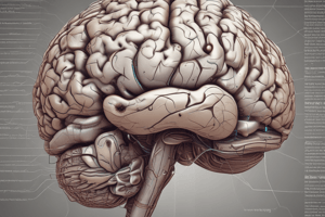
Brain Anatomy: Lateral View
- SULCUS is a "furrow/valley," with the plural form being SULCI ("furrows/valleys").
- GYRUS is a “hill”, with the plural form being GYRI ("hills”).
- The brain's anatomy as seen from the side includes the frontal, parietal, occipital, and temporal lobes.
Brain Anatomy: Sulci, Gyri, and Fissures
- The central sulcus divides the frontal and parietal lobes.
- The lateral sulcus separates the temporal lobe from the frontal and parietal lobes.
- The parieto-occipital sulcus separates the parietal and occipital lobes.
- The transverse fissure separates the cerebrum from the cerebellum.
Functions of Cerebral Lobes (Cerebral Cortex)
- The frontal lobe is associated with motor control, language, and personality.
- The parietal lobe is associated with somatosensory processing.
- The temporal lobe is associated with memory and hearing.
- The occipital lobe is associated with vision.
Brain Divisions: Medial View
- The cerebral cortex (aka cerebrum) is a major brain division.
- The corpus callosum (white matter) is a major brain division.
- The diencephalon, including the thalamus and hypothalamus is a major brain division.
- The brainstem, including the midbrain, pons, and medulla oblongata is a major brain division.
- The cerebellum is a major brain division.
Brainstem Anatomy: Lateral Aspect
- The thalamus is part of the diencephalon and not the brainstem.
- The brainstem includes the midbrain, pons, and medulla oblongata
Brainstem Anatomy: Ventral Aspect
- The thalamus, part of the diencephalon, is not part of the brainstem.
- The brainstem consists of the midbrain, pons, and medulla oblongata.
- The spinal cord is not part of the brainstem.
- The level of the foramen magnum is shown.
Brain Internal Structures: Coronal View
- The cerebral cortex (gray matter) is a key component.
- White matter (3 types) constitute a significant portion.
- Deep nuclei are present.
White Matter: Commissural Tracts (#1)
- Axons cross from side to side in both directions.
- The corpus callosum is an example.
White Matter: Projection Tracts (#2)
- Axons extend between the cortex and other CNS areas outside the cerebrum.
- The corticospinal tract (somatic motor pathway) is an example.
White Matter: Association Tracts (#3)
- Axons are located on the same side within the cerebral cortex.
- Communication occurs between brain areas over short or long distances.
Cortical Areas for Motor Control / Somatosensory Perception
- The central sulcus separates the frontal lobe and parietal lobe.
- The pre-central gyrus is the primary motor cortex.
- The post-central gyrus is the primary somatosensory cortex.
Somatic Efferent (Motor) Division
- Voluntary movement is controlled by this division.
- Information flows AWAY from the CNS (efferent).
- The pathway includes two neurons between the brain and effector: an upper motor neuron and a lower motor neuron.
- Axons are myelinated.
- Acetylcholine (ACh) serves as the neurotransmitter.
- The effector is a skeletal muscle.
- The upper motor neuron cell body is in the primary motor cortex (precentral gyrus).
Motor Cortex Organization
- The precentral gyrus functions as the primary motor cortex.
- Specific regions of the motor cortex control specific body regions.
- More cortical area is devoted to sensitive and/or precision areas like hands and mouth/tongue.
- Relatively little area is devoted to regions like the trunk/abdomen.
Corticospinal Pathway
- It is a somatic efferent pathway.
- Involves two neurons: upper and lower motor neurons.
- The upper motor neuron's cell body is in the primary motor cortex (precentral gyrus).
- The upper motor neuron axon extends from the motor cortex to the spinal cord on the opposite side and makes a synapse on the lower motor neuron.
- The lower motor neuron's cell body is in the ventral horn (grey matter) of the spinal cord.
- The lower motor neuron axon extends out of the spinal cord (ventral root) into the body and makes a synapse on skeletal muscle.
Motor Cortex Damage
- Damage to the motor cortex results in muscle weakness and paralysis.
- These symptoms occur in the body region corresponding to the location of damage, but on the opposite side.
Somatosensory Cortex
- The postcentral gyrus functions as the primary somatosensory cortex.
- Specific regions of the somatosensory cortex receive sensory information from specific body regions.
Dorsal/Posterior Column Pathway
- It mediates fine touch, vibration, pressure, and proprioception.
- This pathway involves three neurons between the sensory receptor (in the body) and the somatosensory neuron in the postcentral gyrus.
- Neuron #1: cell body in the dorsal root ganglion; the peripheral fiber is from a sensory receptor in the skin; the central fiber ascends toward the brain in the dorsal columns; it makes a synapse on neuron #2 in the medulla oblongata.
- Neuron #2: cell body in the medulla oblongata; the axon crosses to the opposite side and ascends; it makes a synapse on neuron #3 in the thalamus.
- Neuron #3: cell body in the thalamus; the axon ascends to the somatosensory cortex and makes a synapse on the cell body of a somatosensory cortex neuron resulting in the perception of light touch.
Spinothalamic Tract
- It mediates pain and temperature senses.
- Involves three neurons between the sensory receptor (in the body) and the somatosensory neuron in the postcentral gyrus.
- Neuron #1: cell body in dorsal root ganglion; makes synapse on neuron #2 in the spinal cord.
- Neuron #2: cell body in the spinal cord, axon crosses to the opposite side via the anterior white commissure and the cell is called a tract cell; makes a synapse on neuron #3 in the thalamus.
- Neuron #3: cell body in the thalamus; its axon ascends to the somatosensory cortex, making a synapse on the cell body of a somatosensory cortex neuron.
Damage to Primary Somatosensory Cortex
- Occurs when a cell in the somatosensory cortex that receives information from the dorsal column pathway dies.
- This means ascending information has no place to go.
- This lack of processing leads to a lack of perception of touch in that body area on the opposite side.
Cortex Damage: Agnosias and Aphasias
- Agnosia: Damage to a region involved with sensory perception, causes changes in perception.
- Aphasia: Damage to regions governing language, causes changes in speech perception or production.
Agnosias
- Prosopagnosia: "Face blindness," is a type of agnosia with two kinds:
- Apperceptive: inability to recognize facial expressions/cues.
- Associative: inability to recognize an individual from their facial features.
Aphasias
- Wernicke's aphasia: individuals can produce words and even whole sentences, but the meaning is lost, resulting in a "word salad."
- Broca's aphasia: individuals can understand words, but cannot form the motor patterns to produce whole speech.
Blindsight
- Blindsight Damage to V1, where the brain recieves primary visual information.
- Individuals experience no conscious perception of "sight” but will still react to visual stimuli.
Blindsight Visual Pathway
- Retina → LGN → PN/SC → Posterior Parietal
- Vision is processed in the Posterior Parietal Cortex . Pulvinar Nucleus (PN) and Superior Colliculus (SC).
Studying That Suits You
Use AI to generate personalized quizzes and flashcards to suit your learning preferences.