Podcast
Questions and Answers
What is the primary consequence of demyelination on neuronal function?
What is the primary consequence of demyelination on neuronal function?
- Increased conduction velocity
- Enhanced neurotransmitter release
- Impaired neuronal survival
- Decreased synaptic transmission (correct)
Which of the following is NOT a characteristic of an active plaque in MS?
Which of the following is NOT a characteristic of an active plaque in MS?
- Demyelination and inflammation
- Normal myelin sheath appearance (correct)
- Axonal damage and remyelination
- Presence of macrophages and T-cells
What is the primary mechanism of neurological damage in nerve compression?
What is the primary mechanism of neurological damage in nerve compression?
- Axonal degeneration and regeneration
- Demyelination and remyelination
- Ischemia and inflammation (correct)
- Neurotransmitter depletion
Which of the following is a characteristic of Guillain-Barre syndrome?
Which of the following is a characteristic of Guillain-Barre syndrome?
What is the term for a disorder affecting multiple nerves?
What is the term for a disorder affecting multiple nerves?
What is the primary reason why septic arthritis must be managed urgently?
What is the primary reason why septic arthritis must be managed urgently?
Which condition is synovial fluid analysis particularly useful in distinguishing between?
Which condition is synovial fluid analysis particularly useful in distinguishing between?
What is the primary indication for performing synovial fluid analysis?
What is the primary indication for performing synovial fluid analysis?
What is analyzed in the 'three C's' of synovial fluid analysis?
What is analyzed in the 'three C's' of synovial fluid analysis?
What is the significance of crystals seen under microscopy in synovial fluid analysis?
What is the significance of crystals seen under microscopy in synovial fluid analysis?
What is the role of Fc receptor protein in structural defenses?
What is the role of Fc receptor protein in structural defenses?
Which type of toxin produced by Staphylococcus aureus is responsible for producing large holes in the lining of blood vessels?
Which type of toxin produced by Staphylococcus aureus is responsible for producing large holes in the lining of blood vessels?
What is the name of the toxin responsible for Toxic Shock Syndrome?
What is the name of the toxin responsible for Toxic Shock Syndrome?
Which of the following diseases is NOT caused by Staphylococcus aureus?
Which of the following diseases is NOT caused by Staphylococcus aureus?
What is the primary mechanism of Staphylococcus aureus to avoid phagocytosis?
What is the primary mechanism of Staphylococcus aureus to avoid phagocytosis?
What is the mode of inheritance of hereditary angioedema?
What is the mode of inheritance of hereditary angioedema?
What is the primary molecule responsible for the pathogenesis of hereditary angioedema?
What is the primary molecule responsible for the pathogenesis of hereditary angioedema?
What is the treatment for hereditary angioedema?
What is the treatment for hereditary angioedema?
What is the increased risk of infections with systemic use of glucocorticoids?
What is the increased risk of infections with systemic use of glucocorticoids?
What is the name of the fungal infection of the lung that can occur with systemic use of glucocorticoids?
What is the name of the fungal infection of the lung that can occur with systemic use of glucocorticoids?
What is the effect of H1-receptors on appetite?
What is the effect of H1-receptors on appetite?
What is the mechanism of TCA overdose leading to life-threatening cardiac arrhythmias?
What is the mechanism of TCA overdose leading to life-threatening cardiac arrhythmias?
What is the effect of Venlafaxine on dopamine reuptake?
What is the effect of Venlafaxine on dopamine reuptake?
Which of the following adverse effects of TCA is NOT related to M-block?
Which of the following adverse effects of TCA is NOT related to M-block?
What is the role of serotonin in SSRI's?
What is the role of serotonin in SSRI's?
What is a common symptom of benzodiazepine withdrawal after short-term use and/or low-doses?
What is a common symptom of benzodiazepine withdrawal after short-term use and/or low-doses?
What is a consequence of taking a second pill during the same sleep cycle when experiencing early morning rebound insomnia?
What is a consequence of taking a second pill during the same sleep cycle when experiencing early morning rebound insomnia?
What is the primary mechanism behind benzodiazepine tolerance?
What is the primary mechanism behind benzodiazepine tolerance?
What is a characteristic of benzodiazepine dependence?
What is a characteristic of benzodiazepine dependence?
What is the reason behind the increased likelihood of tolerance, dependence, and withdrawal symptoms when using benzodiazepines for more than 14-21 nights in a row?
What is the reason behind the increased likelihood of tolerance, dependence, and withdrawal symptoms when using benzodiazepines for more than 14-21 nights in a row?
What is the primary mode of transmission of West Nile Virus?
What is the primary mode of transmission of West Nile Virus?
What percentage of people infected with West Nile Virus experience neuroinvasion?
What percentage of people infected with West Nile Virus experience neuroinvasion?
What is the primary reservoir of the poliovirus?
What is the primary reservoir of the poliovirus?
What is the significance of the connection between modern plumbing and sewer systems in the emergence of polio as an epidemic in the 1900s?
What is the significance of the connection between modern plumbing and sewer systems in the emergence of polio as an epidemic in the 1900s?
What is the current status of polio in the Americas and Europe?
What is the current status of polio in the Americas and Europe?
What is the future site of the mouth indicated by?
What is the future site of the mouth indicated by?
What is the result of the proliferation and movement of epiblast cells to the median plane of the embryonic disc?
What is the result of the proliferation and movement of epiblast cells to the median plane of the embryonic disc?
What is the term for the supporting tissues of the embryo formed by mesenchyme?
What is the term for the supporting tissues of the embryo formed by mesenchyme?
What is the stage of the embryo during which the three germ layers are established?
What is the stage of the embryo during which the three germ layers are established?
What is the name of the narrow groove that develops in the primitive streak?
What is the name of the narrow groove that develops in the primitive streak?
At what day does the neural tube no longer communicate with the amniotic cavity?
At what day does the neural tube no longer communicate with the amniotic cavity?
What is the derivative of the ventricular zone?
What is the derivative of the ventricular zone?
What is the function of the ventral horn cells?
What is the function of the ventral horn cells?
What is the origin of the unipolar neurons in the spinal ganglia?
What is the origin of the unipolar neurons in the spinal ganglia?
What is the result of the fusion of the neural folds in the cranial region?
What is the result of the fusion of the neural folds in the cranial region?
Where do the extra-embryonic blood vessels develop from?
Where do the extra-embryonic blood vessels develop from?
During which stage do the angioblastic cords form in the embryo?
During which stage do the angioblastic cords form in the embryo?
What is the process that links the separate cords/blood islands together to form a linked circulatory system?
What is the process that links the separate cords/blood islands together to form a linked circulatory system?
What is the name of the cavity that develops from the intra-embryonic coelom near the heart tube?
What is the name of the cavity that develops from the intra-embryonic coelom near the heart tube?
By which week do the red blood cells start to develop from the dorsal aorta?
By which week do the red blood cells start to develop from the dorsal aorta?
What is the primary function of T-tubules in skeletal muscle fibers?
What is the primary function of T-tubules in skeletal muscle fibers?
What is the primary source of calcium ions that activates the skeletal muscle sarcomere?
What is the primary source of calcium ions that activates the skeletal muscle sarcomere?
What is the term for the group of muscle fibers innervated by a single alpha-motor neuron?
What is the term for the group of muscle fibers innervated by a single alpha-motor neuron?
What is the result of the binding of calcium ions to troponin in the skeletal muscle sarcomere?
What is the result of the binding of calcium ions to troponin in the skeletal muscle sarcomere?
What is the purpose of the action potential in skeletal muscle fibers?
What is the purpose of the action potential in skeletal muscle fibers?
What is the measure of the amount of work that a muscle can perform in a given period of time?
What is the measure of the amount of work that a muscle can perform in a given period of time?
What is the maximal contractile strength of a weight lifter with a quadriceps muscle with a cross-sectional area of 150 square centimeters?
What is the maximal contractile strength of a weight lifter with a quadriceps muscle with a cross-sectional area of 150 square centimeters?
What is the primary factor that determines muscle endurance?
What is the primary factor that determines muscle endurance?
What is the result of muscle hypertrophy?
What is the result of muscle hypertrophy?
What is the optimal training regimen for muscle development?
What is the optimal training regimen for muscle development?
Which molecule is released from a wide range of tissues and is a receptor expressed by nociceptors?
Which molecule is released from a wide range of tissues and is a receptor expressed by nociceptors?
What is the primary pathway for ascending pain transmission in the spinal cord?
What is the primary pathway for ascending pain transmission in the spinal cord?
What type of pain is carried by A-delta fibers?
What type of pain is carried by A-delta fibers?
What is the effect of bradykinin on TRP receptors?
What is the effect of bradykinin on TRP receptors?
Where do the peripheral afferent fibers of both A-δ and C types have their cell bodies?
Where do the peripheral afferent fibers of both A-δ and C types have their cell bodies?
What is the primary characteristic of Chronic Fatigue Syndrome (CFS)?
What is the primary characteristic of Chronic Fatigue Syndrome (CFS)?
What is the diagnostic criteria for Chronic Fatigue Syndrome (CFS)?
What is the diagnostic criteria for Chronic Fatigue Syndrome (CFS)?
What is the etiology of Chronic Fatigue Syndrome (CFS)?
What is the etiology of Chronic Fatigue Syndrome (CFS)?
Which of the following is a common demographic feature of Chronic Fatigue Syndrome (CFS)?
Which of the following is a common demographic feature of Chronic Fatigue Syndrome (CFS)?
What is the term for Chronic Fatigue Syndrome (CFS)?
What is the term for Chronic Fatigue Syndrome (CFS)?
Which of the following nerves is responsible for the muscles of mastication?
Which of the following nerves is responsible for the muscles of mastication?
What is the origin of the pharyngotympanic tube?
What is the origin of the pharyngotympanic tube?
Which of the following pharyngeal pouches develops into the parathyroid glands?
Which of the following pharyngeal pouches develops into the parathyroid glands?
At what week does the thyroid gland usually locate in its final site in the neck?
At what week does the thyroid gland usually locate in its final site in the neck?
What is the derivative of the part of the tubotympanic recess that contacts the first pharyngeal groove?
What is the derivative of the part of the tubotympanic recess that contacts the first pharyngeal groove?
Flashcards are hidden until you start studying
Study Notes
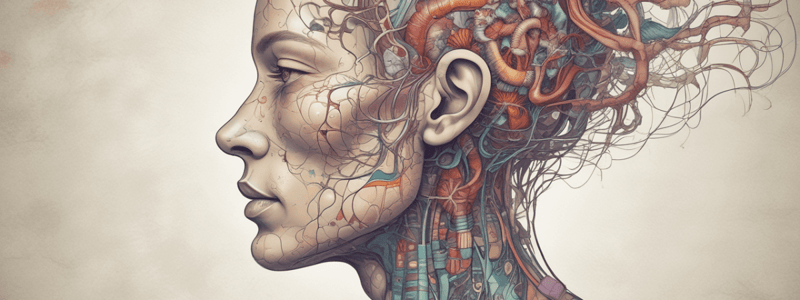
Compressive and Demyelinating Illness in the Central Nervous System
Objectives
- Describe pathophysiologic mechanisms of demyelination and its consequences on neuronal function
- Develop a model of MS pathophysiology, incorporating pathological findings, immunological mechanisms, and known etiological factors
- Describe epidemiology, clinical features, and diagnostic aspects of MS
- Describe MS time course, including:
- Three major patterns of MS progression
- Major signs and symptoms typical of early MS
- Clinical features of flares and periods between flares
- Pathologic appearance of active and inactive plaques
Definitions and Descriptions
Neuropathies
- Define:
- Neuropathy
- Neuralgia
- Neuritis
- Radiculopathy
- Polyneuropathy
- Multiple mononeuropathy
Hereditary Sensory and Motor Neuropathies
- Basic epidemiology
- Pathogenesis
- Major clinical features
- Prognosis
Guillain-Barre Syndrome
- Basic epidemiology
- Pathogenesis
- Major clinical features
- Prognosis
Nerve Compression
- Pathophysiologic mechanisms leading to neurological damage
Bell's Palsy
- Basic epidemiology
- Pathogenesis
- Major clinical features
- Prognosis
Nucleotide Structure
- Nucleosides consist of a sugar (5-carbon ribose in RNA or deoxyribose in DNA) and a base (pyrimidine or purine)
- Nucleotides are nucleosides with one or more phosphate groups attached
Nucleotide Bases
- Bases to know: • Adenine (purine) • Guanine (purine) • Cytosine (pyrimidine) • Thymine (pyrimidine, found in DNA only) • Uracil (pyrimidine, found in RNA only)
Purine Biosynthesis
- Occurs via two main pathways: de novo synthesis and the salvage pathway
- These pathways ensure cells have a sufficient supply of purine nucleotides for DNA and RNA synthesis, energy transfer, and signaling
Pyrimidine Nucleotide Synthesis
- To make CTP: • UMP is phosphorylated to make UTP • UTP is aminated to make CTP using glutamine as the N source
- To make dTMP: • UMP is first phosphorylated to make UDP • UDP is converted to dUMP • dUMP is methylated to dTMP using a folate coenzyme
Nucleotide Catabolism
- Nucleotidases remove phosphates from nucleotides to release nucleosides
- Sugar is removed to release bases
- Pyrimidine bases are degraded to: • Cytosine to uracil and ultimately alanine • Thymine to aminoisobutyrate
- Purine bases are degraded to: • Xanthine, then uric acid • Uric acid is eventually excreted in urine
Gout
- Can be due to underexcretion (most common) or overproduction (less common) of uric acid
- Hyperuricemia can lead to gout
- Gout is characterized by: • Deposition of monosodium urate crystals in the joints • Chronic kidney disease
- Pathophysiology of acute gout: • Monosodium urate crystal precipitation results in acute gouty arthritis • Local anatomical factors increase the likelihood of arthritis • Urate crystals are phagocytosed by macrophages, activating them and releasing chemokines that attract neutrophils • Neutrophils mediate joint inflammation
Calcium Pyrophosphate Crystal Deposition Disease (CPPD)
- Also known as pseudogout
- Epidemiology: • Common, prevalence increases with age • Can be caused by genetic components, hyperparathyroidism, hemochromatosis, diabetes, and hypothyroidism
- Pathology: • Crystals deposit in the matrix of menisci and connective tissue of joints • Rupture, eliciting inflammation as macrophages phagocytose the crystals • Recruit neutrophils, which mediate inflammatory damage
- Clinical features: • Can be asymptomatic, can mimic osteoarthritis or rheumatoid arthritis • Asymmetric, can be monoarticular or polyarticular • Commonly affects knees, less common sites are wrists, shoulders, elbows, and ankles
Synovial Fluid Analysis
- Why it's done: • To distinguish between septic arthritis, gout, pseudogout, hemarthroses, and rheumatic joint diseases • To manage urgent conditions like septic arthritis, hemarthrosis, and gout
- When it's done: • Suspicion of infectious arthritis, flare of crystal arthritis, or hemarthrosis • Monoarthritis (with or without a prior history of arthritis of other joints) • Trauma to a joint with effusion
- Analyzing the three C's: • Crystals (seen under microscopy with gout and pseudogout) • Cells (red blood cells, leukocytes) • Culture (any microorganisms growing in the fluid)
Otitis Media
- Acute Otitis Media (AOM) complications:
- Suppurative complications: acute mastoiditis, meningitis, brain abscesses
- 21,000 deaths per year, 30 per 10,000 individuals experience hearing loss
- Perforation of the tympanic membrane
Otitis Media with Effusion (OME)
- Epidemiology: incidence and prevalence are difficult to establish
Chronic Suppurative Otitis Media (CSOM)
- Epidemiology: incidence and prevalence are difficult to establish
Pathogenesis of Otitis Media
- Eustachian tube anatomy plays a role
- Common otopathogens:
- Streptococcus pneumoniae (Pneumococcal conjugate vaccine)
- Haemophilus influenzae
- Moraxella catarrhalis
- Pseudomonas aeruginosa
Haemophilus influenzae
- Virulence factors:
- Adhesins
- Polysaccharide capsule
- Lipid A chains/lipooligosaccharides
- Fimbriae
- IgA protease
- Biofilms
- Requires X factor (Hemin) and V factor (NAD+)
Moraxella catarrhalis
- Virulence factors:
- Antibiotic resistance (β-lactamase resistance)
- Outer membrane proteins (uspA1-A2, pili)
- Iron-regulated proteins (transferrin-binding proteins, lactoferrin-binding proteins)
- Lipid A chains/lipooligosaccharides
Streptococcus pneumoniae
- Virulence factors:
- Polysaccharide capsule
- Fimbriae
- Surface proteins that inhibit complement activation
Pseudomonas aeruginosa
- Virulence factors:
- Fimbriae and adhesins
- Biofilm formation
- Elastase production (breaks down elastic fibers, degrades the complement system, cleaves IgG and IgA antibodies)
- Pyocyanin production (triggers free radical accumulation, causing tissue damage)
- Epidemiology: found in soil, not part of regular microbiota; common nosocomial infection agent (10% of hospital infections)
Staphylococcus aureus
- Virulence factors:
- Enzymes (coagulase, hyaluronidase, staphylokinase)
- Structural defenses (capsule/slime layer glycocalyx, preventing phagocytosis)
- Binding IgG antibody stem regions (preventing opsonization)
- Toxins (cytolytic toxins, leukocydin, epidermal cell differentiation inhibitors, exfoliative toxins)
- Causes various diseases, including skin diseases, reproductive system diseases, systemic infections, cardiovascular diseases, respiratory system diseases, and gastrointestinal system diseases.
Immunodeficiencies
- Defined as any defect in the immune response that renders an individual more susceptible to infectious diseases
- Patients with immunodeficiency diseases are more prone to contracting infections, which are more likely to end in long-term debilitation or death
- They also have a higher risk of developing autoimmunity or cancer
Primary Immunodeficiencies
- Mostly inborn (genetic) and often detected in infancy or childhood
- Can be detected in adulthood
- Include B cell deficiencies, T cell deficiencies, and innate immunodeficiencies
B Cell Deficiencies
- Isolated IgA deficiency
- Common in Caucasians
- Pathogenesis: defects in a receptor for a B-cell activating cytokine
- Reduced amounts of IgA in serum and secretions, but normal levels of other antibodies and lymphocytes
- Lymphatic tissues look normal under a microscope
- Clinical Features:
- Most are asymptomatic
- History of recurrent otitis media, sinusitis, bronchitis, pneumonia, and GI tract infections
- Increased incidence of autoimmunity, particularly lupus and RA
- A potentially deadly complication is life-threatening anaphylaxis post-blood transfusion
- Prognosis is good
- Hyper IgM syndrome
- Patients make IgM but have difficulty producing IgG, IgA, and IgE
- Inability of helper T-cells to activate B-cells and macrophages due to CD40L deficit or lack of function
- Very uncommon, with an incidence of 1/1,000,000 live births
- Clinical Features:
- Recurrent pyogenic infections
- Many viral infections
- These people are very immunodeficient
- Neutrophil counts are also decreased for unknown reasons
- Treatment and Prognosis:
- IVIG and intense antibiotic prophylaxis
- Prognosis is guarded, with a 20% survival rate in those 25 and older
- X-linked agammaglobulinemia (Bruton's)
- Not mentioned in detail in the text
T Cell Deficiencies
- DiGeorge syndrome (22q11 deletion syndrome)
- T-cell deficiency
- 3rd and 4th pharyngeal pouches don't develop, resulting in:
- Shortage of thymus
- Shortage of parathyroid glands
- Some thyroid tissue
- Heart and great vessel defects
- Hypoparathyroidism, resulting in hypocalcemia
- Relatively common, with an incidence of 1-2/2000 population
- Clinical Features:
- Immunodeficiency: thymic hypoplasia results in variable loss of T-lymphocytes
- Increased fungal and viral infections
- Increased autoimmunity (RA, thyroiditis)
- Cardiac abnormalities
- Craniofacial abnormalities
- Developmental delay
- Treatment:
- Avoid blood products, as they can result in graft-vs-host disease
- Infectious disease specialist for immunotherapy and antibiotic prophylaxis
- Prognosis varies greatly
Severe Combined Immunodeficiencies (SCID)
- A multitude of etiologies, defined by:
- Recurrent, severe infections by a wide range of pathogens
- Defects in both cell-mediated and humoral immunity
- Occurs in ~1/75,000 live births
- 50-60% of SCID is X-linked, due to a mutation in the gamma-chain of a variety of cytokine receptors
- Remainder is autosomal recessive, due to a mutation in adenosine deaminase
- Pathological and clinical features:
- Pathology: small thymus with very few lymphocytes, depletion of T-cell areas of other lymphatic tissues
- Signs and symptoms: presents very early in life with recurrent severe infections, occasionally with graft-versus-host disease ( GVHD)
Innate Immunodeficiencies
- Complement defects:
- C2 deficiency: most common deficiency, increased bacterial or viral infections, though many have no increased incidence of infection
- Increased risk of SLE
- Deficiencies of other components: properdin, factor D, C3, and others, are uncommon, but result in more severe immunosuppression
- Hereditary angioedema:
- Autosomal dominant disorder, less common than the complement deficiency disorders (1 in 50,000)
- Deficit in C1 inhibitor, resulting in:
- Unchecked activation of the classical complement pathway
- Increased bradykinin production
- Increased activation of certain components of the clotting cascade
- Clinical features:
- Episodic, attacks usually become progressively more severe
- Symptoms: severe abdominal pain, vomiting, diarrhea, swelling of face, hands, legs, groin, and potentially life-threatening airway involvement
- Treatment and Prognosis:
- Can treat with C1 inhibitor from blood products, greatly improving prognosis
- Mortality used to range between 20-30%
Infectious Diseases Risk of Systemic Glucocorticoid Use
- Systemic use of glucocorticoids can increase risk of infections
- Increased risk with higher doses (e.g., 15-30g prednisone) and for greater than 2-4 weeks
- Specific infections:
- Pneumocystis jiroveci pneumonia
- Fungal infection of the lung
- Combination antibiotics prophylactically or treatment
Bipolar Disorders
- Epidemiology: 1.5% of Canada's population experiences bipolar disorder.
- Clinical Features:
- Mania
- Hypomania
- Depression (most of the time spent in depressive state)
- Cyclical changes between these states are common in the disorder
- Manic psychosis, which may include delirium
- Bipolar 1:
- At least 1 manic episode and usually depressive episodes
- Bipolar 2:
- Major depressive episodes with at least 1 hypomanic episode
Mania Symptoms
- 3 or more symptoms (representing significant change from normal) of mania:
- Inflated self-esteem or grandiosity
- Decreased need for sleep (feels rested after 3 hours of sleep)
- More talkative than usual or pressure to keep talking
- Flight of ideas or subjective experience that thoughts are racing
- Distractibility (i.e., attention too easily drawn to unimportant/external stimuli)
- Increase in goal-directed activity (purposeful) or psychomotor agitation (purposeless)
- Excessive involvement in activities that have a high potential for painful consequences (i.e., foolish business investments, unrestrained buying sprees)
Pathophysiology of Bipolar Disorders
- Circadian Rhythm Dysfunction: Changes in melatonin levels, melatonin receptor expression with CNS, and cortisol profiles.
- Metabolic Dysfunction:
- Various metabolic abnormalities, such as increased risk of obesity, type 2 diabetes, and reduced longevity due to increased cardiovascular problems.
- Increased amount of leptin secreted in obese patients with bipolar compared to obesity alone.
- Mitochondria Dysfunction:
- Impairment in mitochondrial function, resulting in brain metabolism shifting towards glycolysis.
- Observations: polymorphisms within mitochondrial DNA linked to BD, reduced levels of phosphocreatine within frontal cortex, and evidence of impaired oxidative phosphorylation.
- Glutamate Excitotoxicity:
- Increased glutamate in frontal cortex of patients with BD.
- Mood stabilizers appear to return glutamate levels to normal.
Antidepressants
- Mechanism Overview:
- Review: select theories of depression, including low levels of neurotransmitters (NTs), leading to receptor upregulation.
- Possible mechanisms of antidepressant action:
- Reuptake inhibitors: immediate increase in NT levels, delayed down-regulation of NT receptors.
- Reuptake enhancers: decreased NT levels (approved in Europe).
- Antidepressant Suicide Risk:
- Increased suicide risk in children, adolescents, and young adults up to 24 years of age when taking antidepressants.
- TCA: Common Receptor-block Adverse Effects:
- M-block: dry mouth, constipation, urinary retention, mydriasis, blurred vision, and confusion.
- Alpha 1-block: orthostatic hypotension, sedation, and sexual dysfunction.
- H1-block: weight gain, sedation, and drowsiness.
- SNRI's: Venlafaxine:
- Also weakly inhibits dopamine reuptake.
- No significant alpha 1, H1, or M-block, making it less likely to cause related adverse effects.
- SSRI's:
- Therapeutic mechanism: reuptake inhibition.
- Adverse effects often related to M, α1, H1-block, but typically more mild, except for sexual dysfunction.
Anxiety Disorders
- Fear is considered "pathologic" when:
- Out of proportion to risk/severity of threat
- Response lasts beyond the duration of the threat
- Response becomes generalized to other situations
- Social or occupational functioning is impaired
Types of Anxiety Disorders
- "True" anxiety disorders:
- Panic disorder
- Agoraphobia
- Specific phobia
- Generalized anxiety disorder
- "Anxiety-like" disorders (no longer strictly considered as part of the anxiety disorder spectrum):
- Obsessive-compulsive disorder
- Post-traumatic stress disorder
Panic Disorder
- Characterized by recurrent panic attacks
- To be diagnosed with panic disorder, need to have periods in between attacks where patient:
- Fears another attack
- Does maladaptive things to avoid another attack
- Need at least 1 month history of avoidance or fear of another panic attack
- The panic can't be due to a particular phobia or another anxiety-related disorder
- The panic symptoms cannot be due to an underlying medical disorder
Panic Attacks - Features
- Four or more of the following:
- Palpitations, pounding heart, or accelerated heart rate
- Sweating
- Trembling or shaking
- Sensations of shortness of breath or smothering
- Feelings of choking
- Chest pain or discomfort
- Nausea or abdominal distress
- Feeling dizzy, unsteady, light-headed, or faint
- Chills or heat sensations
- Paresthesias (numbness or tingling sensations)
- Derealization (feelings of unreality) or depersonalization (being detached from oneself)
- Fear of losing control or "going crazy"
- Fear of dying
Generalized Anxiety Disorder
- GAD - generalized worry that occurs more days than not that is disproportionate to the severity of the event that is feared
- Prevalence: 3-8%
- Can be difficult to treat
- Diagnostic criteria:
- Excessive anxiety for more days than not for 6 months
- Individual has difficulty controlling the anxiety
- Is accompanied by typical symptoms (see next slide)
Generalized Anxiety Disorder - Symptoms
- 3 of 6 of the following must be present for diagnosis:
- Restlessness or feeling "keyed up" or on edge
- Being easily fatigued
- Difficulty concentrating or mind going blank
- Irritability
- Muscle tension
- Sleep disturbance
- As with all psychiatric diagnoses, the anxiety, worry, or physical symptoms must:
- Cause clinically significant distress
- Impairment in social, occupational, or other important areas of function
Agoraphobia
- In general, unreasonable fear of being out-of-doors or being in a crowd or being in a place where they can't escape from or may suffer embarrassment
- The anxiety and its symptoms are typically present almost all of the time… even when the patient is somewhere comfortable to them
- Avoidance is prominent
- Very common disease, but again has a good prognosis if well-treated
- Prevalence: about 1%
Agoraphobia - Symptoms
- Examples of situations that cause worry or fear in agoraphobia:
- Using public transportation
- Being in open spaces… or enclosed spaces
- Standing in line, being in a crowd, or being in other social situations
- Being outside of the home
- These situations should almost always provoke fear or anxiety
- The fear or anxiety needs to be present for > 6 months and needs to cause significant distress or impairment in social or occupational function
Specific Phobias
- Fears of specific objects or situations that go beyond the true threat of the stimulus and cause avoidance and functional impairment
- Prevalence: 12-16%
- Diagnostic criteria:
- Exposure to stimulus provokes an immediate fear/anxiety response; may present as a panic attack
- Phobic object/situation is actively avoided or endured with intense anxiety
- Fear/anxiety out of proportion to actual danger/sociocultural context
- Person recognizes fear as excessive or unreasonable
- Significant distress or impact on daily routine, occupational/social functioning and/or marked distress
- Must be present > 6 months
Social Anxiety Disorder
- Marked and persistent (>6 mo) fear of social or performance situations in which:
- One is exposed to unfamiliar people or to possible scrutiny by others
- One is afraid that fearing he/she will act in a way that may be humiliating or embarrassing
- Out-of-proportion fear that they will be harshly judged by their interpersonal interactions
- Prevalence: 2-7%
Post-traumatic Stress Disorder
- When does it happen?
- Exposure to actual death, threatened death, physical or sexual violence, serious injury
- Could have witnessed or received the violent act
- Can occur in people that are repetitively exposed to disturbing or violent events
- Neurobiology:
- Involves the hippocampus, amygdala, and prefrontal cortex
- The serotonin-releasing nucleus in the brainstem (the raphe nucleus) can also have a more "global" modulatory effect on mood, memory, and also fear + stress responses
Neurobiology of Anxiety Disorders
- Noradrenergic pathway:
- Neurotransmitter: Noradrenaline (also known as norepinephrine)
- Origin: The cell bodies of noradrenergic neurons are located in the locus coeruleus in the brainstem
- Function: Plays a key role in regulating mood, attention, arousal, and stress response
- Clinical significance: Dysregulation of noradrenergic pathways is implicated in various psychiatric disorders such as depression, anxiety disorders, and attention deficit hyperactivity disorder (ADHD)
- Serotonergic pathway:
- Neurotransmitter: Serotonin (also known as 5-hydroxytryptamine or 5-HT)
- Origin: The cell bodies of serotonergic neurons are located in the raphe nuclei in the brainstem
- Function: Regulates mood, emotion, appetite, sleep, and various cognitive functions
- Clinical significance: Imbalances in serotonin levels are associated with mood disorders such as depression and anxiety, as well as obsessive-compulsive disorder (OCD) and eating disorders
- Dopaminergic pathway:
- Neurotransmitter: Dopamine
- Origin: Dopaminergic neurons have several nuclei in the brain, including the substantia nigra and the ventral tegmental area (VTA)
- Function: Plays a crucial role in reward, motivation, pleasure, movement, and reinforcement learning
- Clinical significance: Dysregulation of dopamine pathways is implicated in various psychiatric disorders such as schizophrenia, addiction, and Parkinson's disease
Benzodiazepine Adverse Effects
- Selected adverse effects (most common with hypnotic use):
- Hangover effects
- Early morning rebound insomnia
- Tolerance
- Dependence
- Withdrawal symptoms
- Withdrawal symptoms:
- Mild symptoms can occur after short-term use and/or low-doses, including:
- Extra-sensory awareness
- Muscle twitching or tremors
- Rebound excitation
- Severe symptoms usually occur on abrupt discontinuation after long-term use and/or high doses, and include:
- Increased blood pressure, temperature, and pulse
- Rage
- Hallucinations and paranoia
- Seizures
- Mild symptoms can occur after short-term use and/or low-doses, including:
Bacterial Meningitis
- Inflammatory bacterial infections of the meninges, particularly the pia and arachnoid mater, causing meningial swelling and restricting CSF flow.
- Symptoms: nausea, pain, vomiting, reduced brain function, stiff neck, and decreased motor control.
- If the infection is in the spinal cord, muscles of the neck will become stiff and motor control will be reduced.
- If the infection is in the brain (encephalitis), sensory perceptions are decreased, behavioral changes occur, and coma or death may result.
Testing for Bacterial Meningitis
- Cloudy CSF and positive meningitis test indicate bacterial meningitis.
- Lumbar puncture (aka spinal tap) is used to collect CSF for testing.
Causes of Bacterial Meningitis
- Opportunistic members of normal microbiota: Staphylococcus aureus, Streptococcus pyogenes, Klebsiella pneumoniae.
- Regular disease-causing bacteria: Streptococcus pneumoniae, Haemophilus influenzae, Neisseria meningitidis, Listeria monocytogenes.
Neisseria Meningitidis
- Causes meningococcal meningitis.
- Virulence factors: fimbriae, polysaccharide capsules, lipooligosaccharide (with Lipid A/Endotoxin), and various factors to prevent digestion in phagocytes.
Streptococcus Pneumoniae
- Leading cause of meningitis.
- Virulence factors: capsule, secretory IgA protease, pneumolysin (inactivator of lysosomal enzymes).
- Primary virulence factor: phosphorylcholine (attachment to cells of lungs, meninges, blood vessels – and triggers endocytosis).
Listeria Monocytogenes
- Gram +ve coccobacillus found in soil, water, and many animals.
- Obtained through contaminated food/drink.
- Causes meningitis in immunocompromised individuals, but only mild flu in healthy adults.
- Avoids immune system detection by dividing inside macrophages and epithelial cells.
Pathogenesis of Bacterial Meningitis
- N.meningitidis, H.influenzae, S.pneumoniae – inhaled in respiratory droplets.
- Listeria – unpasteurized milk, cheese, meat.
- Bacteria usually spreads to meninges from infections of lungs, sinuses, or inner ear.
- Head or neck trauma may expose meninges directly.
- Bacteria ferment glucose in CSF for energy.
Prevention
- Susceptible individuals should avoid undercooked veggies, unpasteurized milk, undercooked meat, and soft cheese.
- People living in dormitories should receive vaccinations.
Hansen's Disease (Leprosy)
- Causative agent: Mycobacterium leprae.
- Optimal growth – 30°C – in the chilly parts of the body (peripheral nerve endings, earlobes, nose, tips of fingers and toes).
- Signs of disease may not be present for 10-30 years, but when the population becomes big enough, the immune system will aggressively attack them.
Botulism
- Causative agent: Clostridium botulinum toxin (not an infection).
- 3 types of botulism: foodborne, infant, and wound.
- Foodborne/Wound Botulism symptoms: paralysis of all voluntary muscles, blurred vision, nausea (death from respiratory paralysis).
- Infant botulism: not ingested, but C.botulinum can infect GI tract due to absence of microbiota.
Tetanus
- Causative agent: Clostridium tetani.
- Portal of entry: endospores enter through breaks in skin.
- Signs/symptoms: tightening of jaw and neck muscles, difficulty swallowing, fever, spasms.
- Treatment: aggressive treatment of wound, antibiotics.
- Prevention: Vaccination.
West Nile Fever
- Absent from N.America until 1999.
- Virus infects 100s of bird, 37 mosquito, 18 other vertebrate (including humans/horses) species.
- Transmitted by mosquito bite.
- Incubation period of 3-14 days.
- 20-30% get flu-like illness called West Nile Fever.
- 80% - NO symptoms.
- 1/150 experience neuroinvasion.
- Headache, ocular manifestations, muscle weakness, cognitive impairment, polio-like flaccid paralysis, and 10% mortality.
Poliomyelitis (Polio)
- Causative agent: Poliovirus.
- Spread by fecal-oral transmission.
- Peaks during warm months in temperate climates.
- Complication: post-polio syndrome.
- 30-40 year interval.
- 25-40% not an infectious process.
- Humans are the only known reservoir.
- Epidemic in the 1900s:
- Polio has only minor symptoms for infants and adults (seems like a mild cold).
- Early in the 1900s, white, wealthy children started getting paralytic polio.
- Connection to modern plumbing, sewer systems, etc.
Gastrulation and Early Embryology
- Definition of Gastrulation: The process by which the three germ layers of the embryo are established: Ectoderm, Mesoderm, and Endoderm.
- Bilaminar Embryonic Disc: The stage where the embryo consists of two layers: Epiblast and Hypoblast.
- Trilaminar Embryonic Disc: The stage where the embryo consists of three layers: Ectoderm, Mesoderm, and Endoderm.
Female Reproductive Anatomy
- Ovaries: Produce oocytes (female haploid gametes) and hormones such as progesterone and estrogens.
- Uterine Tube (Fallopian Tube): Receives oocytes from ovaries and site of fertilization.
- Uterus: Site of embryo development and placenta and membrane formation.
Ovulation and Fertilization
- Ovulation: Release of a secondary oocyte from the ovarian follicle.
- Fertilization: Fusion of the pronucleus of the two gametes (sperm and oocyte) resulting in a diploid zygote.
- Zygote: A fertilized, diploid oocyte that has not yet divided.
- Meiosis: A process in which a diploid cell (germ cell) undergoes reduction division to produce a unique haploid gamete.
Early Embryology
- Week 1: Begins with fertilization, and the embryo develops from a zygote to a blastocyst.
- Zona Pellucida: A protein coat that surrounds the oocyte and early embryo.
- Blastocyst: A spherical mass of cells that is composed of a trophoblast, inner cell mass (embryoblast), and blastocoel.
- Cleavage: Cell division in the early embryo, resulting in smaller cells.
- Blastomere: A cell that is totipotential and is present during early development.
- Implantation: The process by which the embryo contacts and becomes surrounded by the endometrium of the uterus.
Implantation
- Zona Hatching: The process by which the blastocyst escapes from the zona pellucida.
- Syncytiotrophoblast: A layer of cells that invades the endometrium and forms the placenta.
- Cytotrophoblast: A layer of cells that forms the embryoblast and later the embryo.
Week 2 and 3
- Epiblast and Hypoblast: The epiblast differentiates into amnioblasts, and the hypoblast extends around the entire interior surface of the blastocoel.
- Gastrulation: The process by which the three germ layers of the embryo are established.
- Primitive Streak: A thickened linear band in the median plane of the dorsal aspect of the embryonic disc.
- Primitive Node: A proliferation of cells at the cephalad end of the primitive streak.
- Primitive Groove and Pit: A narrow groove and depression in the primitive node.
Key Terms
- Gamete: A haploid germ cell.
- Morula: A 12-32 cell stage of an embryo.
- Blastocoel: A fluid-filled cavity within the blastocyst.
- Trochoblast: A layer of cells on the outside of the blastocyst.
- Embryoblast: A group of cells that develop into the embryo.
- Coelom: A fluid-filled cavity.
- Ectoderm: A germ layer that is typically found on the exterior of the organism.
- Endoderm: A germ layer that is typically found on the interior of the organism.
- Mesoderm: A germ layer found between the ectoderm and endoderm.
Embryology: Neurulation, Folding, and Development of the Nervous System
The Notochord
- During week 3, the notochordal process forms from mesenchymal cells diving into the primitive pit and migrating cephalad.
- The notochordal process develops a lumen known as the notochordal canal.
- After the notochordal process approaches the prechordal plate, the floor of the process fuses with the endoderm, forming the notochordal plate.
- The notochordal plate cells proliferate and fold inwards, forming the fully-developed notochord with no canal present.
- The notochordal plate to notochord transition starts cranially and progresses caudally.
Intraembryonic Mesoderm
- During week 3, intraembryonic mesoderm proliferates to form a thick column of mesoderm on either side of the notochord.
- The paraxial mesoderm is located beside the axis of the organism, while the intermediate mesoderm is found just lateral to the paraxial mesoderm.
- The lateral mesoderm is lateral to the intermediate mesoderm.
Somites
- During weeks 3-5, somites develop adjacent to the neural tube.
- Somites are cuboidal masses of mesoderm on either side of the notochord, formed from the paraxial mesoderm.
- Somites give rise to most of the axial skeleton and associated musculature, as well as the dermis in those areas.
Intraembryonic Coelom
- The primordium of the intraembryonic coelom appears as isolated spaces in the lateral mesoderm and cardiogenic mesoderm.
- The coelomic spaces soon coalesce and form a single, horseshoe-shaped intraembryonic coelom.
- The intraembryonic coelom divides the lateral mesoderm into two layers: somatic and splanchnic mesoderm.
Embryonic Folding
- Embryonic folding is the process by which a relatively flat embryonic disk becomes cylindrical in shape.
- Folding occurs in two general planes: the median plane and the horizontal plane.
- During week 3, the neural folds fuse to form the neural tube, which eventually closes and no longer communicates with the amniotic cavity.
Spinal Cord Development
- The lateral walls of the caudal portion of the neural tube thicken, forming the ventricular zone, intermediate zone, and marginal zone.
- The ventricular zone gives rise to all neurons and macroglia.
- The intermediate zone becomes populated with primordial neuroblasts, and the outer marginal zone develops into white matter tracts.
- The alar and basal plates of the developing spinal cord are separated by a groove, the sulcus limitans.
- The cell bodies in the alar plates form the dorsal gray horns, while the cell bodies in the basal plates form the ventral and lateral gray horns.
Neural Crest Cell Derivatives
- The neural crest cells give rise to the peripheral nervous system (PNS), including the spinal ganglia, sympathetic ganglia, and adrenal medulla.
Brain Development
- The neural folds in the cranial region fusion and closure of the rostral neuropore form three primary brain vesicles: forebrain, midbrain, and hindbrain.
- The forebrain partially divides into two secondary brain vesicles: telencephalon and diencephalon.
- The rhombencephalon also partially divides into two secondary brain vesicles: metencephalon and myelencephalon.
- The optic vesicles, telencephalic vesicles, and thalamus, hypothalamus, and epithalamus develop from the forebrain.
Early Fetal-Maternal Circulation
- By the end of the second week, the embryo is surrounded by:
- An amniotic cavity at the dorsal surface, lined by amnioblasts and surrounded by extraembryonic somatic mesoderm
- The umbilical vesicle at the ventral surface, which will eventually become smaller as the embryo develops and undergoes folding
- The chorion, consisting of extraembryonic somatic mesoderm on the inside and trophoblastic cells on the outside
Trophoblast
- The trophoblast consists of cytotrophoblast and syncytiotrophoblast
- The syncytiotrophoblast:
- Invades the endometrial stroma and induces the apoptotic death of decidual cells, releasing energy-rich glycogen and lipids that can freely diffuse to the embryo
- Secretes hCG, which allows the ovary to continue secreating high levels of progesterone, preventing the endometrium (and embryo) from being lost during menstruation
- The cytotrophoblast begins to form primary chorionic villi that project into the lacuna
Maternal-Fetal Circulation
- By the 4th week, the embryo and chorion have grown large enough that diffusion of nutrients between the embryo and endometrium is inadequate for metabolic demands
- A circulation that exchanges between the maternal and fetal tissues is necessary
- The embryonic heart begins to beat at day 21, with blood vessels and red blood cells present in the fetus, and gas/nutrient/electrolyte/waste exchange between mother and embryo increases
Chorionic Villi
- Primary chorionic villi: present in week 2, composed of cytotrophoblasts surrounded by a syncytiotrophoblastic shell
- Secondary chorionic villi: present in week 3, composed of an extraembryonic mesenchymal core surrounded by cytotrophoblastic and syncytiotrophoblastic shell
- Tertiary chorionic villi: present by the end of week 3, with blood vessels (capillaries) within the mesenchymal core, allowing fetal blood to exchange substances with the maternal lacuna through the membrane formed by the tertiary villus
Maternal-Fetal Circulation - Week 3
- Day 16: the syncytiotrophoblast invades the endometrial stroma and induces the apoptotic death of decidual cells
- Day 21: the embryonic heart begins to beat, and blood vessels and red blood cells are present in the fetus
Development of Fetal Membranes
- The decidua is the endometrium in a pregnant woman, consisting of three separate areas:
- Decidua basalis: the decidua that forms the placenta, connected to the embryo by the umbilical cord
- Decidua capsularis: the decidua that does not include the basalis, but is still associated with/covers the embryo
- Decidua parietalis: formed by the endometrium that is not part of the embryo, far from the site of implantation
- As the embryo develops, the villi within the capsularis deteriorate and the amniotic cavity grows larger, eventually filling the uterus and contacting the decidua parietalis
Development of the Fetal Cardiovascular System
- Begins in the umbilical vesicle and neighbouring connecting stalk/chorion, arising from the extra-embryonic mesoderm, starting early in the 3rd week
- Embryonic vessels develop from mesoderm about 2 days after the extra-embryonic vessels develop
- Divided into vasculogenesis and angiogenesis
- Vasculogenesis: development of brand new blood vessels from mesoderm
- Angiogenesis: "sprouting" of blood vessels formed by vasculogenesis, connecting blood vessels to each other
Vasculogenesis and Angiogenesis
- Angioblasts derived from mesoderm develop in specialized regions known as blood islands
- The blood islands develop lumens and become blood vessels, which is vasculogenesis
- The angioblasts also give rise to red blood cells within and outside the embryo
- The blood cells are derived from the inner lining of the new vessels (endothelium)
- Extra-embryonically, blood cells develop from the endothelium in the allantois and the umbilical vesicle
- Intra-embryonically, blood cells develop from the endothelium of the dorsal aorta
Early Embryo - 3rd and 4th Week
- The intra-embryonic coelom near the heart tube develops into the pericardial cavity
- The paired heart tubes are connected with the extra-embryonic vessels once the heart starts to beat at day 21
- The red blood cells develop first in the extra-embryonic vessels
- By the 5th week, RBCs arise from the dorsal aorta
Skeletal Muscle Tissue
- Skeletal muscles are composed of bundles of fascicles
- Each fascicle is composed of linearly aligned muscle fibers (also known as myofibers)
- Each muscle fiber is a single, multi-nucleated, elongated cell
- Muscle fibers are composed of many sarcomeres, arranged linearly
- Sarcomeres are composed of myofibrils (organelles)
Connective Tissue Sheath
- The connective tissue sheath surrounding the whole muscle and extending from the tendons is called the epimysium
- The sheath surrounding each fascicle is called a perimysium
- The sheath surrounding each individual muscle fiber is called the endomysium
- Below the endomysium is the sarcolemma – the cell membrane of the muscle cell
Muscle Fibers
- Muscle fibers are large, multinucleated cells
- Contain 1000 – 2000 myofibrils
- Each myofibril is composed of many myofilaments
- Myofilaments are composed of:
- Contractile proteins (actin and myosin)
- Regulatory proteins (tropomyosin and troponin)
- Additional accessory proteins (actinin, dystrophin, titin, nebulin)
Contractile Proteins - Sarcomeres
- Myofibrils are arranged into a series of sarcomeres, which form the contractile unit of skeletal muscle
- Sarcomeres consist of interdigitating myofilaments, composed of:
- Thin filaments (made of actin)
- Thick filaments (made of myosin)
- The striated appearance of skeletal muscle is due to the overlapping of thick and thin filaments
Sarcomeres - Actin
- Structure: monomer (G-actin) and polymer (F-actin)
- Thin filaments are composed of two strands of F-actin wound together
- Each G-actin monomer has a binding site for myosin
- F-actin is the major constituent of the thin filament
Sarcomeres - Myosin
- Structure: arranged into thick filaments composed of many myosin units
- Each myosin unit is composed of head and tail regions
- Head region forms cross-bridges that interact with adjacent actin filaments
- Many myosin units are arranged in a staggered position into a thick filament
Sarcomeres - Myosin (closer look)
- Myosin head region has 3 important biochemical features:
- ATPase activity
- Actin-binding region
- ATP binding region
Regulatory Proteins
- Tropomyosin: a regulatory protein that associates with actin
- When a skeletal muscle is in a relaxed state, tropomyosin molecules cover the myosin-binding site on G-actin monomers
- Prevents cross-bridge formation between actin and myosin
- Troponin: another regulatory protein that associates with actin
- Forms a complex with 3 subunits: Troponin I, Troponin T, and Troponin C
- Function: when calcium binds to Troponin C, the troponin complex undergoes a conformational change and Troponin T "pulls" tropomyosin and Troponin I off of the myosin-binding site of G-actin subunits
Sarcolemma
- The plasma membrane of the muscle fiber is called the sarcolemma
- Contains invaginations called transverse Tubules (T-tubules)
- Allows the action potential to be carried deep into the muscle fiber
- Continuous with the extracellular fluid
Muscle Triad
- The junction between T-tubules and sarcoplasmic reticulum cisterna is called the muscle triad
- Volume of T-tubule is large compared to the SR cisterna
- Review: the concentration of calcium in the extracellular fluid is low
Neuro-Muscular Junction
- A motor nerve axon contacts each muscle fiber near the middle of the fiber, forming a synapse called the neuromuscular junction
- The region of the sarcolemma in closest contact with the presynaptic nerve terminal is called the motor end plate
- The motor nerve terminal releases acetylcholine (ACh), which binds to the nicotinic receptor on the sarcolemma
- This gives rise to a graded, depolarizing end-plate potential
Action Potential Propagation
- The action potential propagates along the surface of the skeletal muscle fiber and penetrate deeper into the muscle fiber via the T-tubules
- This is the purpose of the T-tubule – to "bring" the action potential deep within the very large muscle fibers
- The action potential then signals to the sarcoplasmic reticulum
Excitation-Contraction (EC) Coupling
- Action potential propagates along the T-tubules and activates L-type Ca2+ channels in the sarcolemma
- Activation of the L-type Ca2+ channels also triggers mechanical activation of Ryanodine receptors (RYR) on the surface of the SR terminal cisterna
- Opening of L-type Ca2+ channels and RYR allows Ca2+ to flow down its concentration gradient into the cytosol of the muscle fiber
Whole Muscle Contraction
- Typical muscle receives input from ~ 100 alpha-motor neurons
- The number of muscle fibers that each motor neuron innervates varies widely
- All the muscle fibers innervated by a single nerve fiber is called a motor unit
- Review: what is the alpha-motor neuron-muscle fiber complex called?
Muscle Fibers
- Muscle fibers can be classified into two main types: slow-twitch (Type I) and fast-twitch (Type II)
- Slow-twitch fibers have a high mitochondrial density, high oxidative capacity, and minimal glycolytic capacity, making them suitable for endurance activities
- Fast-twitch fibers have a low mitochondrial density, low oxidative capacity, and high glycolytic capacity, making them suitable for high-intensity, short-duration activities
Muscle Fiber Differences
- Mitochondrial density: high in slow-twitch fibers, some in fast-twitch fibers, and none in fast-twitch (IIB) fibers
- Oxidative capacity: high in slow-twitch fibers, low in fast-twitch fibers, and none in fast-twitch (IIB) fibers
- Glycolytic capacity: minimal in slow-twitch fibers, medium in fast-twitch fibers, and high in fast-twitch (IIB) fibers
- Major storage fuel: fat in slow-twitch fibers, glycogen in fast-twitch fibers
Skeletal Muscle Energy
- Three different metabolic systems responsible for recycling AMP and ADP back into ATP: phosphagen system, glycogen-lactic acid system, and aerobic system
- Phosphagen system: provides 8-10 seconds of maximal power, uses phosphocreatine and ATP, and replenishes ATP and phosphocreatine
- Glycogen-lactic acid system: provides 60-90 seconds of maximal power, uses glycogen and ATP, and replenishes ATP and phosphocreatine
- Aerobic system: provides unlimited duration of energy, uses oxygen, and replenishes ATP, phosphocreatine, and glycogen-lactic acid system
Skeletal Muscle Recovery
- Energy systems must be replenished: phosphocreatine, ATP, and glycogen-lactic acid system
- Oxygen debt: after exercise, stored oxygen must be replenished by breathing extra amounts of oxygen, totaling 11.5 liters
Skeletal Muscles in Action
- Muscle strength: determined by muscle size, with a maximum contractile force of 3-4 kg/cm2 of muscle cross-sectional area
- Muscle power: measures the amount of work performed in a given period, determined by strength, distance, and rate of contraction
- Muscle endurance: depends on nutritive support, glycogen storage, and type and size of muscle fiber
Skeletal Muscle Remodeling
- Muscle hypertrophy: results from an increase in the number of actin and myosin filaments in each muscle fiber, enlarging individual muscle fibers
- Enzyme systems providing energy also increase, especially those for glycolysis
Pain - Introduction
- Pain is an unpleasant sensation and emotional experience associated with actual or potential tissue damage, or described in terms of such damage.
- The emotional aspect of pain makes it unique, impacting mood and quality of life.
- Pain perception is very subjective, and people can suffer serious injuries without feeling pain or have little to no tissue damage but still report pain.
Types of Pain and Abnormal Sensation
- Dysesthesia: Any abnormal sensation described by a patient as unpleasant.
- Paresthesia: A sensation typically described as "pins-and-needles" but not notably unpleasant.
- Analgesia: Reduction or loss of pain perception.
- Anaesthesia: Reduced perception of all touch and pain sensation.
- Hypoalgesia: Decreased sensation and raised threshold to painful stimuli.
- Hyperalgesia: Exaggerated pain response from a normally painful stimulus.
- Allodynia: Abnormal perception of pain from a normally non-painful mechanical or thermal stimulus.
- Hyperesthesia: Exaggerated perception of a touch stimulus.
- Causalgia: Burning pain in the distribution of a peripheral nerve.
Pain Pathophysiology
- Pain does not exhibit adaptation, unlike almost all other perceptions.
- The central nervous system regulates perception and transmission of pain from lower levels, using multiple different molecules and pathways.
- Pain transmission can cause inflammation in peripheral tissues.
Physiology of Nociception
- Nociceptors are widely distributed through multiple depths in the skin and visceral organs.
- Types of nociceptors:
- Thermal nociceptors: Activated by temperatures > 45°C or < 5°C.
- Mechanical nociceptors: Activated by intense pressure applied to a structure.
- Polymodal nociceptors: Activated by high-intensity mechanical, chemical, or thermal stimuli.
- Silent nociceptors: Receptors that do not normally transmit pain information but are "awakened" in a setting of continuous damage or inflammation.
Nociceptor Characteristics
- C fibres:
- Unmyelinated axons with cell body in the dorsal root ganglia.
- Conduction velocity: 0.5 – 2 m/sec.
- Responsible for conducting slow pain and thermoception (temperature).
- Often dull, poorly-localized pain (large receptive fields).
- C-fibres also carry itching sensations.
- A-delta fibres:
- Myelinated axons with cell body in the dorsal root ganglia.
- Conduction velocity: 12 – 30 m/sec.
- Responsible for conducting sharp, "pricking" pain as well as some thermoception (temperature).
- Usually well-localized (smaller receptive fields).
What can Nociceptors Detect?
- Very wide range of ion channels expressed in the free nerve endings, allowing them to detect a wide range of stimuli associated with tissue damage.
- Transient-receptor potential receptors (TRP) recognize a wide range of tissue insults, including cold and heat, low pH, and free radicals.
- ASIC receptors detect low pH (typical of ischemia or tissue damage).
- Nociceptors also express receptors for molecules released during inflammatory processes, such as prostaglandins, bradykinin, histamine, substance P, serotonin, and ATP.
Neuroanatomical Pain Pathways
- Spinothalamic tract is the major nociceptive sensory pathway.
- Peripheral afferent pain fibres of both A-δ and C types have their cell bodies in the dorsal root ganglia.
- Fibres of the spinothalamic tract usually cross over (2nd order neurons) two or three levels superior to where the 1st-order neurons enter the spinal cord.
- The paleospinothalamic pathway travels a somewhat different route, synapsing in different thalamic nuclei and brainstem areas.
Ascending Pain Pathways
- Visceral pain sensation likely ascends along the anterior spinothalamic tract.
- Better-localized skin-associated pain sensation likely ascends via the lateral spinothalamic tract.
Fast and Slow Pain
- Fast pain: Well-localized, sharp pain carried by A-delta fibres.
- Slow pain: Poorer-localized, duller pain carried by C-fibres, tends to last longer.
Muscular Dystrophy
Duchenne Muscular Dystrophy
- Most common muscular dystrophy: 1 per 3500 live male births
- Pathogenesis: Dystrophin is a key component of the dystrophin glycoprotein complex, which provides mechanical stability to the muscle fiber and its cell membrane during muscle contraction
- Pathophysiology:
- Defects in the complex can lead to sarcolemma tears, causing calcium influx from the extracellular fluid, ultimately triggering muscle fiber necrosis
- Muscle fiber necrosis leads to chronic muscle damage that outpaces the capacity for repair
- Pathology:
- Muscle biopsies show segmental muscle fiber degeneration and regeneration, atrophic muscle fibers, and eventually replacement of muscle tissue with collagen and fat cells
- Muscle fascicular architecture is preserved initially but becomes abnormal over time with muscle remodeling
- Clinical Features:
- Normal at birth, but early motor milestones are met, and walking is often delayed
- Clumsiness and inability to keep up with peers are first indications of muscle weakness
- Weakness begins in pelvic girdle muscles and extends to shoulder girdle muscles
- (+) Gower's sign: patient often has to push off the thighs to stand from sitting
- Pseudohypertrophy of lower leg
- Cognitive impairment and learning disability are common
- Clinical feature progression & prognosis:
- Eventual progression to wheelchair dependence at an average age of 9.5 years
- Patients develop joint contractures, scoliosis, worsening respiratory reserve, and sleep hypoventilation
- Development of cardiomyopathies and arrhythmias can occur in older patients due to the impact of dystrophin on cardiac muscle
- Female carriers have an increased risk of cardiomyopathy
- Mean age of death is 25-30 years of age, commonly from respiratory insufficiency, pulmonary infection, or heart failure
Becker Muscular Dystrophy
- Etiology: X-linked mutation in dystrophin, resulting in a truncated dystrophin protein that retains partial function
- Epidemiology: Less common than Duchenne muscular dystrophy, with an incidence of 1 per 30,000 live male births
- Clinical Features:
- Onset later in childhood, adolescence, or adulthood
- Generally similar to Duchenne muscular dystrophy, but with a less severe phenotype and slower progression
- Patients are still at risk of developing dilated cardiomyopathy, but this is rare compared to Duchenne muscular dystrophy
- Prognosis: Near-normal life expectancy
Myotonic Dystrophy
- Etiology: Autosomal dominant disorder caused by expansion of triplet repeats
- Type I: expansion of CGT trinucleotide repeat within the myotonic dystrophy protein kinase (DMPK) gene
- Type II: CCTG repeat expansion within the gene nucleic acid-binding protein (CNBP) gene
- Epidemiology: Affects 1 in 8,000 individuals
- Pathogenesis: Not well understood, but may involve a "toxic" gain-of-function effect by the triplet repeat expansion, disrupting splicing of mRNA transcripts of other proteins, including the transcript of a chloride channel (CLC1)
- Clinical Features:
- Onset begins in adolescence or young adulthood
- Type I: myotonia, weakness, and wasting of peripheral muscles and facial muscles
- Can also include cardiomyopathy, intellectual disability, cataracts, and endocrine disorders
- Patients with type I will also experience myotonia of involuntary muscles (e.g., MG)
Myasthenia Gravis (MG)
- Pathogenesis and pathophysiology:
- The majority of patients have auto-antibodies to the post-synaptic acetylcholine receptors (nicotinic receptors)
- Ab-Ach receptor complexes lead to a variety of problems that ultimately limit the ability of the myofiber to respond to Ach at the neuromuscular junction
- Clinical Features:
- Fluctuating proximal muscle weakness
- Ptosis and diplopia are common initial findings
- Proximal muscle involvement: diaphragm, neck, facial muscles, and dysphagia
- Weakness worsens with activity/exercise and improves with rest (fatiguability)
- Weakness seems to come and go initially, but eventually, symptoms become more consistent
- Diagnosis and treatment:
- Based on clinical history, physical exam findings, (+) antibody screen, and electrophysiologic studies
- Acetylcholinesterase inhibitors (first line of treatment) increase the half-life of Ach in the neuromuscular junction
- Plasmapheresis and immunosuppressive drugs
- Thymectomy for patients with a thymoma
Skeletal Muscle Tumors
- Rhabdomyosarcoma:
- Malignant mesenchymal tumor with skeletal muscle differentiation
- 4 subtypes: alveolar, embryonal, pleomorphic, and spindle cell/sclerosis
- Epidemiology: most common soft tissue sarcoma of childhood, with an incidence of 6/1,000,000/year in children under 15 years
- Etiology: chromosomal translocations commonly found, typically involving transcription factor (PAX) involved in skeletal muscle differentiation
- Clinical features: tumor is detected as a mass, sometimes painful, with metastases tend to cause respiratory difficulties, bone pain, and bone marrow failure
- Synovial cell sarcoma:
- Epidemiology: most common soft tissue tumor in adolescents and young adults, with an incidence of 2.75 per 100,000
- Etiology: chromosomal translocation has been found in >90% of cases, with a fusion of SYT and SSX genes
- Clinical features: usually a history of long-standing nodule, with a rapid increase in size over a few months, and a painless and deep mass
Fibromyalgia
- Syndrome of persistent widespread pain, stiffness, fatigue, disrupted sleep, and cognitive difficulties
- Characterized by unexplained and profound fatigue that is worsened by exertion
- Etiology and pathogenesis: unknown, but implications of a number of viruses have been studied, and environmental factors have also been suspected as a trigger
- Epidemiology: true prevalence is unknown, but most common in young to middle-aged adults and more common in females
- Diagnostic criteria: presence of 3 or more of the following symptoms for at least 6 months, with symptoms severity being moderate or severe at least 50% of the time
- Fatigue - unrelated to exertion and not relieved by rest
- Post-exertional malaise (PEM)
- Unrefreshing sleep – fatigue despite a night's sleep
- AND at least 1 of the following:
- Cognitive impairment
- Orthostatic intolerance
Pharyngeal Arches
- Pharyngeal arch = a core of mesenchyme (embryonic connective tissue) covered externally by ectoderm and internally by endoderm
- Mesenchyme is derived from mesoderm in the third week, and from neural crest cells that migrate into the pharyngeal arches during the fourth week
- Migration of neural crest cells into the arches and their differentiation into mesenchyme produces the maxillary and mandibular prominences
Pharyngeal Arch Components
- A pharyngeal arch contains:
- A pharyngeal arch artery that arises from the truncus arteriosus of the primordial heart and passes around the primordial pharynx to enter the dorsal aorta
- A cartilaginous rod that forms the skeleton of the arch
- A muscular component that differentiates into muscles in the head and neck
- Sensory and motor nerves that supply the mucosa and muscles derived from the arch
Pharyngeal Arch Development
- The pharyngeal arches are the main formative elements of the face, nasal cavities, mouth, larynx, pharynx, and neck
- During the fifth week, the second pharyngeal arch enlarges and overgrows the third and fourth arches, forming an ectodermal depression-the cervical sinus
- By the end of the seventh week, the second to fourth pharyngeal grooves and the cervical sinus have disappeared, giving the neck a smooth contour
Pharyngeal Grooves and Pouches
- The primordial pharynx, derived from the foregut, widens cranially where it joins the stomodeum, and narrows caudally where it joins the esophagus
- The pharyngeal endoderm lines the internal aspects of the pharyngeal arches and passes into diverticula (outpouchings) = pharyngeal pouches
- There are four pairs of pharyngeal pouches; the fifth pair is rudimentary or absent
Oropharyngeal Apparatus
- The pharyngeal arches begin to develop early in the fourth week
- Neural crest cells migrate into the ventral parts of the future head and neck regions
- The first pair of pharyngeal arches, the primordium of the jaws, appears as surface elevations lateral to the developing pharynx
- By the end of the fourth week, four pairs of pharyngeal arches are visible externally
Bony and Cartilaginous Derivatives
- The first pharyngeal arch (mandibular arch) separates into two prominences:
- The maxillary prominence gives rise to the maxilla, zygomatic bone, and a portion of the vomer
- The mandibular prominence forms the mandible and the squamous temporal bone
- The second and third pharyngeal arches form the hyoid bone
- The first and second pharyngeal cartilages give rise to the ossicles of the middle ear and the styloid process of the temporal bone
- The fourth and sixth pharyngeal arches give rise to the laryngeal cartilage
Muscular Derivatives
- Muscles of mastication, muscles of the middle ear, and other muscles are derived from the pharyngeal arches
- The specific muscles derived from each pharyngeal arch are listed in the table
Pharyngeal Pouches
- The first pharyngeal pouch expands into a tubotympanic recess, which contributes to the formation of the tympanic membrane (eardrum) and the tympanic cavity (where the bones of the middle ear are found)
- The second pharyngeal pouch eventually gives rise to parts of the palatine tonsils
- The third and fourth pouches develop into parathyroid glands and thymus
Development of the Thyroid
- The thyroid gland is the first endocrine gland to develop in the embryo
- It begins to form 24 days after fertilization from a median endodermal thickening in the floor of the primordial pharynx
- The developing thyroid gland descends in the neck, passing ventral to the developing hyoid bone and laryngeal cartilages
- By 7 weeks, the thyroid gland is usually located in its final site in the neck
Studying That Suits You
Use AI to generate personalized quizzes and flashcards to suit your learning preferences.


