Podcast
Questions and Answers
How should the slide be positioned when placing a drop of blood on it?
How should the slide be positioned when placing a drop of blood on it?
- Flat on the surface with the non-frosted edge held steady (correct)
- Upright with the frosted area pointed downwards
- On a tilted surface with the frosted area at the top
- Horizontally with the frosted area facing left
What size should the drop of blood be when placed on the slide?
What size should the drop of blood be when placed on the slide?
- 1 mm in diameter
- 5 mm in diameter
- 2 mm in diameter (correct)
- 3 mm in diameter
Where should the drop of blood be placed on the slide?
Where should the drop of blood be placed on the slide?
- Near the corners of the slide
- An inch from the frosted area (correct)
- At the edge of the slide
- Directly in the center of the frosted area
Which fingers should hold the non-frosted edge of the slide?
Which fingers should hold the non-frosted edge of the slide?
What is the purpose of positioning the slide correctly while placing blood?
What is the purpose of positioning the slide correctly while placing blood?
What is the recommended time frame to make smears from EDTA blood specimens to prevent distortion of cell morphology?
What is the recommended time frame to make smears from EDTA blood specimens to prevent distortion of cell morphology?
Which method is used to create blood smears directly from blood specimens?
Which method is used to create blood smears directly from blood specimens?
What storage condition is specified for EDTA specimens before making smears?
What storage condition is specified for EDTA specimens before making smears?
What is a consequence of not making blood smears within the recommended time frame?
What is a consequence of not making blood smears within the recommended time frame?
What is the main purpose of making blood smears from EDTA specimens?
What is the main purpose of making blood smears from EDTA specimens?
What type of conditions can be investigated through the examination of thin blood films?
What type of conditions can be investigated through the examination of thin blood films?
Which cellular count is notably assessed during the examination of blood smears?
Which cellular count is notably assessed during the examination of blood smears?
Why is it important to examine changes in blood cell appearance?
Why is it important to examine changes in blood cell appearance?
What could be a consequence of improper blood smear preparation?
What could be a consequence of improper blood smear preparation?
What role do thin blood films serve in the management of health conditions?
What role do thin blood films serve in the management of health conditions?
What is the required fill level for the capillary tube when preparing a wedge blood smear?
What is the required fill level for the capillary tube when preparing a wedge blood smear?
Which equipment is necessary for creating a wedge blood smear?
Which equipment is necessary for creating a wedge blood smear?
What is the correct diameter for the drop of blood to be placed on the slide?
What is the correct diameter for the drop of blood to be placed on the slide?
What is the appropriate distance from the frosted area that the drop of blood should be placed?
What is the appropriate distance from the frosted area that the drop of blood should be placed?
What is the primary purpose of using anticoagulated blood in the preparation of a blood smear?
What is the primary purpose of using anticoagulated blood in the preparation of a blood smear?
Which of the following is NOT mentioned as equipment required for a wedge blood smear?
Which of the following is NOT mentioned as equipment required for a wedge blood smear?
How should the slide be held while preparing the smear?
How should the slide be held while preparing the smear?
What is the impact of using a capillary tube that is less than three-quarters full during the preparation of a blood smear?
What is the impact of using a capillary tube that is less than three-quarters full during the preparation of a blood smear?
What type of surface should the slide be placed on while preparing a blood smear?
What type of surface should the slide be placed on while preparing a blood smear?
What should be the focus when placing a drop of blood on the slide?
What should be the focus when placing a drop of blood on the slide?
What is the first step in the process of making a blood film?
What is the first step in the process of making a blood film?
Which process follows the preparation in the making of a blood film?
Which process follows the preparation in the making of a blood film?
Which of the following steps is involved in creating a blood smear?
Which of the following steps is involved in creating a blood smear?
What is the primary aim of a blood smear?
What is the primary aim of a blood smear?
What is the last step in making a blood film?
What is the last step in making a blood film?
What is one of the primary reasons for examining thin blood films?
What is one of the primary reasons for examining thin blood films?
Which of the following conditions is NOT typically associated with the examination of thin blood films?
Which of the following conditions is NOT typically associated with the examination of thin blood films?
Which factor is considered important for the effective management of health conditions through blood smear examination?
Which factor is considered important for the effective management of health conditions through blood smear examination?
What is a key benefit of examining changes in blood cell appearance during a blood smear analysis?
What is a key benefit of examining changes in blood cell appearance during a blood smear analysis?
What aspect of blood smear examination is particularly important in the management of anemia?
What aspect of blood smear examination is particularly important in the management of anemia?
What is the maximum recommended fill level for a capillary tube when preparing a wedge blood smear?
What is the maximum recommended fill level for a capillary tube when preparing a wedge blood smear?
Which equipment is not essential for making a wedge blood smear?
Which equipment is not essential for making a wedge blood smear?
What is the primary function of using anticoagulated blood in a blood smear?
What is the primary function of using anticoagulated blood in a blood smear?
What aspect of the slide preparation process is crucial for maintaining sample quality?
What aspect of the slide preparation process is crucial for maintaining sample quality?
What is an appropriate volume of blood to use when preparing a smear?
What is an appropriate volume of blood to use when preparing a smear?
Flashcards
Blood Drop Placement
Blood Drop Placement
A small, circular area of blood placed on a microscope slide.
Frosted Area
Frosted Area
The frosted area of a slide is the non-smooth, roughened surface.
Narrow Edge
Narrow Edge
The narrow edge of the slide is the thinner side, opposite the frosted area.
Flat Surface
Flat Surface
Signup and view all the flashcards
Blood Drop Distance
Blood Drop Distance
Signup and view all the flashcards
Blood smear
Blood smear
Signup and view all the flashcards
EDTA Specimen
EDTA Specimen
Signup and view all the flashcards
1 hour
1 hour
Signup and view all the flashcards
Room temperature
Room temperature
Signup and view all the flashcards
Finger sticking
Finger sticking
Signup and view all the flashcards
Blood Smear Preparation
Blood Smear Preparation
Signup and view all the flashcards
Importance of Thin Blood Films
Importance of Thin Blood Films
Signup and view all the flashcards
Conditions Diagnosed by Blood Smears
Conditions Diagnosed by Blood Smears
Signup and view all the flashcards
Differential White Cell Count
Differential White Cell Count
Signup and view all the flashcards
Wedge blood smear
Wedge blood smear
Signup and view all the flashcards
Blood capillary tube
Blood capillary tube
Signup and view all the flashcards
Micropipette
Micropipette
Signup and view all the flashcards
Anticoagulated Specimen
Anticoagulated Specimen
Signup and view all the flashcards
Creating a Wedge Blood Smear
Creating a Wedge Blood Smear
Signup and view all the flashcards
Blood Smear Creation
Blood Smear Creation
Signup and view all the flashcards
Narrow Edge Hold
Narrow Edge Hold
Signup and view all the flashcards
What is a blood smear?
What is a blood smear?
Signup and view all the flashcards
What are the three basic steps for making a blood smear?
What are the three basic steps for making a blood smear?
Signup and view all the flashcards
What is a wedge blood smear?
What is a wedge blood smear?
Signup and view all the flashcards
Why is an anticoagulant used in blood smear preparation?
Why is an anticoagulant used in blood smear preparation?
Signup and view all the flashcards
What is the purpose of a blood smear?
What is the purpose of a blood smear?
Signup and view all the flashcards
What is a blood capillary tube?
What is a blood capillary tube?
Signup and view all the flashcards
What is a micropipette?
What is a micropipette?
Signup and view all the flashcards
How is a wedge blood smear created?
How is a wedge blood smear created?
Signup and view all the flashcards
What is EDTA and why is it used in blood smears?
What is EDTA and why is it used in blood smears?
Signup and view all the flashcards
Study Notes
Blood Smear Preparation
- Blood smear preparation is crucial for diagnosing anemia, infections, and other conditions affecting blood cells.
- A blood film report provides rapid, low-cost information about a patient's condition.
- The aim of a blood smear is to examine thin blood films to identify changes in blood cells' appearance and differential white cell count.
Steps to Make a Blood Film
- Preparation of the blood smear
- Fixation of the blood smear
- Staining of the blood smear
Methods for Making Blood Smears
- Cover glass smear
- Wedge smear
Specimen Requirements
- Peripheral blood smear must be made from EDTA-anticoagulated blood.
- Smears should be made within 1 hour of collection from EDTA specimens stored at room temperature to avoid distorting cell morphology.
- Blood smears can be prepared by directly placing a drop of blood onto a slide.
Equipment
- Spreaders
- Clean slides
- Blood capillary tube or micropipette (10µL)
Procedure
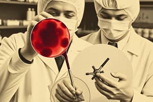
- Fill a capillary tube three-quarters full of anticoagulated blood.
- Place a drop of blood (approximately 2 mm diameter) on a slide, near the frosted edge.
- Hold the narrow edge of the slide while a second slide (spreader) is placed over the blood drop.
- Push the spreader slide forward, maintaining a smooth, light motion, to spread blood across the entire slide evenly. The blood should spread to almost the edges of the slide in the shape of a bullet with a feathered edge.
- Allow the blood film to air dry totally before staining to prevent RBC artifacts. Avoid blowing on the slide to dry it as this may introduce moisture causing artifacts.
- Label the slide with patient name, ID number, and date.
- Place the slide at a 30-degree angle against the drop of blood and move it back and forth creating the blood film, ensuring a smooth, even distribution.
Characteristics of Good Blood Smears
- Should be thick at drop end and thin at opposite end.
- Occupy the center of the slide, and leave margins free.
- Do not touch the edges of the slide (except at point of application).
Common Causes of Poor Blood Smears
- Blood drop too large or too small.
- Spreader slide pushed across slide in a jerky manner.
- Spreader slide does not cover the entire slide's edge when making the smear.
- Spreader slide not held at a 30° angle.
- Failure to push the spreader slide fully across the slide.
- Irregular spread, such as ridges and long tails.
- Dusty or chipped slides.
- Slides contaminated with fat, grease, or other substances.
- Cellular degenerative changes, caused by insufficient fixing time, methanol contamination, or insufficient drying time.
Fixing Blood Smears
- Fixing preserves cell morphology.
- Fix films quickly after drying to prevent water contact.
- Use methyl alcohol (methanol); ethyl alcohol (absolute alcohol) is also an acceptable alternative.
- Methylated spirit (95% ethanol) should not be used, as it contains water.
Staining Blood Smears
- Stain thin films using Leishman's stain.
- Flood the smear with stain.
- Stain for 1-5 minutes (optimum time varies based on experience).
- Mix in an equal amount of buffer solution and blow to form an eddy in the stain.
- Leave the mixture on the slide for 10-15 minutes.
- Wash away the stain by running water from the center of the slide to prevent residue.
- Stand the slide on end to air dry.
Studying That Suits You
Use AI to generate personalized quizzes and flashcards to suit your learning preferences.