Podcast
Questions and Answers
What is a characteristic feature of neutrophils in blood smears?
What is a characteristic feature of neutrophils in blood smears?
- Multilobulated nuclei with thick strands
- Multilobulated nuclei with very thin strands (correct)
- No visible nucleus
- Monolobulated nuclei with thin strands
What is another name for neutrophils?
What is another name for neutrophils?
- Multilobulated cells
- Polymorphonuclear leukocytes (correct)
- Mononuclear leukocytes
- Lymphoid cells
What happens to the nuclear shape of neutrophils?
What happens to the nuclear shape of neutrophils?
- It changes frequently (correct)
- It remains constant
- It becomes smaller
- It becomes larger
What is the term used to describe the connection between the lobules of neutrophils' nuclei?
What is the term used to describe the connection between the lobules of neutrophils' nuclei?
What is the characteristic of neutrophils that allows them to be identified in blood smears?
What is the characteristic of neutrophils that allows them to be identified in blood smears?
What is a distinctive feature of the cytoplasm of eosinophils?
What is a distinctive feature of the cytoplasm of eosinophils?
What is a characteristic of the nucleus of basophils?
What is a characteristic of the nucleus of basophils?
What is a function of monocytes?
What is a function of monocytes?
What is a characteristic of platelets?
What is a characteristic of platelets?
What is a feature of lymphocytes?
What is a feature of lymphocytes?
Which of the following lymphoid organs is classified as primary?
Which of the following lymphoid organs is classified as primary?
What is a common feature among lymph nodes, spleen, and mucosa associated lymphoid tissue?
What is a common feature among lymph nodes, spleen, and mucosa associated lymphoid tissue?
Bone marrow is classified as which type of lymphoid organ?
Bone marrow is classified as which type of lymphoid organ?
Which of the following is NOT a lymphoid organ?
Which of the following is NOT a lymphoid organ?
What is a common function among thymus, bone marrow, spleen, and lymph nodes?
What is a common function among thymus, bone marrow, spleen, and lymph nodes?
What is a major component of sections of red bone marrow?
What is a major component of sections of red bone marrow?
What type of cells can be distinguished at higher magnification in red bone marrow?
What type of cells can be distinguished at higher magnification in red bone marrow?
What is the location of hemopoietic cells in red bone marrow?
What is the location of hemopoietic cells in red bone marrow?
What is a challenge in identifying cells in routinely stained sections of red bone marrow?
What is a challenge in identifying cells in routinely stained sections of red bone marrow?
What is the function of blood-filled sinusoids in red bone marrow?
What is the function of blood-filled sinusoids in red bone marrow?
What is the function of the septa that extend into the thymus?
What is the function of the septa that extend into the thymus?
What type of cells are found in the cortex of the thymus?
What type of cells are found in the cortex of the thymus?
What is a characteristic feature of thymic epithelial cells (TECs)?
What is a characteristic feature of thymic epithelial cells (TECs)?
What is a characteristic of the medulla of the thymus?
What is a characteristic of the medulla of the thymus?
What is a type of structure found in the medulla of the thymus?
What is a type of structure found in the medulla of the thymus?
What is a characteristic of the young thymus?
What is a characteristic of the young thymus?
What is the primary function of the blood-thymus barrier?
What is the primary function of the blood-thymus barrier?
Which of the following components is NOT part of the blood capillary wall in the blood-thymus barrier?
Which of the following components is NOT part of the blood capillary wall in the blood-thymus barrier?
What is the function of the epithelioreticular cell layer in the blood-thymus barrier?
What is the function of the epithelioreticular cell layer in the blood-thymus barrier?
Which type of collagen is present in the perivascular connective tissue of the blood-thymus barrier?
Which type of collagen is present in the perivascular connective tissue of the blood-thymus barrier?
What is the purpose of the basal lamina of the epithelial reticular cells in the blood-thymus barrier?
What is the purpose of the basal lamina of the epithelial reticular cells in the blood-thymus barrier?
What is the approximate number of lymph nodes in the human body?
What is the approximate number of lymph nodes in the human body?
Which part of the lymph node contains the nodules?
Which part of the lymph node contains the nodules?
Where do lymphocytes and macrophages pass easily between the sinuses and the tissue of the lymph node?
Where do lymphocytes and macrophages pass easily between the sinuses and the tissue of the lymph node?
What is the role of macrophages in the lymph node sinuses?
What is the role of macrophages in the lymph node sinuses?
What is the order of lymph flow through a lymph node?
What is the order of lymph flow through a lymph node?
What is the primary function of a lymph node?
What is the primary function of a lymph node?
What type of vessels drain the capillary beds in a lymph node?
What type of vessels drain the capillary beds in a lymph node?
What is the function of the red pulp in a lymph node?
What is the function of the red pulp in a lymph node?
What is the role of high endothelial venules (HEVs) in a lymph node?
What is the role of high endothelial venules (HEVs) in a lymph node?
What is the significance of the hilus in a lymph node?
What is the significance of the hilus in a lymph node?
What is the entry point of blood into the spleen?
What is the entry point of blood into the spleen?
What is the function of penicillar arterioles?
What is the function of penicillar arterioles?
Which structure does blood percolate through after entering the splenic cords?
Which structure does blood percolate through after entering the splenic cords?
What is the final destination of blood after passing through the splenic sinuses?
What is the final destination of blood after passing through the splenic sinuses?
What surrounds the central arteries in the spleen?
What surrounds the central arteries in the spleen?
What is the primary component of the white pulp in the spleen?
What is the primary component of the white pulp in the spleen?
Where are B-cells primarily located in the spleen's white pulp?
Where are B-cells primarily located in the spleen's white pulp?
What is the function of the periarterial lymphatic sheath in the spleen's white pulp?
What is the function of the periarterial lymphatic sheath in the spleen's white pulp?
What is the primary function of the red pulp in the spleen?
What is the primary function of the red pulp in the spleen?
What is the location of T-cells in the spleen's white pulp?
What is the location of T-cells in the spleen's white pulp?
What occurs in the white pulp of the spleen as the body is exposed to antigens?
What occurs in the white pulp of the spleen as the body is exposed to antigens?
What is the significance of lymph nodules with germinal centers in the spleen?
What is the significance of lymph nodules with germinal centers in the spleen?
What type of immune response leads to the appearance of lymph nodules with germinal centers in the spleen?
What type of immune response leads to the appearance of lymph nodules with germinal centers in the spleen?
Where are lymph nodules with germinal centers located in the spleen?
Where are lymph nodules with germinal centers located in the spleen?
What is the significance of germinal centers in lymph nodules?
What is the significance of germinal centers in lymph nodules?
Flashcards are hidden until you start studying
Study Notes
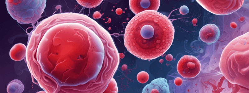
Neutrophils
- Identified in blood smears by their multilobulated nuclei
- Nuclei consist of lobules connected by very thin strands
- Also known as polymorphonuclear leukocytes (PMNs) or polymorphs due to their multilobulated nuclei
- Cells are dynamic, with nuclear shape changing frequently
Eosinophils
- Characterized by bilobed nuclei and abundant coarse cytoplasmic granules
- Cytoplasm contains brightly eosinophilic specific granules and some azurophilic granules
Basophils
- Distinguished by large, strongly basophilic specific granules
- Nucleus is often obscured by granules and has 2-3 irregular lobes
Lymphocytes
- Have a single nucleus that occupies most of the cell
- Thin rim of basophilic cytoplasm surrounds the nucleus
- Contain azurophilic granules
Monocytes
- Large agranulocytes that mature into macrophages and other cells of the mononuclear phagocyte system
- Nuclei are eccentric, indented, kidney-shaped, or U-shaped
Platelets
- Often found in aggregates
- Consist of a lightly stained hyalomere region surrounding a darker central granulomere containing membrane-enclosed granules
Lymphoid Organs
- Lymphoid organs are classified into two categories: primary and secondary lymphoid organs.
Primary Lymphoid Organs
- Thymus is a primary lymphoid organ.
- Bone marrow is a primary lymphoid organ.
Secondary Lymphoid Organs/Tissue
- Spleen is a secondary lymphoid organ.
- Lymph nodes are secondary lymphoid organs.
- Lymphoid tissue is also classified as a secondary lymphoid organ.
Components of Lymphoid Organs
- Lymphoid organs include lymphocytes.
- Thymus is a component of lymphoid organs.
- Mucosa associated lymphoid tissue (MALT) is a component of lymphoid organs.
- Lymph nodes are components of lymphoid organs.
- Spleen is a component of lymphoid organs.
Bone Marrow Structure
- Red bone marrow consists of trabeculae of cancellous bone, adipocytes, and blood-filled sinusoids.
- Sinusoids are located between hemopoietic cords or islands of developing blood cells.
Component Cells
- Sinusoidal endothelial cells have flattened nuclei.
- Hemopoietic cells are densely packed in the cords between sinusoids and adipocytes.
- Stromal cells and specific cells of the hemopoietic lineages are difficult to identify with certainty in routinely stained sections of marrow.
Thymus Structure
- The thymus is composed of vascularized connective tissue with a capsule that extends septa into the parenchyma, dividing the organ into many incompletely separated lobules.
- The thymus consists of two main parts: the cortex and the medulla.
Cortex of Thymus
- The cortex is composed of newly arrived lymphoblasts, macrophages, and thymic epithelial cells (TECs).
- TECs have characteristics of both epithelial and reticular cells and are morphologically and functionally diverse.
- The cortex is divided into three layers.
Medulla of Thymus
- The medulla contains fewer and larger, mature lymphocytes.
- The medulla has three related types of medullary TECs that form:
- A second layer of the boundary between the cortex and medulla.
- A cytoreticulum that supports less densely packed T lymphocytes, dendritic cells, and macrophages, and expresses many specialized proteins specific to cells of other organs.
- Large aggregates of TECs, called Hassall corpuscles, sometimes arranged concentrically.
Characteristics of Young Thymus
- The young thymus is surrounded by a connective tissue (CT) capsule.
- The cortex has a large number of lymphocytes, while the medulla has fewer.
- The thymus does not have germinal centers.
Blood-Thymus Barrier
- The education of T-cells requires a highly controlled environment where antigens are presented exclusively by epithelial reticular cells.
- The Blood-Thymus Barrier is crucial to ensure the absence of other cells and free antigens in this environment.
Components of the Blood-Thymus Barrier
- The blood capillary wall consists of: • Endothelial cells • Endothelial cell basal laminae • Pericytes
Perivascular Connective Tissue
- Composed of: • Type III collagen • Macrophages
Epithelioreticular Cell Layer
- Consists of: • Basal lamina of the epithelial reticular cells (type I ERCs) • Epithelial reticular cells
Lymph Nodes
- Bean-shaped and encapsulated structures
- Human body has approximately 400-450 lymph nodes
- Each lymph node has three major regions:
- Outer cortex containing nodules
- Deeper paracortex region lacking nodules
- Medulla with prominent draining sinusoids adjacent to the hilum
Lymphatic Circulation through a Lymph Node
- Lymph nodes filter lymph
- Lymph flow:
- Afferent lymphatic vessels → Subcapsular Sinus
- Subcapsular Sinus → Trabecular sinuses
- Trabecular sinuses → Medullary sinuses
- Lymphocytes and macrophages can easily pass between sinuses and lymph node tissue
- Macrophages in sinuses monitor fluids, phagocytose antigenic material, and present it to T- and B-cells
Blood Circulation Through a Lymph Node
- Blood circulation into a lymph node begins with the entry of blood through an artery at the hilus.
- The hilar artery branches into arterioles, which then feed into capillary beds.
- The capillary beds are drained by high endothelial venules (HEVs), which are specialized sites where lymphocytes can leave the bloodstream and enter the lymph node tissue.
Functions of the Spleen
- The spleen filters the blood.
- It destroys old red blood cells.
- It serves as an immune organ.
- The spleen is divided into two main parts: the Red Pulp and the White Pulp.
Red Pulp
- Responsible for RBC/hemoglobin recycling.
White Pulp
- Responsible for immune functions.
Splenic Circulation
- Blood enters the spleen via the splenic artery at the hilus
- The splenic artery branches into trabecular arteries, which are surrounded by connective tissue trabeculae
- Trabecular arteries give off central arteries, which leave the trabeculae and enter the spleen's substance, covered by a peri-arterial lymphatic sheath
- Central arteries branch into penicillar arterioles, which pierce the lymphatic sheath and spill into splenic cords
- Blood percolates through splenic cords and across the walls of splenic sinuses
- Splenic sinuses drain into pulp veins
- Pulp veins drain into trabecular veins, which then drain into the splenic vein at the hilus
Blood and Lymphoid Tissue
- Neutrophils: • Identified by their multilobulated nuclei, with lobules held together by very thin strands • Also called polymorphonuclear leukocytes, PMNs, or polymorphs • Dynamic cells with frequently changing nuclear shape
- Eosinophils: • Bilobed nuclei • Abundant coarse cytoplasmic granules • Cytoplasm filled with brightly eosinophilic specific granules and azurophilic granules
- Basophils: • Large, strongly basophilic specific granules • Granules usually obstruct the appearance of the nucleus, which has two or three irregular lobes
- Lymphocytes: • Single nucleus occupying most of the cell • Thin rim of cytoplasm with fine azurophilic granules
- Monocytes: • Large agranulocytes • Precursors to macrophages and other cells of the mononuclear phagocyte system • Eccentric nuclei, indented, kidney-shaped, or U-shaped
- Platelets: • Often found in aggregates • Individual platelets have a lightly stained hyalomere region surrounding a more darkly stained central granulomere containing membrane-enclosed granules
The Immune System and Lymphoid Organs
- Lymphoid organs: • Classified as primary and secondary (effector) lymphoid organs/tissues • Include thymus, bone marrow, spleen, lymph nodes, mucosa-associated lymphoid tissue (MALT)
- Primary lymphoid organs: • Thymus • Bone marrow
- Secondary (effector) lymphoid organs/tissues: • Spleen • Lymph nodes • Mucosa-associated lymphoid tissue (MALT)
Bone Marrow
- Sections of red bone marrow: • Trabeculae of cancellous bone • Adipocytes (A) • Blood-filled sinusoids between hemopoietic cords or islands of developing blood cells
- At higher magnification: • Flattened nuclei of sinusoidal endothelial cells can be distinguished • Variety of densely packed hemopoietic cells in the cords between the sinusoids and adipocytes
Thymus
- Vascularized connective tissue capsule • Extends septa into paranchyme, dividing the organ into many incompletely lobules
- Cortex: • Composed of newly arrived lymphoblasts, macrophages, and thymic epithelial cells (TECs) • TECs have both epithelial and reticular cell characteristics • Divided into three groups (layers)
- Medulla: • Contains fewer and larger, mature lymphocytes • Three related types of medullary TECs form a boundary layer, cytoreticulum, and Hassall corpuscles
Blood-Thymus Barrier
- Education of T-cells must occur in a controlled environment where antigens are only presented by epithelial reticular cells
- The blood-thymus barrier ensures that no other cells or free antigens are present
- Consists of: • Blood capillary wall (endothelial cells, endothelial cell basal laminae, pericytes) • Perivascular connective tissue (type III collagen, macrophages) • Epithelioreticular cell layer (basal lamina of the epithelial reticular cells, epithelial reticular cells)
Lymph Nodes
- Bean-shaped, encapsulated structures
• Approximately 400-450 lymph nodes
• Three major regions within each lymph node:
- Outer cortex containing nodules
- Deeper extension of cortex called the paracortex, which lacks nodules
- Medulla with prominent draining sinusoids adjacent to the hilum
Lymphatic Circulation through a Lymph Node
- Lymph nodes filter lymph • Afferent lymphatic vessels drain lymph into the Subcapsular Sinus • Lymph then passes to the Trabecular sinuses • From there, the lymph goes to the Medullary sinuses • Lymphocytes and macrophages pass easily between these sinuses and the tissue of the lymph node
Blood Circulation through a Lymph Node
- Blood enters through an artery at the hilus • Arterioles branch from hilar artery to feed into capillary beds • Capillary beds are drained by high endothelial venules (HEVs) • HEVs drain into hilar vein • HEVs are sites where lymphocytes can leave the bloodstream to enter the lymph node tissue bed
Spleen
- Functions: • Filters the blood • Destroys old red blood cells • Serves as an immune organ • Divided into Red Pulp (RBC/hemoglobin recycling) and White Pulp (responsible for immune functions)
Splenic Circulation
- Blood enters via splenic artery at hilus • Splenic artery branches into trabecular arteries (which travel within connective tissue trabeculae) • Trabecular arteries give off branches known as central arteries which leave the trabecula and enter the substance of the spleen (covered by a peri-arterial lymphatic sheath) • Central arteries branch into penicillar arterioles that piece through the lymphatic sheath and spill into splenic cords • Blood percolates through splenic cords and across walls of splenic sinuses • Splenic sinuses drain into pulp veins • Pulp veins drain into trabecular veins • Trabecular veins drain into splenic vein at the hilus
Studying That Suits You
Use AI to generate personalized quizzes and flashcards to suit your learning preferences.


