Podcast
Questions and Answers
What is the most common staining technique used in the UK for blood films?
What is the most common staining technique used in the UK for blood films?
- Hematoxylin and eosin
- May-Grunwald Giemsa (correct)
- Crystal violet
- Wright's stain
What is the proper pH for the buffered water used in the staining protocol?
What is the proper pH for the buffered water used in the staining protocol?
- 5.5
- 6.8 (correct)
- 7.4
- 7.0
Which of the following is NOT a morphological feature that can be observed in red blood cells?
Which of the following is NOT a morphological feature that can be observed in red blood cells?
- Hypochromic cells
- Anisocytosis
- Polychromasia
- Eosinophilia (correct)
What is the appropriate myeloid to erythroid ratio in normal bone marrow?
What is the appropriate myeloid to erythroid ratio in normal bone marrow?
When examining a blood smear, what indicates that the red blood cells are in the right area for detail observation?
When examining a blood smear, what indicates that the red blood cells are in the right area for detail observation?
What can occur if a blood smear is too thin during examination?
What can occur if a blood smear is too thin during examination?
What is the typical duration for fixing a blood sample in methanol for staining?
What is the typical duration for fixing a blood sample in methanol for staining?
Which cell type is NOT found in normal bone marrow?
Which cell type is NOT found in normal bone marrow?
What is the purpose of fixing a blood film in methanol?
What is the purpose of fixing a blood film in methanol?
What characteristic indicates that a blood smear is too thick?
What characteristic indicates that a blood smear is too thick?
What should be the appropriate range for neutrophils in normal bone marrow?
What should be the appropriate range for neutrophils in normal bone marrow?
Which of the following describes polychromasia in red blood cells?
Which of the following describes polychromasia in red blood cells?
In the examination of a blood smear, what does a spherocytic appearance indicate?
In the examination of a blood smear, what does a spherocytic appearance indicate?
What is the significance of the myeloid to erythroid ratio in bone marrow examination?
What is the significance of the myeloid to erythroid ratio in bone marrow examination?
How long should a bone marrow sample be fixed in methanol?
How long should a bone marrow sample be fixed in methanol?
What is the appearance of the red blood cells that indicates they are in the right area for detailed examination?
What is the appearance of the red blood cells that indicates they are in the right area for detailed examination?
Flashcards
Blood film preparation
Blood film preparation
A thin blood sample spread on a slide for microscopic examination.
May-Grunwald-Giemsa stain
May-Grunwald-Giemsa stain
Common blood stain used in the UK for visualizing blood cells.
Blood smear examination
Blood smear examination
Microscopic view of a blood film to assess cell types and morphology.
Ideal blood smear area
Ideal blood smear area
Signup and view all the flashcards
Bone marrow
Bone marrow
Signup and view all the flashcards
Myeloid to erythroid ratio
Myeloid to erythroid ratio
Signup and view all the flashcards
Normal bone marrow
Normal bone marrow
Signup and view all the flashcards
Blood cell types (bone marrow)
Blood cell types (bone marrow)
Signup and view all the flashcards
Hypochromic cells
Hypochromic cells
Signup and view all the flashcards
Polychromasia
Polychromasia
Signup and view all the flashcards
Anisocytosis
Anisocytosis
Signup and view all the flashcards
Lymphocytes
Lymphocytes
Signup and view all the flashcards
Neutrophils
Neutrophils
Signup and view all the flashcards
Monocytes
Monocytes
Signup and view all the flashcards
Eosinophils
Eosinophils
Signup and view all the flashcards
Basophils
Basophils
Signup and view all the flashcards
Study Notes
Blood and Bone Marrow
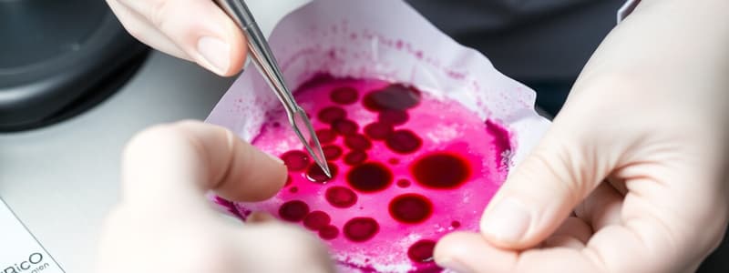
- Blood smears are prepared using a drop of blood, air-dried, and fixed with methanol.
- The smears are then stained using May-Grunwald Giemsa stain to visualize cell features.
Blood Film Preparation
- Different quality blood films are shown, from well-made films to films that are too thick, too thin, or have inconsistent pressure.
- Proper technique is crucial for clear cell visualization.
- Inconsistent pressure and uneven spreading during blood film preparation can lead to problems like elongated tails in the film.
Blood Film Staining
- Fixation in methanol, crucial for cells, for 5 minutes is standard procedure.
- Bone marrow fixation requires 15-20 minutes.
- May-Grunwald is diluted 50:50 in buffered water (pH 6.8)
- Giemsa stain is diluted 1:10 in buffered water (pH 6.8)
- A final step is rinsing in buffered water (pH 6.8).
Blood Smear Examination
- Oil immersion microscopy is used to examine the blood smear.
- The appropriate area shows red blood cells just touching, enabling detailed visualization.
- The smear is examined systematically from side-to-side, or from the thick to thin area.
Blood Smear Quality Considerations
- Too thick films obstruct cell detail.
- Too thin films make RBCs appear spherocytic.
Red Cell Morphology
- Important parameters for red cells include hypochromia and polychromasia, and anisocytosis.
White Cell Morphology
- Different types of white blood cells (WBCs), like lymphocytes, neutrophils, monocytes, eosinophils, basophils, metamyelocytes, myelocytes, and promyelocytes, are examined for their morphology.
- Slides of each WBC type are presented.
Bone Marrow
- Normal bone marrow has a myeloid to erythroid ratio of 3:1 (with variations from 2:1 to 4:1).
- Cell types present are both mature and immature.
- The cellular and fatty components are distributed evenly.
Bone Marrow - Trephine Biopsies
- Fixation using 10% formal saline is performed up to 48 hours.
- Decalcification methods depend on the laboratory process.
- EDTA is used as one type of preservation method.
- Staining techniques are dependent on the laboratory procedure
- H&E, iron (Perls' reaction), immunohistochemistry, and reticulin (silver impregnation) stains are examples.
Required Samples
- A full blood count (FBC) is collected at the same time.
- Trephine biopsies are not always necessary.
- Bone marrow aspirates are sometimes required.
- Multiple slides are typically made of the samples depending on the need for various stains and tests. (e.g., MGG (May-Grunwald-Giemsa) and cytochemistry)
- An EDTA blood sample is crucial for cytogenetic analysis and usually is obtained at 2 x 5 ml.
Normal Bone Marrow - Quantitative Data
- Cell counts for different bone marrow types are shown in a table.
Studying That Suits You
Use AI to generate personalized quizzes and flashcards to suit your learning preferences.