Podcast
Questions and Answers
What is the primary function of osteoblasts in bone tissue?
What is the primary function of osteoblasts in bone tissue?
- To form new bone tissue (correct)
- To provide structural support to ligaments
- To maintain the bone matrix
- To break down bone tissue
What distinguishes cancellous bone from compact bone?
What distinguishes cancellous bone from compact bone?
- Cancellous bone contains a dense arrangement of osteons.
- Cancellous bone has a porous structure that facilitates lightweight support. (correct)
- Cancellous bone is less vascularized than compact bone.
- Cancellous bone does not contain bone cells.
Which cell type is primarily responsible for the resorption of bone tissue?
Which cell type is primarily responsible for the resorption of bone tissue?
- Osteoprogenitor cells
- Osteoblasts
- Osteoclasts (correct)
- Osteocytes
What is a characteristic feature of the Harversian system in compact bone?
What is a characteristic feature of the Harversian system in compact bone?
Which process describes the development of bone from a fibrous membrane?
Which process describes the development of bone from a fibrous membrane?
What is the primary function of cancellous bone?
What is the primary function of cancellous bone?
Which of the following statements about compact bone is true?
Which of the following statements about compact bone is true?
What connects the lacunae in compact bone?
What connects the lacunae in compact bone?
What is the role of Volkmann's canals in compact bone?
What is the role of Volkmann's canals in compact bone?
How are nutrients supplied to cancellous bone?
How are nutrients supplied to cancellous bone?
Which structure of compact bone acts as a conduit for blood supply?
Which structure of compact bone acts as a conduit for blood supply?
What is the composition of concentric lamellae in compact bone?
What is the composition of concentric lamellae in compact bone?
Where do bones begin their development?
Where do bones begin their development?
What is the term used for the structure that forms around the fracture site during bone repair?
What is the term used for the structure that forms around the fracture site during bone repair?
Which stage follows fracture hematoma formation in the bone repair process?
Which stage follows fracture hematoma formation in the bone repair process?
How often does the distal end of the femur undergo repair?
How often does the distal end of the femur undergo repair?
What will likely happen to a limb that is in a cast in terms of bone mass?
What will likely happen to a limb that is in a cast in terms of bone mass?
Which of the following best describes the complete process of bone remodeling?
Which of the following best describes the complete process of bone remodeling?
What factors might affect bone growth and repair?
What factors might affect bone growth and repair?
What is a factor that could prolong healing time in bone repair?
What is a factor that could prolong healing time in bone repair?
Which part of the bone is primarily formed by secondary ossification centers?
Which part of the bone is primarily formed by secondary ossification centers?
What is the primary function of the periosteum?
What is the primary function of the periosteum?
Which part of the long bone is primarily hollow and forms the central structure?
Which part of the long bone is primarily hollow and forms the central structure?
What is the role of the endosteum?
What is the role of the endosteum?
Where are the secondary ossification centers typically found?
Where are the secondary ossification centers typically found?
What is the main characteristic of the metaphysis during bone growth?
What is the main characteristic of the metaphysis during bone growth?
What is primarily found inside the epiphysis of long bones?
What is primarily found inside the epiphysis of long bones?
What substance mainly composes the epiphyseal plate?
What substance mainly composes the epiphyseal plate?
What is a key function of osteoblasts found in the endosteum?
What is a key function of osteoblasts found in the endosteum?
What occurs during the fracture hematoma formation stage?
What occurs during the fracture hematoma formation stage?
What type of tissue is primarily formed during the fibrocartilaginous callus formation?
What type of tissue is primarily formed during the fibrocartilaginous callus formation?
Which cells specifically invade the callus during the formation of the bony callus?
Which cells specifically invade the callus during the formation of the bony callus?
What is the primary role of phagocytic cells in the fracture healing process?
What is the primary role of phagocytic cells in the fracture healing process?
How long does it typically take for the hematoma to form at a fracture site?
How long does it typically take for the hematoma to form at a fracture site?
What characterizes the fibrocartilaginous callus?
What characterizes the fibrocartilaginous callus?
Which stage follows the fibrocartilaginous callus formation in bone repair?
Which stage follows the fibrocartilaginous callus formation in bone repair?
What happens to the fibrocartilaginous callus over time?
What happens to the fibrocartilaginous callus over time?
What is the primary function of osteoclasts during the bone remodeling process?
What is the primary function of osteoclasts during the bone remodeling process?
Which type of bone replaces spongy bone or the medullary cavity during bone remodeling?
Which type of bone replaces spongy bone or the medullary cavity during bone remodeling?
What does synovial fluid primarily provide to the cartilage in a synovial joint?
What does synovial fluid primarily provide to the cartilage in a synovial joint?
What role do bursae serve in relation to joints?
What role do bursae serve in relation to joints?
Which structure covers the ends of bones in a synovial joint?
Which structure covers the ends of bones in a synovial joint?
What is included in synovial fluid?
What is included in synovial fluid?
How are ligaments important in joints?
How are ligaments important in joints?
What leads to the remodeling of bone architecture to correct alignment?
What leads to the remodeling of bone architecture to correct alignment?
Flashcards
Compact Bone Histology
Compact Bone Histology
Compact bone is dense and strong. It's structure is organized in units called osteons (Haversian systems).
Cancellous Bone
Cancellous Bone
Cancellous bone is spongy and lightweight. It has a lattice-like structure.
Bone Composition & Function
Bone Composition & Function
Bone is mostly made up of collagen and calcium phosphate. Its functions include support, protection, movement, and blood cell production.
Osteoprogenitor Cells
Osteoprogenitor Cells
Signup and view all the flashcards
Osteoblasts
Osteoblasts
Signup and view all the flashcards
Osteocytes
Osteocytes
Signup and view all the flashcards
Osteoclasts
Osteoclasts
Signup and view all the flashcards
Bone Growth
Bone Growth
Signup and view all the flashcards
Intramembranous Ossification
Intramembranous Ossification
Signup and view all the flashcards
Endochondral Ossification
Endochondral Ossification
Signup and view all the flashcards
Axial Skeleton
Axial Skeleton
Signup and view all the flashcards
Appendicular Skeleton
Appendicular Skeleton
Signup and view all the flashcards
Bone's functions
Bone's functions
Signup and view all the flashcards
Lamellar bone
Lamellar bone
Signup and view all the flashcards
Cancellous bone
Cancellous bone
Signup and view all the flashcards
Haversian system/Osteon
Haversian system/Osteon
Signup and view all the flashcards
Cancellous bone structure
Cancellous bone structure
Signup and view all the flashcards
Compact bone structure
Compact bone structure
Signup and view all the flashcards
Concentric lamellae
Concentric lamellae
Signup and view all the flashcards
Lacunae
Lacunae
Signup and view all the flashcards
Canalliculi
Canalliculi
Signup and view all the flashcards
Volkmann's canals
Volkmann's canals
Signup and view all the flashcards
Trabeculae
Trabeculae
Signup and view all the flashcards
Primary Ossification Centre
Primary Ossification Centre
Signup and view all the flashcards
Secondary Ossification Centre
Secondary Ossification Centre
Signup and view all the flashcards
Periosteum
Periosteum
Signup and view all the flashcards
Endosteum
Endosteum
Signup and view all the flashcards
Diaphysis
Diaphysis
Signup and view all the flashcards
Medullary Cavity
Medullary Cavity
Signup and view all the flashcards
Metaphysis
Metaphysis
Signup and view all the flashcards
Epiphysis
Epiphysis
Signup and view all the flashcards
Epiphyseal plate
Epiphyseal plate
Signup and view all the flashcards
Primary ossification center
Primary ossification center
Signup and view all the flashcards
Fracture Hematoma Formation
Fracture Hematoma Formation
Signup and view all the flashcards
Secondary ossification centers
Secondary ossification centers
Signup and view all the flashcards
Diaphysis
Diaphysis
Signup and view all the flashcards
Fibrocartilaginous Callus Formation
Fibrocartilaginous Callus Formation
Signup and view all the flashcards
Epiphysis
Epiphysis
Signup and view all the flashcards
Bony Callus Formation
Bony Callus Formation
Signup and view all the flashcards
Articular cartilage
Articular cartilage
Signup and view all the flashcards
Hematoma
Hematoma
Signup and view all the flashcards
Epiphyseal plate
Epiphyseal plate
Signup and view all the flashcards
Granulation tissue
Granulation tissue
Signup and view all the flashcards
Bone Repair Stages
Bone Repair Stages
Signup and view all the flashcards
Epiphyseal closure
Epiphyseal closure
Signup and view all the flashcards
Bone remodeling
Bone remodeling
Signup and view all the flashcards
Bone repair stages
Bone repair stages
Signup and view all the flashcards
Factors affecting bone growth
Factors affecting bone growth
Signup and view all the flashcards
Bone Remodelling
Bone Remodelling
Signup and view all the flashcards
Synovial Joint
Synovial Joint
Signup and view all the flashcards
Joint Capsule
Joint Capsule
Signup and view all the flashcards
Hyaline Cartilage
Hyaline Cartilage
Signup and view all the flashcards
Synovial Membrane
Synovial Membrane
Signup and view all the flashcards
Synovial Fluid
Synovial Fluid
Signup and view all the flashcards
Bursae
Bursae
Signup and view all the flashcards
Ligaments
Ligaments
Signup and view all the flashcards
Tendons
Tendons
Signup and view all the flashcards
Study Notes
Basic Osteology Lecture Notes
- The lecture covers compact and cancellous bone histology, bone composition, bone cell types (osteoprogenitor cells, osteoblasts, osteocytes, osteoclasts), bone growth processes (intramembranous and endochondral ossification), factors affecting bone growth, and types of skeletons (endoskeleton, exoskeleton).
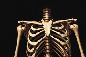
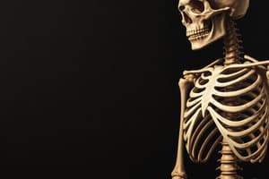
- The human skeleton is comprised of approximately 206 bones, divided into the axial skeleton (skull/face, vertebral column, and thorax) and the appendicular skeleton (shoulder girdle, upper extremities, pelvic girdle, and lower extremities).
- Bones (skull/cranium) are categorized into 8 cranial bones, 14 facial bones, 1 hyoid, and 6 ossicles.
- The vertebral column includes 7 cervical, 12 thoracic, 5 lumbar vertebrae, 1 sacrum, and 1 coccyx bone.
- The thorax contains 25 bones (1 sternum, 24 ribs).
- The shoulder girdle consists of 4 bones (2 clavicles, 2 scapulae).
- The upper extremities consist of 60 bones (2 humerus, 2 radius, 2 ulna, 16 carpals, 10 metacarpals, 28 phalanges).
- The pelvic girdle consists of 2 innominate bones (each comprising the ilium, ischium, and pubis).
- The lower extremities include 60 bones (2 femurs, 2 fibulas, 2 tibias, 2 patellas, 14 tarsals, 10 metatarsals, and 28 phalanges).
- Bone types include long bones, short bones, flat bones, irregular bones, and sesamoid bones.
- Bone microstructure involves: Woven bone – fragile in development and repair; Lamellar bone- compact/cortical covering spongy bone, a stronger structure; Cancellous or spongy- trabecular bone found in the ends of long bones, provides support.
- Compact bone contains Haversian systems/osteons (structural units) arranged regularly; the structures consist of a Haversian canal, lamellae (calcified rings), lacunae (spaces for osteocytes), canaliculi (tiny canals radiating out connecting lacunae), and Volkmann’s canals (channels running at 90° to the central canal connecting blood vessels to the periosteum).
- Cancellous bone is unorganized extensions of bone tissue surrounding bone marrow spaces.
- Bone growth and development includes primary and secondary ossification (the process by which bones form from embryonic tissue). Primary ossification begins in the womb, usually around the shaft of the long bone. Secondary ossification happens later in different times of the child’s development. Bone development stages include cartilage template (surrounded by perichondrium), bony collar formation, primary centre formation, blood vessels entering, marrow cavity formation, collar thickening and lengthening forming the diaphysis, and secondary ossification centres forming the epiphysis.
- The epiphyseal growth plate (hyaline cartilage) separates the epiphysis from the rest of the bone & bone lengthens.
- The metaphysis is the region proximal to the growth plate where ossification occurs
- The periosteum (fibrous membrane covering the bone) and the endosteum (inner membrane facing the medullary cavity) are key parts of bone structure. The periosteum protects and nourishes the bone; endosteum has blood supply & contains osteoblasts & osteoclasts.
- Bony repair involves fracture hematoma formation, fibrocartilaginous callus formation, formation of bony callus, and bone remodelling.
- Synovial joints have a joint cavity containing synovial fluid for nourishing and lubricating cartilage. Pouches of fluid (bursae) cushion the joint. The capsule and membranes and the joint cavity are lined by synovial membrane, cartilage, and fluid.
Studying That Suits You
Use AI to generate personalized quizzes and flashcards to suit your learning preferences.