Podcast
Questions and Answers
Which nerve innervates all muscles in the posterior compartment of the forearm?
Which nerve innervates all muscles in the posterior compartment of the forearm?
- Radial nerve (correct)
- Ulnar nerve
- Median nerve
- Musculocutaneous nerve
How many muscles are located in the superficial layer of the posterior forearm?
How many muscles are located in the superficial layer of the posterior forearm?
- 7 (correct)
- 8
- 6
- 5
With which action are muscles of the posterior compartment of the forearm primarily associated?
With which action are muscles of the posterior compartment of the forearm primarily associated?
- Extension of the wrist joint (correct)
- Adduction of the shoulder
- Flexion of the elbow joint
- Pronation of the forearm
Which of the following muscles does not extend as tendons into the hand?
Which of the following muscles does not extend as tendons into the hand?
From the lateral to medial side, how are extensors typically arranged?
From the lateral to medial side, how are extensors typically arranged?
What is the primary action of the brachioradialis muscle?
What is the primary action of the brachioradialis muscle?
Which nerve supplies the extensor carpi radialis longus muscle?
Which nerve supplies the extensor carpi radialis longus muscle?
What is the insertion point of the extensor carpi ulnaris muscle?
What is the insertion point of the extensor carpi ulnaris muscle?
Through which structure does the posterior interosseous nerve emerge to lie between the superficial and deep layers of the posterior compartment?
Through which structure does the posterior interosseous nerve emerge to lie between the superficial and deep layers of the posterior compartment?
What is the action of the extensor digitorum muscle on the wrist joint?
What is the action of the extensor digitorum muscle on the wrist joint?
The superficial palmar arch is formed by the terminal end of which artery?
The superficial palmar arch is formed by the terminal end of which artery?
In the context of the extensor expansion, what is the destiny of the intermediate slip?
In the context of the extensor expansion, what is the destiny of the intermediate slip?
Which of the following are boundaries of the anatomical snuffbox?
Which of the following are boundaries of the anatomical snuffbox?
What is the function of the extensor retinaculum at the wrist?
What is the function of the extensor retinaculum at the wrist?
Which artery gives rise to the posterior interosseous artery?
Which artery gives rise to the posterior interosseous artery?
What action does the anconeus muscle perform?
What action does the anconeus muscle perform?
After originating from the common interosseous artery, where does the posterior interosseous artery pass?
After originating from the common interosseous artery, where does the posterior interosseous artery pass?
Which nerve supplies the extensor indicis muscle?
Which nerve supplies the extensor indicis muscle?
At what location does posterior interosseous artery terminates?
At what location does posterior interosseous artery terminates?
What is the origin of the deep (ulnar) layer of the supinator muscle?
What is the origin of the deep (ulnar) layer of the supinator muscle?
What is characteristic of the anterior interosseous artery in relation to the posterior compartment?
What is characteristic of the anterior interosseous artery in relation to the posterior compartment?
Which specific muscles are supplied by the radial nerve before it divides in the cubital fossa?
Which specific muscles are supplied by the radial nerve before it divides in the cubital fossa?
What is the functional consequence of the posterior interosseous nerve passing between the two layers of the supinator muscle?
What is the functional consequence of the posterior interosseous nerve passing between the two layers of the supinator muscle?
What are the attachments described in the text for the extensor retinaculum?
What are the attachments described in the text for the extensor retinaculum?
Which structure does not pass through the compartments deep to the extensor retinaculum?
Which structure does not pass through the compartments deep to the extensor retinaculum?
What structures pass superficial to the extensor retinaculum?
What structures pass superficial to the extensor retinaculum?
What is the origin of the extensor carpi radialis brevis?
What is the origin of the extensor carpi radialis brevis?
Which of the following muscles is innervated by the radial nerve before its division into superficial and deep branches?
Which of the following muscles is innervated by the radial nerve before its division into superficial and deep branches?
Name the muscles among the superficial group of posterior forearm muscles that are part of the posterior group?
Name the muscles among the superficial group of posterior forearm muscles that are part of the posterior group?
What is the function of the anterior interosseous artery in the posterior compartment?
What is the function of the anterior interosseous artery in the posterior compartment?
If the posterior interosseous nerve is compressed within the supinator muscle, which movement would be LEAST affected?
If the posterior interosseous nerve is compressed within the supinator muscle, which movement would be LEAST affected?
A patient exhibits weakened wrist extension and impaired finger abduction, but maintains normal supination strength. Where is the most probable site of nerve lesion?
A patient exhibits weakened wrist extension and impaired finger abduction, but maintains normal supination strength. Where is the most probable site of nerve lesion?
A surgeon is performing a procedure to repair the ulnar collateral ligament of the elbow. To minimize the risk of iatrogenic injury to the structures of the posterior compartment of the forearm, what is the MOST important anatomical consideration?
A surgeon is performing a procedure to repair the ulnar collateral ligament of the elbow. To minimize the risk of iatrogenic injury to the structures of the posterior compartment of the forearm, what is the MOST important anatomical consideration?
How many compartments is the extensor retinaculum divided into, and how many septa facilitate this division?
How many compartments is the extensor retinaculum divided into, and how many septa facilitate this division?
Which best describes the difference between superficial and deep muscles of the posterior forearm?
Which best describes the difference between superficial and deep muscles of the posterior forearm?
Which of the following is NOT a muscle of the superficial layer of the posterior compartment of the forearm?
Which of the following is NOT a muscle of the superficial layer of the posterior compartment of the forearm?
What is the general action of the deep muscles of the posterior compartment?
What is the general action of the deep muscles of the posterior compartment?
What muscles are associated with the lateral group of superficial extensors?
What muscles are associated with the lateral group of superficial extensors?
Which of the following choices best describes the innervation by the posterior interosseous nerve?
Which of the following choices best describes the innervation by the posterior interosseous nerve?
Flashcards
Forearm Muscle Layers
Forearm Muscle Layers
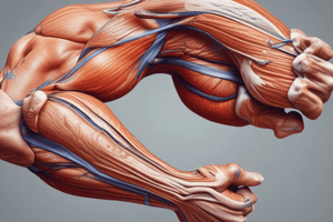
The muscles in the posterior compartment of the forearm are differentiated into two layers: superficial (7 muscles) and deep (5 muscles).
Functions of Forearm Muscles
Functions of Forearm Muscles
Muscles associated with wrist joint movement, finger/thumb extension, and supination.
Innervation of Forearm Muscles
Innervation of Forearm Muscles
Innervated by the radial nerve, superficial muscles cross the elbow joint, but deep muscles don't.
Superficial Forearm Muscles
Superficial Forearm Muscles
Signup and view all the flashcards
Origin of Superficial Forearm Muscles
Origin of Superficial Forearm Muscles
Signup and view all the flashcards
Lateral Group of Superficial Extensors
Lateral Group of Superficial Extensors
Signup and view all the flashcards
Posterior Group of Superficial Extensors
Posterior Group of Superficial Extensors
Signup and view all the flashcards
Brachioradialis Origin & Insertion
Brachioradialis Origin & Insertion
Signup and view all the flashcards
Brachioradialis Nerve & Action
Brachioradialis Nerve & Action
Signup and view all the flashcards
ECRL Origin & Insertion
ECRL Origin & Insertion
Signup and view all the flashcards
ECRL Action
ECRL Action
Signup and view all the flashcards
Extensor Carpi Radialis Brevis
Extensor Carpi Radialis Brevis
Signup and view all the flashcards
Extensor Digitorum Muscle
Extensor Digitorum Muscle
Signup and view all the flashcards
Extensor Digitorum Muscle
Extensor Digitorum Muscle
Signup and view all the flashcards
Extensor carpi ulnaris
Extensor carpi ulnaris
Signup and view all the flashcards
Extensor carpi ulnaris
Extensor carpi ulnaris
Signup and view all the flashcards
Anconeus Muscle
Anconeus Muscle
Signup and view all the flashcards
Deep Group Muscles
Deep Group Muscles
Signup and view all the flashcards
Supinator Muscle
Supinator Muscle
Signup and view all the flashcards
Supinator Action
Supinator Action
Signup and view all the flashcards
Abductor Pollicis Longus
Abductor Pollicis Longus
Signup and view all the flashcards
Tendon of Extensor Pollicis Brevis
Tendon of Extensor Pollicis Brevis
Signup and view all the flashcards
Extensor pollicis longus
Extensor pollicis longus
Signup and view all the flashcards
Extensor indicis muscle
Extensor indicis muscle
Signup and view all the flashcards
Radial Nerve
Radial Nerve
Signup and view all the flashcards
Deep Branch Radial
Deep Branch Radial
Signup and view all the flashcards
interosseous artery
interosseous artery
Signup and view all the flashcards
Extensor Retinaculum
Extensor Retinaculum
Signup and view all the flashcards
Reduces Friction
Reduces Friction
Signup and view all the flashcards
Study Notes
- Muscles in the posterior compartment of the forearm are differentiated into two layers: superficial (7 muscles) and deep (5 muscles).
- These muscles facilitate wrist joint movement, finger and thumb extension, and supination.
- All posterior compartment muscles are innervated by the radial nerve.
- Only superficial muscles cross the elbow joint. Deep muscles do not cross the elbow joint.
Superficial Group of Back Forearm Muscles
- These muscles share a common origin point at the supracondylar ridge and lateral epicondyle of the humerus.
- Most extend as tendons into the hand, except for the brachioradialis and anconeus.
- The order from lateral to medial is: brachioradialis, extensor carpi radialis longus, extensor carpi radialis brevis, extensor digitorum, extensor digiti minimi, extensor carpi ulnaris, and anconeus.
- Can also be further divided into lateral (brachioradialis, extensor carpi radialis longus, extensor carpi radialis brevis) and posterior (extensor digitorum, extensor digiti minimi, extensor carpi ulnaris, anconeus) groups.
Brachioradialis (BR)
- It originates from the proximal part (upper 2/3) of the lateral supracondylar ridge of the humerus and the adjacent intermuscular septum.
- Insertion point is the lateral side of the distal end of the radius, just above the styloid process.
- It is supplied by the Radial nerve C5, C6.
- It flexes the elbow joint and acts most efficiently when the forearm is in a midprone position, forming a prominent bulge against resistance.
- Can pronate a supinated forearm and supinate a pronated forearm to the relaxed mid-prone position.
Extensor Carpi Radialis Longus (ECRL)
- Originates from the distal part (lower 1/3) of the lateral supracondylar ridge and adjacent intermuscular septum, and the lateral epicondyle of the humerus (common extensor origin).
- Inserts into the dorsal surface of the base of the second metacarpal bone.
- It is supplied by the radial nerve (C6,7).
- It facilitates extension and abduction of the wrist joint.
Extensor Carpi Radialis Brevis (ECRB)
- Originates from the common extensor origin (front of the lateral epicondyle of humerus) and adjacent intermuscular septum.
- Inserts into the dorsal surface of the base of the second and third metacarpal bones.
- Supplied by the posterior interosseous nerve C7,8.
- It facilitates extension and abduction of the wrist joint.
Extensor Digitorum (ED)
- Originates from the common extensor origin (C.E.O) and adjacent intermuscular septum.
- By 4 tendons, it inserts via a triangular-shaped connective tissue aponeurosis (extensor hood or dorsal extensor expansion) into the base of the dorsal surfaces of the middle and distal phalanges of the medial 4 fingers.
- Expansion supplements tendons in the one lumbrical and 1 or 2 interossei muscles.
- Supplied by the posterior interosseous nerve (C7, 8).
- It extends the interphalangeal and metacarpophalangeal joints of the medial 4 fingers and helps in wrist extension.
Extensor expansions of the medial four fingers
- They are tendinous aponeurosis (flattend tendons) formed on the dorsum of the proximal phalanges of the medial 4 fingers by the 4 tendons of the dorsal extensor digitorum and are supplemented by the tendons of one lumbrical and one or two interossei muscles.
- The expansion of the index finger receives the tendon of extensor indicis, two interossei tendons (one palmar (1st) and other dorsal (1st) muscles and one lumbrical.
- The expansion of the little finger receives the tendon of extensor digiti minimi and tendon of the 4th palmar interossus and one lumbrical (4th).
- The expansion of middle finger receives the 2 tendons of the 2nd and 3rd dorsal interossei and one lumbrical.
- These consist of a middle slip (attached to the base of the middle phalanges) and two collateral slips that join each other to attach to the bases of the terminal phalanges of the medial 4 fingers.
Extensor Digiti Minimi (EDM)
- Originates from the common extensor origin (C.E.O.) and adjacent intermuscular septum, along with the extensor digitorum.
- Its tendon joins the extensor expansion of the little finger.
- Supplied by the posterior interosseous nerve (C7,8).
- It extends all joints of the little finger and helps in wrist extension.
Extensor Carpi Ulnaris (ECU)
- Originates from the common extensor origin (C.E.O.) and the posterior border of the ulna.
- Inserts onto the tubercle on the medial side of the base of the 5th metacarpal bone.
- It is supplied by the posterior interosseous nerve (C7,8).
- Facilitates wrist extension and adduction.
Anconeus
- Originates at the back of the lateral epicondyle (narrow origin).
- Insertion point is lateral side of the olecranon process and upper part of the back of the shaft of ulna.
- Supplied by the radial nerve (C6, C7, C8) (via branch to medial head of triceps brachii).
- Facilitates elbow joint extension and abduction of the ulna in pronation.
Deep Group of Back Forearm Muscles
- The deep muscles include the supinator, abductor pollicis longus, extensor pollicis brevis, extensor pollicis longus, and extensor indicis.
- None of the deep muscles cross the elbow joint.
- All arise from the radius, ulna, and interosseous membrane.
- All are supplied by the posterior interosseous nerve (deep branch of the radial nerve).
Supinator
- Superficial (humeral) layer: lateral epicondyle of the humerus and the related anular and radial collateral ligaments.
- Deep (ulnar) layer: supinator fossa and crest on the posterolateral surface of the ulna.
- Insertion point is front, lateral, and back of the upper 1/3 of the radius shaft.
- Supplied by the posterior interosseous nerve (C6,7).
- Facilitates supination of the forearm and the posterior interosseous nerve passes between the two layers to reach the back of the forearm.
Abductor Pollicis Longus
- Originates from: Upper part of posterior surface of the ulna, upper part of posterior surface of radius and back of the interosseous membrane
- Inserts on into the lateral side of the base of the first metacarpal bone.
- Supplied by posterior interosseous nerve (C7,8).
- Abduction of the thumb.
- Can feel distinct muscular bulge on the lateral side of distal forearm on abduction of thumb.
Extensor Pollicis Brevis
- Originates from the radius and interosseus membrane.
- Inserts into dorsal surface of the base of the proximal phalanx of the thumb.
- Supplied by posterior interosseous nerve (C7,8).
- Extension of the metacarpalphalangeal joint of the thumb.
Extensor Pollicis Longus
- Originates lower part of ulna.
- Inserts into the dorsal surface of the base of the terminal phalanx of the thumb.
- Supplied by posterior Interosseous nerve (C7, 8)
- Extension of all joints of the thumb.
Extensor Indicis
- Originates lower part of the ulna
- Inserts into the extensor expansion (hood) of the index finger.
- Supplied by posterior Interosseous nerve (C7,8)
- Facilitates Extension of all joints of the index finger and helps in extension of wrist joint.
Nerves in the Back of Forearm
- The radial nerve innervates the posterior compartment of the forearm.
- Most muscles are innervated by the deep branch, which originates from the radial nerve in the lateral wall of the cubital fossa deep to the brachioradialis muscle and becomes the posterior interosseous nerve after emerging from between the superficial and deep layers of the supinator muscle.
- In the cubital fossa's lateral wall, before dividing, the radial nerve innervates the brachioradialis and extensor carpi radialis longus muscles.
- After innervating the extensor carpi radialis brevis, the deep branch passes between the supinator muscle layers and continues as the posterior interosseous nerve.
- The posterior interosseous nerve supplies the remaining posterior compartment muscles and terminates as articular branches that pass deep to the extensor pollicis longus muscle to reach the wrist.
- The posterior interosseous nerve innervates the supinator, extensor carpi radialis brevis, extensor digitorum, extensor digiti minimi, extensor carpi ulnaris, abductor pollicis longus, extensor pollicis longus, extensor pollicis brevis, and extensor indicis, and may occasionally supply the anconeus.
Arteries in the Back of Forearm
- Arterial supply through branches of the radial, posterior interosseous, and anterior interosseous arteries.
- The posterior interosseous artery originates in the anterior compartment from the common interosseous branch of the ulnar artery, passing posteriorly over the proximal margin of the interosseous membrane and contributes the recurrent interosseous artery branch to the elbow joint's vascular network.
- The posterior interosseous artery joints ends of the anterior interosseous artery, then terminates by joining the dorsal carpal arch of the wrist.
- The branches are muscular and generally accompany the nerves.
Anterior Interosseous Artery
- Branch of common interosseous of the ulnar artery.
- It has numerous perforating branches to the posterior compartment.
- The terminal end passes posteriorly to join posterior interosseous.
Radial Artery
- Muscular branches contributing to supply of the extensor muscles.
Extensor Retinaculum
- A thickened band of deep fascia located one inch behind the wrist joint on the lower ends of the radius and ulna's dorsum.
- It attaches to the lower part of the anterior border of the radius laterally, and the styloid process of ulna, triquetral, and pisiform bones medially.
- It holds extensor tendons in place and prevents bowstringing when the hand extends at the wrist joint.
- The space deep to it is divided into six compartments by five septa.
- Numbered I to VI from lateral to medial.
- Tendons are enclosed within synovial sheaths to reduce friction as they pass across the dorsum of the wrist.
Structures deep to the Extensor Retinaculum
- Abductor pollicis longus (APL).
- Extensor pollicis brevis (APB)
- Extensor carpi radialis longus (ECRL) and Extensor carpi radialis brevis (ECRB).
- Extensor pollicis longus (EPL).
- Extensor digitorum (ED) and Extensor indicis (EI).
- Posterior interosseous nerve and Anterior interosseous artery
- Extensor digiti minimi (EDM)
- Extensor carpi ulnaris (EUC).
Structures superficial to Extensor Retinaculum
- Medially (Basilic vein and Dorsal cutaneous branch of the ulnar nerve)
- Laterally (Cephalic vein and Superficial terminal branch of the radial nerve).
Studying That Suits You
Use AI to generate personalized quizzes and flashcards to suit your learning preferences.