Podcast
Questions and Answers
During the stance phase of gait, what specific action occurs involving the tibia and fibula?
During the stance phase of gait, what specific action occurs involving the tibia and fibula?
- Increased shock absorption due to bone compression
- A mechanism for rotation of the tibia and fibula
- Lateral gliding of the fibula on the tibia to maintain stability (correct)
- Medial rotation of the tibia relative to the fibula
Ankle dorsiflexion and plantarflexion occur in what plane and around which axis?
Ankle dorsiflexion and plantarflexion occur in what plane and around which axis?
- Frontal plane; Sagittal axis
- Vertical plane; Transverse axis
- Sagittal plane; Frontal axis (correct)
- Transverse plane; Vertical axis
What combination of motions describes ankle supination?
What combination of motions describes ankle supination?
- Dorsiflexion, eversion, abduction
- Plantar flexion, eversion, abduction (correct)
- Plantar flexion, inversion, adduction
- Dorsiflexion, inversion, adduction
In ankle arthrokinematics, during dorsiflexion, how does the talus move in relation to the tibia?
In ankle arthrokinematics, during dorsiflexion, how does the talus move in relation to the tibia?
Which of the following is a fibrous joint that allows for slight movement?
Which of the following is a fibrous joint that allows for slight movement?
What motions primarily occur at the subtalar joint?
What motions primarily occur at the subtalar joint?
Which of the following describes the position for finding subtalar neutral?
Which of the following describes the position for finding subtalar neutral?
During goniometry of ankle dorsiflexion, where is the fulcrum of the goniometer placed?
During goniometry of ankle dorsiflexion, where is the fulcrum of the goniometer placed?
Why is it important for the knee to be flexed to 90 degrees when measuring talocrural joint dorsiflexion?
Why is it important for the knee to be flexed to 90 degrees when measuring talocrural joint dorsiflexion?
In a normal ankle dorsiflexion, which of the following structures contribute to a capsular/firm end feel?
In a normal ankle dorsiflexion, which of the following structures contribute to a capsular/firm end feel?
When measuring ankle dorsiflexion with the knee extended, what limitation may occur, impacting the range of motion?
When measuring ankle dorsiflexion with the knee extended, what limitation may occur, impacting the range of motion?
Which of the following describes the standard end feel expected during talocrural plantar flexion?
Which of the following describes the standard end feel expected during talocrural plantar flexion?
For goniometric measurements of composite ankle inversion, what is the typical range?
For goniometric measurements of composite ankle inversion, what is the typical range?
What bony landmarks does the stationary arm of the goniometer align with when measuring composite ankle inversion?
What bony landmarks does the stationary arm of the goniometer align with when measuring composite ankle inversion?
A patient exhibits excessive composite ankle inversion. What structures may be contributing to the limitation?
A patient exhibits excessive composite ankle inversion. What structures may be contributing to the limitation?
What is the typical ROM for composite ankle eversion?
What is the typical ROM for composite ankle eversion?
What serves as the fulcrum for goniometric measurement of composite ankle eversion?
What serves as the fulcrum for goniometric measurement of composite ankle eversion?
The normal end feel for composite ankle eversion can present as either capsular/firm or bony. What anatomical structure creates a bony end feel?
The normal end feel for composite ankle eversion can present as either capsular/firm or bony. What anatomical structure creates a bony end feel?
What is the approximate normal range of motion for subtalar inversion?
What is the approximate normal range of motion for subtalar inversion?
When measuring subtalar eversion, what bony landmark is used as the fulcrum?
When measuring subtalar eversion, what bony landmark is used as the fulcrum?
When measuring MTP flexion of the first toe, where is the fulcrum of the goniometer placed?
When measuring MTP flexion of the first toe, where is the fulcrum of the goniometer placed?
What is considered the normal range of motion for MTP flexion of the great toe?
What is considered the normal range of motion for MTP flexion of the great toe?
When assessing MTP extension of the great toe, what structures contribute to the normal end-feel?
When assessing MTP extension of the great toe, what structures contribute to the normal end-feel?
What is the typical ROM for MTP extension of the great toe?
What is the typical ROM for MTP extension of the great toe?
In a capsular pattern, which range of motion is more limited at the talocrural joint?
In a capsular pattern, which range of motion is more limited at the talocrural joint?
According to Cyriax, which range of motion is more limited in the subtalar joint?
According to Cyriax, which range of motion is more limited in the subtalar joint?
According to Cyriax, which range of motion is more limited in the 1st MTP joint?
According to Cyriax, which range of motion is more limited in the 1st MTP joint?
According to Cyriax, which range of motion is more limited in the 2-5 MTP and IP joints?
According to Cyriax, which range of motion is more limited in the 2-5 MTP and IP joints?
According to Cyriax, which range of motion is more limited in the Talonavicular/calcaneocuboid?
According to Cyriax, which range of motion is more limited in the Talonavicular/calcaneocuboid?
During level walking, what range of motion is required for dorsiflexion?
During level walking, what range of motion is required for dorsiflexion?
During gait on level surfaces, what is the typical range of plantar flexion?
During gait on level surfaces, what is the typical range of plantar flexion?
In the MMT grading system, what does a grade of 3/5 indicate?
In the MMT grading system, what does a grade of 3/5 indicate?
Which muscles are responsible for plantar flexion at the ankle?
Which muscles are responsible for plantar flexion at the ankle?
What is the testing position against gravity for the gastrocnemius?
What is the testing position against gravity for the gastrocnemius?
What is the action for testing the Tibialis Posterior?
What is the action for testing the Tibialis Posterior?
What ankle motions are assessed when testing the tibialis anterior muscle?
What ankle motions are assessed when testing the tibialis anterior muscle?
Loss of what movement indicates weakness in the peroneus longus and brevis muscles?
Loss of what movement indicates weakness in the peroneus longus and brevis muscles?
What action primarily tests the extensor hallucis longus and brevis muscles?
What action primarily tests the extensor hallucis longus and brevis muscles?
When performing a manual muscle test of the flexor digitorum longus, the patient is positioned supine with the foot in neutral. The therapist stabilizes the dorsal metatarsals. Which of the following instructions would appropriately isolate the muscle?
When performing a manual muscle test of the flexor digitorum longus, the patient is positioned supine with the foot in neutral. The therapist stabilizes the dorsal metatarsals. Which of the following instructions would appropriately isolate the muscle?
During a muscle test of the lumbricals, a patient demonstrates hyperextension of the MTPs and flexion of the DIPs. Which statement best explains this?
During a muscle test of the lumbricals, a patient demonstrates hyperextension of the MTPs and flexion of the DIPs. Which statement best explains this?
Which term describes a quick manual muscle test to get a basic awareness of strength?
Which term describes a quick manual muscle test to get a basic awareness of strength?
Why might a gross MMT be chosen over a standard MMT?
Why might a gross MMT be chosen over a standard MMT?
What primary function of the foot relies on the rotation of the tibia and fibula during the stance phase of gait?
What primary function of the foot relies on the rotation of the tibia and fibula during the stance phase of gait?
Which of the following structures is NOT part of the lateral ligament complex of the ankle?
Which of the following structures is NOT part of the lateral ligament complex of the ankle?
What action at the ankle is facilitated by contraction of the tibialis anterior muscle?
What action at the ankle is facilitated by contraction of the tibialis anterior muscle?
In the closed kinetic chain, what combination of movements describes pronation?
In the closed kinetic chain, what combination of movements describes pronation?
Which ligament primarily resists excessive eversion of the ankle?
Which ligament primarily resists excessive eversion of the ankle?
During normal gait, in which phase is foot support primarily on the forefoot and toes?
During normal gait, in which phase is foot support primarily on the forefoot and toes?
When measuring talocrural dorsiflexion, why is the subtalar joint held in neutral?
When measuring talocrural dorsiflexion, why is the subtalar joint held in neutral?
When performing a muscle length test of the plantar flexors, limitation in dorsiflexion with the knee extended suggests tightness in which muscle?
When performing a muscle length test of the plantar flexors, limitation in dorsiflexion with the knee extended suggests tightness in which muscle?
When performing goniometry for composite ankle inversion, what is the optimal patient position?
When performing goniometry for composite ankle inversion, what is the optimal patient position?
During subtalar joint eversion, what is the motion of the calcaneus relative to the talus?
During subtalar joint eversion, what is the motion of the calcaneus relative to the talus?
Which of the following best describes finding subtalar neutral position in a patient?
Which of the following best describes finding subtalar neutral position in a patient?
Which of the following muscle actions will be weakened with damage to the S1-S2 nerve roots?
Which of the following muscle actions will be weakened with damage to the S1-S2 nerve roots?
During gait, which functional task is supported by the soleus and tibialis posterior activity acting concentrically?
During gait, which functional task is supported by the soleus and tibialis posterior activity acting concentrically?
If a patient presents with limited ankle dorsiflexion and a capsular end feel is noted, which of the following structures is most likely contributing to the limitation?
If a patient presents with limited ankle dorsiflexion and a capsular end feel is noted, which of the following structures is most likely contributing to the limitation?
Which of the following muscles contributes to plantar flexion at the ankle joint and assists in everting the foot?
Which of the following muscles contributes to plantar flexion at the ankle joint and assists in everting the foot?
What is the typical end feel expected during composite ankle inversion?
What is the typical end feel expected during composite ankle inversion?
In midstance during gait, if adequate foot strength and range of motion are important for what task?
In midstance during gait, if adequate foot strength and range of motion are important for what task?
A patient is unable to extend their great toe at the MTP joint, but has no difficulty with dorsiflexion. Which muscle is MOST likely weak?
A patient is unable to extend their great toe at the MTP joint, but has no difficulty with dorsiflexion. Which muscle is MOST likely weak?
Why does adaptive shortening of the gastrocnemius alter the end feel of ankle dorsiflexion when the knee is extended?
Why does adaptive shortening of the gastrocnemius alter the end feel of ankle dorsiflexion when the knee is extended?
Where is resistance applied during MMT of the peroneus longus and brevis muscles, testing eversion with plantar flexion?
Where is resistance applied during MMT of the peroneus longus and brevis muscles, testing eversion with plantar flexion?
In the weight bearing plantar flexor extensibility test (Kendall), how is dorsiflexion measured, if the patient has knees in extension?
In the weight bearing plantar flexor extensibility test (Kendall), how is dorsiflexion measured, if the patient has knees in extension?
During MMT, if a patient’s DIP joints hyperextend while attempting toe flexion, but MTP flexion remains weak, this implicates what condition?
During MMT, if a patient’s DIP joints hyperextend while attempting toe flexion, but MTP flexion remains weak, this implicates what condition?
According to Cyriax, if a patient has greater limitation in flexion than extension at the MTP joint, this would indicate
According to Cyriax, if a patient has greater limitation in flexion than extension at the MTP joint, this would indicate
When measuring plantar flexion (talocrural joint) with a goniometer where is the fulcrum placed?
When measuring plantar flexion (talocrural joint) with a goniometer where is the fulcrum placed?
Which muscles action are being tested when assessing inversion of the foot with plantar flexion?
Which muscles action are being tested when assessing inversion of the foot with plantar flexion?
What combination of information is essential to accurately document a sensory examination of the foot and ankle?
What combination of information is essential to accurately document a sensory examination of the foot and ankle?
When performing a 1 vs 2-joint plantar flexor muscle length test, what result suggests gastrocnemius tightness?
When performing a 1 vs 2-joint plantar flexor muscle length test, what result suggests gastrocnemius tightness?
Which of the following best describes how to apply resistance during manual muscle testing(MMT) of the lumbricals?
Which of the following best describes how to apply resistance during manual muscle testing(MMT) of the lumbricals?
Which statement is most consistent with the capsular pattern limitation of the talocrural joint?
Which statement is most consistent with the capsular pattern limitation of the talocrural joint?
During gait analysis on a level surface, what plantar flexion range would correspond to a typical maximum?
During gait analysis on a level surface, what plantar flexion range would correspond to a typical maximum?
During gait, what is occurring at the Subtalar Joint?
During gait, what is occurring at the Subtalar Joint?
When testing strength across gravity for plantar flexors, a patient can rise onto their toes, but is unable to perform 8 repetitions of the heel raise, is most likely graded a:
When testing strength across gravity for plantar flexors, a patient can rise onto their toes, but is unable to perform 8 repetitions of the heel raise, is most likely graded a:
What do you need for an achilles tendon reflex test?
What do you need for an achilles tendon reflex test?
During manual muscle testing, which movement is assessed when testing the Tibialis Posterior muscle?
During manual muscle testing, which movement is assessed when testing the Tibialis Posterior muscle?
What is the main function of the foot that requires the support for upright posture?
What is the main function of the foot that requires the support for upright posture?
What part of the foot angled away from midline?
What part of the foot angled away from midline?
What four groups are parts of the Deltoid Ligament?
What four groups are parts of the Deltoid Ligament?
Which of the following the standard position using the Goniometry: Talocrural Dorsiflexion?
Which of the following the standard position using the Goniometry: Talocrural Dorsiflexion?
What part of the Talocrural Joint must you consider when testing ankle dorsiflexion with the knee extended?
What part of the Talocrural Joint must you consider when testing ankle dorsiflexion with the knee extended?
What structure can cause a Bony Normal End Feel during plantar Flexion Normal?
What structure can cause a Bony Normal End Feel during plantar Flexion Normal?
What is a key feature in the Muscle length testing for the Plantar Flexor Group?
What is a key feature in the Muscle length testing for the Plantar Flexor Group?
How is the Goniometer positioned during Joint Plantar measurements?
How is the Goniometer positioned during Joint Plantar measurements?
In the 1 vs 2 Joint Plantar Flexor Muscle Length Test, what position should the patient be in?
In the 1 vs 2 Joint Plantar Flexor Muscle Length Test, what position should the patient be in?
When measuring Subtalar joints, what is a good range for Hindfoot/subtalar inversion?
When measuring Subtalar joints, what is a good range for Hindfoot/subtalar inversion?
When measuring the Subtalar Eversion what position should the patient be in?
When measuring the Subtalar Eversion what position should the patient be in?
During MTP flexion of great toe what position should the patient be in?
During MTP flexion of great toe what position should the patient be in?
When assessing MTP extension of the great toe what end feel is expected?
When assessing MTP extension of the great toe what end feel is expected?
During ankle plantar flexion goniometry, where is the fulcrum of the goniometer positioned?
During ankle plantar flexion goniometry, where is the fulcrum of the goniometer positioned?
Why is subtalar neutral typically assessed prior to measuring the range of motion for dorsiflexion?
Why is subtalar neutral typically assessed prior to measuring the range of motion for dorsiflexion?
What two-joint muscle’s length is most likely being assessed during a plantar flexor muscle length test via limitation into dorsiflexion with the knee extended?
What two-joint muscle’s length is most likely being assessed during a plantar flexor muscle length test via limitation into dorsiflexion with the knee extended?
During the 1 vs 2 Joint Plantar Flexor Muscle Length Test, how should the patient be positioned?
During the 1 vs 2 Joint Plantar Flexor Muscle Length Test, how should the patient be positioned?
Following plantar flexor muscle length testing, which result suggests gastrocnemius tightness?
Following plantar flexor muscle length testing, which result suggests gastrocnemius tightness?
How many degrees of dorsiflexion are expected during weight bearing assessment with knees in extension?
How many degrees of dorsiflexion are expected during weight bearing assessment with knees in extension?
When assessing composite ankle inversion goniometry, what is the patient's position?
When assessing composite ankle inversion goniometry, what is the patient's position?
During composite ankle eversion goniometry, with what bony landmark does the stationary arm align?
During composite ankle eversion goniometry, with what bony landmark does the stationary arm align?
When performing goniometry of subtalar inversion, what is the correct patient position?
When performing goniometry of subtalar inversion, what is the correct patient position?
When measuring subtalar joint eversion, using goniometry, what placement is used for the stationary arm?
When measuring subtalar joint eversion, using goniometry, what placement is used for the stationary arm?
When performing MTP flexion of the 1st toe, what is the recommended patient position?
When performing MTP flexion of the 1st toe, what is the recommended patient position?
When assessing MTP extension of the great toe, what tissue(s) create the normal end-feel?
When assessing MTP extension of the great toe, what tissue(s) create the normal end-feel?
In a patient with a capsular pattern at the talocrural joint, which range of motion will demonstrate greater limitation?
In a patient with a capsular pattern at the talocrural joint, which range of motion will demonstrate greater limitation?
During level walking, what amount of dorsiflexion is needed for normal gait?
During level walking, what amount of dorsiflexion is needed for normal gait?
To achieve a manual muscle test grade of 3/5, what must the patient be able to do?
To achieve a manual muscle test grade of 3/5, what must the patient be able to do?
To accurately test the gastrocnemius muscle, in what position against gravity should the patient be?
To accurately test the gastrocnemius muscle, in what position against gravity should the patient be?
What combination of ankle actions is being tested when assessing the tibialis posterior?
What combination of ankle actions is being tested when assessing the tibialis posterior?
When assessing the Extensor Digitorum Longus and Brevis, what is considered the movement?
When assessing the Extensor Digitorum Longus and Brevis, what is considered the movement?
During resisted testing of the lumbricals, which of the following correctly describes the movement that should be observed?
During resisted testing of the lumbricals, which of the following correctly describes the movement that should be observed?
During normal gait at the Subtalar Joint, what function occurs?
During normal gait at the Subtalar Joint, what function occurs?
Flashcards
Functions of the Foot
Functions of the Foot
Supports upright posture, provides flexibility for uneven terrain, shock absorption, and acts as a lever during push-off.
Ankle Supination
Ankle Supination
Combination of plantar flexion, inversion, and adduction.
Ankle Pronation
Ankle Pronation
Combination of dorsiflexion, eversion, and abduction.
Calcaneal Valgus
Calcaneal Valgus
Signup and view all the flashcards
Calcaneal Varus
Calcaneal Varus
Signup and view all the flashcards
Lateral Ligament of Ankle
Lateral Ligament of Ankle
Signup and view all the flashcards
Deltoid Ligament
Deltoid Ligament
Signup and view all the flashcards
Superior Tibiofibular Joint
Superior Tibiofibular Joint
Signup and view all the flashcards
Inferior Tibiofibular Joint
Inferior Tibiofibular Joint
Signup and view all the flashcards
Talocrural Joint
Talocrural Joint
Signup and view all the flashcards
Subtalar Joint
Subtalar Joint
Signup and view all the flashcards
MTP Joint
MTP Joint
Signup and view all the flashcards
PIP and DIP Joints
PIP and DIP Joints
Signup and view all the flashcards
Ligaments of Lateral Ankle
Ligaments of Lateral Ankle
Signup and view all the flashcards
Deltoid Ligament
Deltoid Ligament
Signup and view all the flashcards
Finding Subtalar Neutral
Finding Subtalar Neutral
Signup and view all the flashcards
Position of Patient: Talocrural DF
Position of Patient: Talocrural DF
Signup and view all the flashcards
Fulcrum: Talocrural DF
Fulcrum: Talocrural DF
Signup and view all the flashcards
Movable Arm: Talocrural DF
Movable Arm: Talocrural DF
Signup and view all the flashcards
Stationary Arm: Talocrural DF
Stationary Arm: Talocrural DF
Signup and view all the flashcards
Dorsiflexion Normal End Feel
Dorsiflexion Normal End Feel
Signup and view all the flashcards
Patient Position: Plantar Flexion
Patient Position: Plantar Flexion
Signup and view all the flashcards
Plantar Flexion Normal End-Feel
Plantar Flexion Normal End-Feel
Signup and view all the flashcards
Patient Position: Ankle Inversion
Patient Position: Ankle Inversion
Signup and view all the flashcards
Fulcrum: Ankle Inversion
Fulcrum: Ankle Inversion
Signup and view all the flashcards
Stationary Arm: Ankle Inversion
Stationary Arm: Ankle Inversion
Signup and view all the flashcards
Movable Arm: Ankle Inversion
Movable Arm: Ankle Inversion
Signup and view all the flashcards
Composite Inversion Normal End Feel
Composite Inversion Normal End Feel
Signup and view all the flashcards
Patient Position: Ankle Eversion
Patient Position: Ankle Eversion
Signup and view all the flashcards
Capsular/firm: Eversion
Capsular/firm: Eversion
Signup and view all the flashcards
Goni Subtalar Inversion
Goni Subtalar Inversion
Signup and view all the flashcards
Subtalar Inversion End Feel
Subtalar Inversion End Feel
Signup and view all the flashcards
Goniometer for Subtalar Eversion
Goniometer for Subtalar Eversion
Signup and view all the flashcards
End Feel-Subtalar Eversion.
End Feel-Subtalar Eversion.
Signup and view all the flashcards
MTP Flexion
MTP Flexion
Signup and view all the flashcards
Normal End-Feel: MTP Flexion
Normal End-Feel: MTP Flexion
Signup and view all the flashcards
MTP Extension
MTP Extension
Signup and view all the flashcards
Normal End-Feel: MTP Extension
Normal End-Feel: MTP Extension
Signup and view all the flashcards
Grade Strength 5/5
Grade Strength 5/5
Signup and view all the flashcards
Grade Strength 2+/5 Prone Muscle Grading
Grade Strength 2+/5 Prone Muscle Grading
Signup and view all the flashcards
Flexor Digitorum Longus Postion Grading
Flexor Digitorum Longus Postion Grading
Signup and view all the flashcards
Achilles Reflex Steps
Achilles Reflex Steps
Signup and view all the flashcards
Measurements Notes
Measurements Notes
Signup and view all the flashcards
Study Notes
- Therapeutic Measurement and Testing (PTA 1008) focuses on the assessment of the ankle and foot.
Objectives
- Review the bones, joints, ligaments, and musculature of the ankle and foot, including surrounding areas.
- Learn range of motion measurements using goniometry for the ankle and foot.
- Learn manual muscle test positions for ankle and foot musculature against and across gravity.
- Review dermatomes on and near the ankle and foot.
- Discuss when range of motion and manual muscle testing is appropriate or inappropriate.
- Documenting the objective section of a note regarding goniometry and strength testing for the ankle and foot.
- Review normal and abnormal end feels and discuss the capsular patterns of each joint.
Functions of the Foot
- Support for upright posture.
- Acts as a mechanism for rotation of the tibia and fibula during the stance phase of gait.
- Provides shock absorption.
- Facilitates flexibility on uneven terrain.
- Serves as a lever during push-off.
Musculoskeletal Overview
- Joints and motions of the ankle and foot.
- Bones and bony landmarks of the ankle and foot.
- Ligaments of the ankle and foot.
Planes of Motion
- Ankle dorsiflexion/plantarflexion occurs in the sagittal plane around a frontal axis.
- Ankle inversion/eversion occurs in the frontal plane around a sagittal axis.
- Foot abduction/adduction occurs in the transverse plane around a vertical axis.
Terms for Motions
- Ankle supination is a combination of plantarflexion, inversion, and adduction.
- It occurs in non-weight bearing or open chain conditions.
- Ankle pronation combines dorsiflexion, eversion and abduction in non-weight bearing or open chain conditions.
- Calcaneal valgus describes when the distal part of the calcaneus is angled away from the midline.
- Calcaneal varus describes when the distal part of the calcaneus is angled towards the midline.
Arthrokinematics
- During ankle dorsiflexion, the convex talus moves on the concave tibia, gliding posteriorly.
- During plantarflexion, the talus glides anteriorly.
Joints
- Superior Tibiofibular Joint: A planar, synovial joint between the superior tibia and fibula.
- Inferior Tibiofibular Joint: A fibrous joint allowing slight movement.
- Talocrural Joint: The ankle joint, where the tibia and fibula articulate with the talus, motion in sagittal plane
- Subtalar Joint: Motion occurs in an inversion/eversion motion.
- MTP (Metatarsophalangeal Joint): Motion occurs in flexion/extension, abduction/adduction with the midline being the second toe.
- PIP and DIP Joints: Joints between the phalanges of the digits of the foot with motions of flexion and extension.
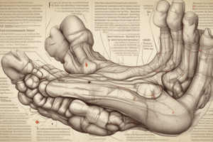
Bones of the Foot
- Key bones include the tibia, fibula, talus, calcaneus, cuboid, navicular, cuneiforms, metatarsals, and phalanges.
- The foot is divided into the forefoot, midfoot, and hindfoot.
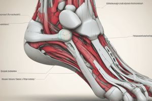
Ligaments of the Lateral Ankle
- The lateral ligament connects the lateral malleolus to the talus and calcaneus and has 3 parts
- Anterior talofibular ligament.
- Posterior talofibular ligament.
- Calcaneofibular ligament.
Ligaments of the Medial Ankle
- Deltoid ligament connects the medial malleolus to the talus, navicular, and calcaneus and has 4 parts
- Posterior tibiotalar ligament.
- Anterior tibiotalar ligament.
- Tibionavicular ligament.
- Tibiocalcaneal ligament.
Ankle and Foot Goniometry
- Subtalar Neutral:
- Position: Prone with foot off the edge, leg crossed behind.
- Technique: Grasp foot at 4th and 5th MT heads, palpate dorsal talus, passively DF to resistance, move through supination/pronation, and identify the "fall off" point which is the non-weight bearing neutral position.
Talocrural Dorsiflexion Goniometry
- Expected ROM: 0-20 degrees.
- Patient Position: Sitting with knee flexed to 90 degrees, foot in subtalar neutral.
- Therapist Position: Beside the patient stabilizing the tibia and fibula.
- Goniometer Placement:
- Fulcrum: Over lateral malleolus.
- Movable Arm: Parallel to the lateral aspect of the 5th metatarsal.
- Stationary Arm: Lateral midline of the fibula (fibular head).
- Normal End Feel: Capsular/firm due to posterior joint capsule tension, soleus muscle, Achilles tendon, deltoid ligament, posterior talofibular ligament, and calcaneofibular ligament.
- It's important to flex the knee to 90 degrees during dorsiflexion measurement
Ankle Dorsiflexion with Knee Extended
- Gastrocnemius: A two-joint muscle crossing the tibiofemoral and talocrural joints.
- Testing with Knee Extended: Gastrocnemius length may limit dorsiflexion range.
- Muscle Length Testing: One versus two joint ankle plantar flexors.
- End Feel: The end feel changes when the knee is extended if the gastrocnemius is shortened.
Talocrural Plantar Flexion Goniometry
- Expected ROM: 0-50 degrees.
- Patient Position: Sitting, knee flexed to 90 degrees, foot in subtalar neutral.
- Therapist Position: Beside the patient, stabilizing the tibia and fibula.
- Goniometer Placement:
- Fulcrum: Over lateral malleolus.
- Movable Arm: Parallel to the lateral aspect of the 5th metatarsal.
- Stationary Arm: Lateral midline of the fibula (fibular head).
- Normal End Feel:
- Capsular/firm: Tension in anterior joint capsule, anterior deltoid ligament, anterior talofibular ligament, tibialis anterior, extensor hallucis longus, and extensor digitorum longus muscles.
- Bony: Contact between posterior tubercles of the talus and posterior margin of the tibia.
Muscle Length Testing for Plantar Flexor Group
- Restriction in Dorsiflexion: It can indicate tightness in muscular tissue or the capsule.
- Elastic Restrictions: Further testing can identify if the restrictions are due to a loss of mobility in the Gastrocnemius, Soleus, or both.
- Intervention: Interventions should be assigned to target the appropriate tissue.
- Testing Positions: Non-weight bearing or weight bearing positions.
- Range of Motion: Weight bearing positions typically find greater ROM.
1 vs 2 Joint Plantar Flexor Muscle Length Test
- Patient Position: Prone with foot hanging off the edge of the plinth.
- Goniometer Alignment: As noted in previous slides (in subtalar neutral).
- Measure Passive DF: Knee extended (should be at least 10 degrees).
- Flex the Knee Passively: Re-measure DF at end range (motion should increase by 10 degrees to a total of 20 degrees).
1 vs 2 Joint Plantar Flexor Muscle Length Test Results
- Normal Length of All Plantar Flexors: DF of 10 degrees or more with leg extended, and 20 degrees once flexed.
- Tight Soleus Muscle: DF of 10 degrees with the leg extended, but it does not increase once the knee is flexed.
- Tight Gastrocnemius Muscle: DF is less than 10 degrees with the knee extended, but it is full (20 degrees) with the knee flexed.
Weight Bearing Assessment (Kendall)
- Test for Two-Joint Plantar Flexor Extensibility: standing with knees in extension
- Dorsiflexion: In this position measures at 10 degrees.
- Test for One-Joint Plantar Flexor Extensibility: Sit forward in a chair, slide feet back until heels rise slightly, push thigh down.
- Dorsiflexion: In this position measures at 20 degrees.
Composite Ankle Inversion Goniometry (Tarsal Joints)
- Expected Range: 0-35 degrees.
- Patient Position: Sitting, knee flexed to 90, hip without add/abd/rot.
- Therapist Position: In front of the patient stabilizing the tibia and fibula.
- Goniometer Placement:
- Fulcrum: Anterior ankle between malleoli.
- Stationary Arm: Anterior midline of the tibia (tibial tuberosity).
- Movable Arm: Anterior midline of the 2nd metatarsal.
- Normal End Feel: Capsular/firm due to tension in joint capsules, anterior and posterior ligaments, including talofibular and calcaneofibular ligaments, talocalcaneal ligaments, calcaneal ligaments, talonavicular ligament, bifurcate ligament, transverse metatarsal ligament, and peroneus longus and brevis muscles.
Composite Ankle Eversion Goniometry
- Expected Range: 0-15 degrees.
- Patient Position: Sitting, knee flexed to 90, hip without abd/add/rot.
- Therapist Position: In front of the patient stabilizing the tibia and fibula.
- Goniometer Placement:
- Fulcrum: Anterior ankle between malleoli.
- Stationary Arm: Anterior midline of the tibia (tibial tuberosity).
- Movable Arm: Anterior midline of the 2nd metatarsal.
- Normal End Feel:
- Capsular/firm: Tension in joint capsules, deltoid ligament, and tibialis posterior muscle.
- Bony: Contact between calcaneus and floor of sinus tarsi.
Subtalar Inversion Goniometry
- Range: 25-30 degrees.
- Patient Position: Prone, knee and hip in neutral with foot over the edge of the table.
- Therapist Position: Standing at the foot of the patient.
- Goniometer Placement:
- Fulcrum: Posterior ankle joint between malleoli.
- Stationary Arm: Posterior midline of the lower leg.
- Movable Arm: Posterior midline of the calcaneus.
- Perform by first DF the patient’s foot and finding subtalar neutral.
- Normal End Feel: Capsular/firm due to tension in lateral joint capsule, anterior and posterior talofibular, calcaneofibular ligament and talocalcaneal ligaments.
Subtalar Eversion Goniometry
- Range: 5-10 degrees
- Patient Position: prone, knee and hip in neutral, foot over the side of the table
- Therapist Position: standing at the foot of the patient
- Goniometer - Place the patient in full DF, then find subtalar neutral, Fulcrum: posterior ankle between the malleoli.
- Stationary arm: posterior midline of the lower leg
- Movable arm: posterior midline of the calcaneus
Calculating Subtalar Inversion and Eversion
- Obtain: measurement of subtalar neutral and end position. Then Document how many degrees of motion occurred
MTP Flexion Goniometry of the 1st Toe
- Normal ROM: 0-45 degrees.
- Patient Position: Supine or seated with leg supported; ankle and foot in neutral; MTP 0 abd/add and IP O flex/ext.
- Therapist Position: Stabilizing MTs (do not hold other MTs in extension).
- Goniometer Placement:
- Fulcrum: Over the dorsal side of the 1st MTP joint.
- Stationary Arm: Over the dorsal midline of the 1st metatarsal.
- Movable Arm: Over the dorsal midline of the proximal phalanx.
- Normal End-Feel: Capsular/firm due to tension in the dorsal joint capsule and collateral ligaments; tension in extensor digitorum brevis muscle may contribute.
MTP Extension Goniometry of 1st Toe
- Normal ROM: 0-70 degrees.
- Patient Position: Supine or seated with leg supported; ankle and foot in neutral; MTP 0 abd/add and IP O flex/ext.
- Therapist Position: Stabilize MTs (do not hold other MTs in flexion).
- Goniometer position:
- Fulcrum: over the dorsal side of 1st MTP joint.
- Stationary arm: over the dorsal midline of the 1st metatarsal.
- Movable arm: over the dorsal midline of the proximal phalanx.
- Normal End-Feel: Capsular/firm, tension in plantar joint capsule.
Examples of Capsular Patterns
- Talocrural: Plantarflexion is more limited than dorsiflexion.
- Subtalar (Talocalcaneal): Inversion is more limited than eversion.
- 1st MTP: Extension is more limited than flexion.
- 2-5 MTP and IP Joints: Flexion is more limited than or equal to extension.
- Talonavicular/calcaneocuboid: Inversion (PF, add, sup) > DF.
Functional Ranges of Ankle and Foot during Locomotion
- Dorsiflexion:
- Gait level surfaces: 0-10 degrees.
- Ascending stairs: 20-24 degrees.
- Descending stairs: 26-36 degrees.
- Plantarflexion:
- Gait level surfaces: 15-30 degrees.
- Ascending Stairs: 24-30 degrees.
- Descending Stairs: 26-31 degrees.
Muscles Responsible for Ankle Plantarflexion
- Gastrocnemius
- Soleus
- Plantaris
- Tibialis Posterior
- Peroneus Longus and Brevis
- Flexor Hallucis Longus and Flexor Digitorum Longus
Ankle Plantar Flexors MMT
- Testing Position Against Gravity: Standing on testing leg.
- Stabilization: Hand on table for support but no pressure through hand.
- Movement: Full heel rise onto toes with body weight upwards.
- Weakness: Unable to perform complete heel rise, leans forward and/or flexes knee.
- Testing across Gravity: Prone with leg supported and foot off the edge of table.
Plantar Flexor Grading in Standing
- 5/5: Heel raised successfully from floor, minimum 25 times, good form, and no fatigue.
- 4/5: Raised successfully from floor 10-24 times.
- 3/5: Raised successfully from floor 1-9 times.
Plantar Flexor Grading in Prone
- 2+/5: Completes PF range and holds against maximal resistance.
- 2/5: Completes PF range but no resistance.
- 2-/5: Completes partial range motion within pronation.
Soleus MMT
- Testing Position Against Gravity: Prone, knee flexed at least 90 degrees.
- Stabilization: Lower leg proximal to the ankle.
- Movement: Plantarflexion lacking inversion/eversion.
- Resistance: Against calcaneus into dorsiflexion.
- Weakness: Unable to plantarflex.
- Testing Position Across Gravity: Side lying, knee bent to 90 degrees.
- Stabilization: Support limb.
Tibialis Posterior MMT
- Testing Position Against Gravity: Supine, leg in lateral rotation.
- Stabilization: Leg by the therapist above the ankle.
- Movement: Inversion of the foot with plantar flexion of the ankle.
- Resistance: On the medial and plantar foot into dorsiflexion and eversion.
- Weakness: Toes flex and inability to perform against resistance.
Tibialis Anterior MMT (DF and Inv)
- Testing Position Against Gravity: Sitting with the knee flexed.
- Stabilization: Leg by therapist proximal to the ankle joint.
- Movement: Into Dorsiflexion and Inversion without great toe extension.
- Resistance: At the medial side, dorsal surface, towards ankle PF and foot eversion.
- Weakness: Inability to flex without eversion.
- Testing Across Gravity: Side lying, testing leg on top.
- Stabilization: Support leg.
Extensor Hallucis Longus and Brevis MMT
Testing Position Against Gravity: Supine or sitting.
- Stabilization: Foot by therapist in slight ankle plantar flexion.
- Movement: Extension of MTP and IP of the great toe.
- Resistance: Against dorsal surface of distal and proximal phalanxes of the great toe into plantar flexion.
- Weakness: Unable to extend great toe/difficulty with DF ankle.
- Testing Position Across Gravity: side lying supporting leg.
Extensor Digitorum Longus and Brevis MMT
- Testing Position Against Gravity: Seated.
- Stabilization: Foot by the therapist in slight plantar flexion.
- Movement: Extension of all joints of 2nd-5th digits (distal IP joint).
- Resistance: Dorsal surface of the toes into flexion.
- Weakness: Decreased toe extension, foot drop, forefoot varus, decreased DF of ankle and eversion of foot.
- Testing Position Across Gravity: Side-lying, foot supported.
Flexor Digitorum Longus MMT (assisted by Quadratus Plantae)
- Testing Position Against Gravity: Supine foot in neutral, Knee flexion to neutralize gastric tightness.
Studying That Suits You
Use AI to generate personalized quizzes and flashcards to suit your learning preferences.