Podcast
Questions and Answers
What primarily characterizes the deep branch of the ulnar nerve?
What primarily characterizes the deep branch of the ulnar nerve?
- It primarily innervates the hypothenar muscles. (correct)
- It innervates the palmaris brevis muscle.
- It provides sensory innervation to the little finger.
- It innervates the three thenar muscles.
Which nerve supplies the skin on the palmar surfaces of the lateral three and one-half digits?
Which nerve supplies the skin on the palmar surfaces of the lateral three and one-half digits?
- Recurrent branch of the median nerve
- Superficial branch of radial nerve
- Deep branch of ulnar nerve
- Palmar digital branches of median nerve (correct)
What is the primary motor function of the superficial branch of the ulnar nerve?
What is the primary motor function of the superficial branch of the ulnar nerve?
- Innervates the interossei
- Assists in wrist joint movement
- Innervates the palmaris brevis muscle (correct)
- Supplies skin over the palm
Which muscle group does the recurrent branch of the median nerve primarily innervate?
Which muscle group does the recurrent branch of the median nerve primarily innervate?
The superficial branch of the radial nerve provides sensory innervation to which areas?
The superficial branch of the radial nerve provides sensory innervation to which areas?
Which muscle is responsible for the abduction of the little finger?
Which muscle is responsible for the abduction of the little finger?
What is the innervation of the first and second lumbricals?
What is the innervation of the first and second lumbricals?
Which muscle originates from the base of the proximal phalanx of the thumb?
Which muscle originates from the base of the proximal phalanx of the thumb?
Which muscles are responsible for adduction of the fingers?
Which muscles are responsible for adduction of the fingers?
Which muscle primarily pulls the thumb medially and forward across the palm?
Which muscle primarily pulls the thumb medially and forward across the palm?
What is the primary function of the flexor pollicis brevis?
What is the primary function of the flexor pollicis brevis?
Which of the following muscles is NOT innervated by the deep branch of the ulnar nerve?
Which of the following muscles is NOT innervated by the deep branch of the ulnar nerve?
Which artery primarily supplies the thumb?
Which artery primarily supplies the thumb?
What is the function of the dorsal interossei muscles?
What is the function of the dorsal interossei muscles?
Which muscle originates from the anterior surface of the shaft of the 5th metacarpal?
Which muscle originates from the anterior surface of the shaft of the 5th metacarpal?
What feature distinguishes the skin of the palm from the dorsal side of the hand?
What feature distinguishes the skin of the palm from the dorsal side of the hand?
Which structure helps to convert the carpal arch into the carpal tunnel?
Which structure helps to convert the carpal arch into the carpal tunnel?
Which of the following is NOT a content of the carpal tunnel?
Which of the following is NOT a content of the carpal tunnel?
What is the main function of the fibrous sheaths surrounding the tendons of the flexor muscles?
What is the main function of the fibrous sheaths surrounding the tendons of the flexor muscles?
Which part of the extensor expansion is attached to the distal phalanx?
Which part of the extensor expansion is attached to the distal phalanx?
The palmar aponeurosis is primarily responsible for covering which area?
The palmar aponeurosis is primarily responsible for covering which area?
How many short muscles are present in the thumb, including the adductor pollicis?
How many short muscles are present in the thumb, including the adductor pollicis?
What is the role of the extensor retinaculum in the hand?
What is the role of the extensor retinaculum in the hand?
Flashcards
What is the main characteristic of the skin on the palm?
What is the main characteristic of the skin on the palm?
The skin on the palm is thick, hairless, and tightly connected to the underlying deep fascia by numerous fibrous bands.
What is the function of the flexor retinaculum?
What is the function of the flexor retinaculum?
The flexor retinaculum is a thickening of the deep fascia that helps convert the carpal arch into the carpal tunnel. It keeps the long flexor tendons in place at the wrist.
What structures are found within the carpal tunnel?
What structures are found within the carpal tunnel?
The carpal tunnel contains the median nerve and the tendons of the flexor digitorum superficialis, flexor digitorum profundus, and flexor pollicis longus muscles.
What is the function of the extensor retinaculum?
What is the function of the extensor retinaculum?
Signup and view all the flashcards
What is the role of fibrous sheaths in the hand?
What is the role of fibrous sheaths in the hand?
Signup and view all the flashcards
What is the main function of the extensor expansion?
What is the main function of the extensor expansion?
Signup and view all the flashcards
What is the role of the palmar aponeurosis?
What is the role of the palmar aponeurosis?
Signup and view all the flashcards
How many short muscles are there in the thumb?
How many short muscles are there in the thumb?
Signup and view all the flashcards
Deep Branch of Ulnar Nerve
Deep Branch of Ulnar Nerve
Signup and view all the flashcards
Superficial Branch of Ulnar Nerve
Superficial Branch of Ulnar Nerve
Signup and view all the flashcards
Recurrent Branch of Median Nerve
Recurrent Branch of Median Nerve
Signup and view all the flashcards
Palmar Digital Branches of Median Nerve
Palmar Digital Branches of Median Nerve
Signup and view all the flashcards
Superficial Branch of Radial Nerve
Superficial Branch of Radial Nerve
Signup and view all the flashcards
What are the 3 short muscles of the little finger?
What are the 3 short muscles of the little finger?
Signup and view all the flashcards
What is the function of the adductor pollicis?
What is the function of the adductor pollicis?
Signup and view all the flashcards
What muscles make up the thenar group?
What muscles make up the thenar group?
Signup and view all the flashcards
What is the function of the lumbricals?
What is the function of the lumbricals?
Signup and view all the flashcards
What is the difference between palmar and dorsal interossei?
What is the difference between palmar and dorsal interossei?
Signup and view all the flashcards
What are the two main blood vessel arches in the palm?
What are the two main blood vessel arches in the palm?
Signup and view all the flashcards
What is the function of the opponens digiti minimi?
What is the function of the opponens digiti minimi?
Signup and view all the flashcards
What is the function of the flexor digiti minimi?
What is the function of the flexor digiti minimi?
Signup and view all the flashcards
What is the function of the abductor digiti minimi?
What is the function of the abductor digiti minimi?
Signup and view all the flashcards
What is the space of Parona?
What is the space of Parona?
Signup and view all the flashcards
Study Notes
Anatomy of the Hand
- The hand's structure is described, focusing on its muscles, skin, and connective tissues.
- Objectives for the study include features of hand skin, attachments, innervations, and actions of hand muscles, the flexor and extensor retinacula, carpal tunnel boundaries and contents, blood vessels and nerves of the hand, palmar aponeurosis, and synovial sheaths.
Skin of the Hand
- Palm: Thick, hairless, bound to underlying fascia by fibrous bands, characterized by creases for movement, and dense sweat gland distribution.
- Dorsum: Thin, hairy, freely mobile over tendons and bones.
Flexor Retinaculum
- Thickening of deep fascia, forming a carpal tunnel.
- Attached to pisiform and hook of hamate medially, and scaphoid and trapezium tubercles laterally.
- Holds long flexor tendons in place at the wrist.
Carpal Tunnel
- Formed by carpal bones (inferiorly) and the flexor retinaculum (superiorly).
- Contains 4 tendons of flexor digitorum profundus, 4 tendons of flexor digitorum superficialis, tendon of flexor pollicis longus, and the median nerve.
Extensor Retinaculum
- Thickening of deep fascia, forming 6 separate tunnels lined with synovial sheaths.
- Holds long extensor tendons in position.
- Attached to pisiform and hook of hamate medially, and distal end of radius laterally.
Synovial Sheaths
- Synovial sheaths surround tendons, reducing friction.
- Variations in sheath arrangement exist (e.g., intermediate bursa).
- Provide lubrication for smooth tendon movement in the hand.
Fibrous Sheaths
- Surround and support tendons of flexor digitorum superficialis and profundus muscles.
- Run anterior to metacarpophalangeal joints, extending to distal phalanges.
- Formed by fibrous arches and cruciate ligaments for support.
- Attached posteriorly to margins of phalanges, and to palmar ligaments.
- Prevent bowing of tendons during flexion.
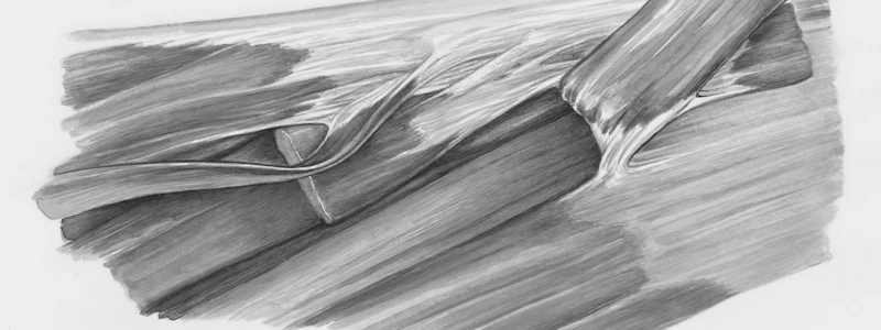
Extensor Expansion
- Triangular attachments over proximal phalanges.
- Supports and connects tendons of extensor digitorum and extensor pollicis longus.
- Attached to distal phalanx, middle/proximal phalanx (thumb).
- Extensor hood wraps around metacarpophalangeal joints.
- Provides attachment for hand muscles.
Palmar Aponeurosis
- Triangular condensation of deep fascia covering the palm.
- Continuous with palmaris longus tendon or flexor retinaculum.
- Fibers radiate to digits, supporting and stabilizing tendons, nerves, and blood vessels that underpin the hand.
Hand Muscles
- Thumb Short Muscles: 4 comprising adductor pollicis and thenar muscles (abductor pollicis brevis, flexor pollicis brevis, opponens pollicis).
- Little Finger Short Muscles: 3 hypothenar muscles (abductor digiti minimi, flexor digiti minimi brevis, opponens digiti minimi).
- Thenar Muscles: Innervated by the median nerve.
- Adductor Pollicis: Innervated by the deep branch of the ulnar nerve.
- Lumbricals: 4 muscles, innervated by median (1st and 2nd) or ulnar (3rd and 4th) nerves.
- Palmar Interossei: 4 muscles; adduct fingers toward the third finger.
- Dorsal Interossei: 4 muscles; abduct fingers away from the third finger.
Movements of Thumb
- Movements include abduction, adduction, extension, flexion, opposition, and reposition.
Blood Vessels of the Hand
- Two interconnected vascular arches in the palm (radial and ulnar arteries): superficial and deep.
- Radial artery supplies thumb and index finger's lateral side.
- Ulnar artery supplies other digits and index medial side.
Nerves of the Hand
- Ulnar Nerve: Deep and superficial branches.
- Deep branch innervates hypothenar muscles, interossei, and medial lumbricals.
- Superficial branch innervates palmaris brevis muscle; skin on ulnar side of hand and medial half of ring finger.
- Median Nerve: Divides into recurrent branch (innervates thenar muscles) and palmar digital branches.
- Radial Nerve: Superficial branch, innervates dorsolateral aspect of palm and digits distally (over terminal interphalangeal joints).
Fascial Spaces of Palm
- Potential spaces filled with loose connective tissue (CT).
- Fibrous septa divide the palm into thenar and midpalmar spaces based on attachments to metacarpals.
- Parona's space is between FDP anteriorly and pronator quadratus/interosseous posteriorly.
Studying That Suits You
Use AI to generate personalized quizzes and flashcards to suit your learning preferences.


