Podcast
Questions and Answers
What is the function of the tapetum lucidum in nocturnal and domestic mammals?
What is the function of the tapetum lucidum in nocturnal and domestic mammals?
- To control the amount of light that passes through the pupil
- To modify the diameter of the pupil
- To absorb light that has passed through the photoreceptors
- To enhance dark-adapted vision under dim light (correct)
What type of muscle is the pupillary dilator muscle?
What type of muscle is the pupillary dilator muscle?
- Striated muscle
- Skeletal muscle
- Smooth muscle (correct)
- Cardiac muscle
What is the function of the suspensory ligaments in the eye?
What is the function of the suspensory ligaments in the eye?
- To control the movement of the eye
- To regulate the amount of light that enters the eye
- To suspend the lens and aid in accommodation (correct)
- To modify the shape of the cornea
What is the function of the vitreous humor in the eye?
What is the function of the vitreous humor in the eye?
What is the term for the constriction of the pupil?
What is the term for the constriction of the pupil?
What type of cells are found in the vitreous humor?
What type of cells are found in the vitreous humor?
What is the function of the ciliary body in the eye?
What is the function of the ciliary body in the eye?
What is the structure that lies behind the vitreous humor in the eye?
What is the structure that lies behind the vitreous humor in the eye?
What is the main difference between the outer segment of cone cells and rod cells?
What is the main difference between the outer segment of cone cells and rod cells?
What is the role of vitamin A in the retina?
What is the role of vitamin A in the retina?
What is the main function of rod cells?
What is the main function of rod cells?
How many types of cones are present in primates?
How many types of cones are present in primates?
Describe the pathway of visual signals from the retina to the visual cortex.
Describe the pathway of visual signals from the retina to the visual cortex.
What is the term for the ability to detect different wavelengths of light?
What is the term for the ability to detect different wavelengths of light?
What is the result of severe vitamin A deficiency?
What is the result of severe vitamin A deficiency?
How many types of cells make up the retina?
How many types of cells make up the retina?
What is the primary function of the retina in the eye?
What is the primary function of the retina in the eye?
What is the point where axons of the retina ganglion cell layer leave the eye on their way to the brain?
What is the point where axons of the retina ganglion cell layer leave the eye on their way to the brain?
What is the structure that arises from the axons leaving the eye at the optic disc?
What is the structure that arises from the axons leaving the eye at the optic disc?
Which of the following statements is true?
Which of the following statements is true?
What is the function of the blood vessels in the retina?
What is the function of the blood vessels in the retina?
What is the structure that surrounds the optic nerve in some animals?
What is the structure that surrounds the optic nerve in some animals?
Where do the arteries and veins enter the retina?
Where do the arteries and veins enter the retina?
What is the comparison between the number of axons in the optic nerves and the dorsal roots of the spinal cord?
What is the comparison between the number of axons in the optic nerves and the dorsal roots of the spinal cord?
What happens to cGMP in the dark?
What happens to cGMP in the dark?
What is the role of the antipoter in rod cells?
What is the role of the antipoter in rod cells?
What is the ratio of rod cells to ganglion cells?
What is the ratio of rod cells to ganglion cells?
Which part of the retina provides the highest visual acuity?
Which part of the retina provides the highest visual acuity?
What is the role of phosphodiesterase 6 (PDE6) in rod cells?
What is the role of phosphodiesterase 6 (PDE6) in rod cells?
What is the equivalent of the film in a photographic camera in the eye?
What is the equivalent of the film in a photographic camera in the eye?
What happens to the light as it passes through the eye?
What happens to the light as it passes through the eye?
What is the effect of the activation of rhodopsin by light?
What is the effect of the activation of rhodopsin by light?
Where is the lens located in relation to the aqueous humor and the vitreous humor?
Where is the lens located in relation to the aqueous humor and the vitreous humor?
What is the purpose of the lens in the eye?
What is the purpose of the lens in the eye?
What is unique about the central retina in most mammals?
What is unique about the central retina in most mammals?
What happens to the light rays as they approach the eye?
What happens to the light rays as they approach the eye?
What is the nature of the images formed on the retina?
What is the nature of the images formed on the retina?
What happens to the images formed on the retina?
What happens to the images formed on the retina?
Where do the optic nerve fibers terminate for visual perception?
Where do the optic nerve fibers terminate for visual perception?
Where do the optic nerve fibers terminate for visual reflexes?
Where do the optic nerve fibers terminate for visual reflexes?
Regarding image formation, the fibers terminate in the midbrain for visual perception or thalamus for visual reflex; respectively.
Regarding image formation, the fibers terminate in the midbrain for visual perception or thalamus for visual reflex; respectively.
Photoreceptors and ganglion cells communicate via receptor potential rather than action potential.
Photoreceptors and ganglion cells communicate via receptor potential rather than action potential.
Which of the following statements regarding the area centralis is false?
Which of the following statements regarding the area centralis is false?
In order to perceive light, ganglion cells must be __________ (activated)
In order to perceive light, ganglion cells must be __________ (activated)
Match to the correct retinal layer
Match to the correct retinal layer
Match the following neurotransmitter(s) to the correct cell of the retina
Match the following neurotransmitter(s) to the correct cell of the retina
Match the correct order of the cells of the retina
Match the correct order of the cells of the retina
In the DARK: photoreceptor cells are _____________ and release __________ (neurotransmitter), which activates bipolar cells. Bipolar cells then inhibit _________ cells which doesn’t allow the optic nerve to perceive no light.
In the DARK: photoreceptor cells are _____________ and release __________ (neurotransmitter), which activates bipolar cells. Bipolar cells then inhibit _________ cells which doesn’t allow the optic nerve to perceive no light.
The activation of light activates ____ and this leads to activation of the G protein _________. Activated ________ split away and activate ________
The activation of light activates ____ and this leads to activation of the G protein _________. Activated ________ split away and activate ________
After PDE6 is activated, it hydrolizes ______ to ______. This causes the ________ to close; thus leading to ___________. This depolarization/hyperpolarization does not release ____________ (neurotransmitter).
After PDE6 is activated, it hydrolizes ______ to ______. This causes the ________ to close; thus leading to ___________. This depolarization/hyperpolarization does not release ____________ (neurotransmitter).
Flashcards
Tapetum Lucidum
Tapetum Lucidum
Reflective material in nocturnal animals enhancing vision in dim light.
Iris Function
Iris Function
Controls the amount of light entering the eye through the pupil.
Mydriasis
Mydriasis
Pupil dilation, increasing the amount of light entering the eye.
Miosis
Miosis
Signup and view all the flashcards
Pupillary Dilator Muscle
Pupillary Dilator Muscle
Signup and view all the flashcards
Pupillary Sphincter Muscle
Pupillary Sphincter Muscle
Signup and view all the flashcards
Lens Function
Lens Function
Signup and view all the flashcards
Ciliary Body
Ciliary Body
Signup and view all the flashcards
Vitreous Humor
Vitreous Humor
Signup and view all the flashcards
Retina Function
Retina Function
Signup and view all the flashcards
Optic Disc
Optic Disc
Signup and view all the flashcards
Rod Cells
Rod Cells
Signup and view all the flashcards
Cone Cells
Cone Cells
Signup and view all the flashcards
Rhodopsin
Rhodopsin
Signup and view all the flashcards
Color Vision
Color Vision
Signup and view all the flashcards
Area Centralis
Area Centralis
Signup and view all the flashcards
Optic Nerve (CN II)
Optic Nerve (CN II)
Signup and view all the flashcards
Outer Segment
Outer Segment
Signup and view all the flashcards
Inner Segment
Inner Segment
Signup and view all the flashcards
Study Notes
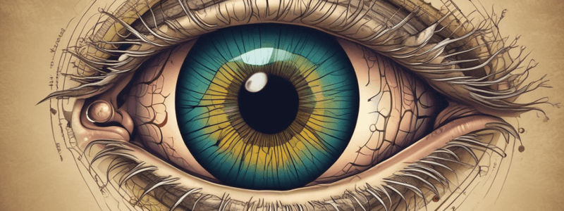
Fundus and Tapetum
- Choroidal vessels are visible throughout the fundus.
- In some diurnal animals, melanocytes absorb light that has passed by photoreceptors without stimulating them.
- Nocturnal and most domestic mammals have a reflective material called tapetum lucidum, which enhances dark-adapted vision under dim light.
Iris and Pupil
- The iris controls the amount of light that passes through the pupil.
- In the dark, the pupil will dilate (mydriasis).
- In the light, the pupil will contract (miosis).
- The iris contains dilator and sphincter muscles.
- Pupillary dilator muscle:
- Radially arranged
- Opposes the action of the sphincter
- Contraction results in pupillary dilation (mydriasis)
- Dilation reflects the general state of sympathetic tone (e.g., pain, fear, anger)
- Pupillary sphincter muscle:
- Circularly arranged near the pupillary margin
- Contraction results in decreased pupillary size (miosis)
- Innervated by parasympathetic fibers
Lens and Accommodation
- Behind the iris is the lens.
- The lens is suspended by suspensory ligaments.
- These fibers are attached to the ciliary body.
- The ciliary body is a muscular structure near the base of the iris.
- The ciliary body helps with accommodation of the lens.
- Increase or decrease in tension on the lens makes the lens curvature more or less convex.
- This allows the lens to focus on near or far objects.
Vitreous Humor and Retina
- Behind the lens is a chamber filled with vitreous humor.
- Vitreous humor is a gelatinous fluid.
- It gives the spherical shape of the eye.
- Contains phagocytic cells.
- Behind the vitreous humor lies the retina.
- The retina is where light is transformed into electrical activity of neurons.
Optic Disc and Optic Nerve
- The retina is interrupted at a point where axons of the retina ganglion cell layer leave the eye on their way to the brain – optic disc or blind spot.
- The axons leaving the eye at the optic disc give rise to the optic nerve (CN II).
- There are more axons in both optic nerves than in all the dorsal roots of the spinal cord.
Photoreceptor Cells
- There are about 130 million photoreceptor cells in the retina.
- There are two types of photoreceptor cells: cone cells and rod cells.
- Each type consists of two portions: outer segment and inner segment.
- Outer segment is the photosensitive region.
- Inner segment is the metabolic region of the photoreceptor.
- Rod cells have rhodopsin, which is responsible for the perception of shades of gray.
- Rhodopsin has a low threshold of excitability, making it easy to stimulate by low-intensity light.
- Rod cells are essential for night vision.
- Cone cells have color pigments or cone pigments, which are responsible for the perception of color.
- There are three different types of cones in primates, each with a different type of color pigment.
- Most mammals have only two types of cones (dichromatic).
Visual System
- Color is the brain's interpretation of differences in the wavelengths of light.
- The visual system involves the activation of rhodopsin, leading to the activation of a G protein, transducin, and the hydrolysis of cGMP to 5'-GMP.
- This leads to hyperpolarization, reducing the release of glutamate, and ultimately, the excitation of bipolar cells.
- The ratio of rods to cone photoreceptor cells, as well as the ratio of photoreceptors cells to ganglion cells, affects the acuity of visual images.
- The area centralis provides the highest visual acuity in the retina.
Eye and Camera
- The eye is optically equivalent to the usual photographic camera.
- The eye catches the light reflected by the objects and guides its passage until the image is formed.
- The lens works by converging the light rays to a certain focal point on the retina.
- The images formed on the retina are real, inverted, and smaller than the object.
Studying That Suits You
Use AI to generate personalized quizzes and flashcards to suit your learning preferences.


