Podcast
Questions and Answers
The ______ ear is divided into the external, middle, and internal ear.
The ______ ear is divided into the external, middle, and internal ear.
human
What is the name of the thin, semitransparent sheet that separates the external ear from the middle ear?
What is the name of the thin, semitransparent sheet that separates the external ear from the middle ear?
tympanic membrane
What are the three tiny bones that connect the tympanic membrane and the inner ear?
What are the three tiny bones that connect the tympanic membrane and the inner ear?
- Hammer, Anvil, Stirrup
- Malleus, Incus, Stapes (correct)
- External Acoustic Meatus, Tympanic Cavity, Auditory Tube
- Cochlea, Utricle, Saccule
What is the name of the fluid that is found within the membranous labyrinth?
What is the name of the fluid that is found within the membranous labyrinth?
The ______ duct is a tube that runs within the cochlea and is filled with endolymph.
The ______ duct is a tube that runs within the cochlea and is filled with endolymph.
High-frequency sounds cause the basilar membrane to vibrate near the oval window.
High-frequency sounds cause the basilar membrane to vibrate near the oval window.
What is the name of the nerve that carries both equilibrium and hearing sensations to the brainstem?
What is the name of the nerve that carries both equilibrium and hearing sensations to the brainstem?
Match the following terms to their correct descriptions.
Match the following terms to their correct descriptions.
A ______ is a false perception of spinning or movement.
A ______ is a false perception of spinning or movement.
Flashcards
What are hair cells?
What are hair cells?
Sensory receptors located in the internal ear, responsible for hearing and balance.
What are stereocilia?
What are stereocilia?
Specialized processes on hair cells that resemble long microvilli. They are present in large numbers, between 80 and 100 per hair cell.
What is a kinocilium?
What is a kinocilium?
A single, large cilium present on hair cells. It is typically larger than stereocilia.
What are hair cells in the internal ear?
What are hair cells in the internal ear?
Signup and view all the flashcards
What is the function of the external ear?
What is the function of the external ear?
Signup and view all the flashcards
What is the middle ear?
What is the middle ear?
Signup and view all the flashcards
What is the eardrum?
What is the eardrum?
Signup and view all the flashcards
What are the auditory ossicles?
What are the auditory ossicles?
Signup and view all the flashcards
What is the function of the auditory tube?
What is the function of the auditory tube?
Signup and view all the flashcards
What is the inner ear?
What is the inner ear?
Signup and view all the flashcards
What is the bony labyrinth?
What is the bony labyrinth?
Signup and view all the flashcards
What is the membranous labyrinth?
What is the membranous labyrinth?
Signup and view all the flashcards
What is perilymph?
What is perilymph?
Signup and view all the flashcards
What is endolymph?
What is endolymph?
Signup and view all the flashcards
What are the semicircular canals?
What are the semicircular canals?
Signup and view all the flashcards
What are the semicircular ducts?
What are the semicircular ducts?
Signup and view all the flashcards
What is the ampulla?
What is the ampulla?
Signup and view all the flashcards
What is the ampullary crest?
What is the ampullary crest?
Signup and view all the flashcards
What is the ampullary cupula?
What is the ampullary cupula?
Signup and view all the flashcards
What are the utricle and saccule?
What are the utricle and saccule?
Signup and view all the flashcards
What is the vestibule?
What is the vestibule?
Signup and view all the flashcards
What are the maculae?
What are the maculae?
Signup and view all the flashcards
What is the otolithic membrane?
What is the otolithic membrane?
Signup and view all the flashcards
What are otoliths?
What are otoliths?
Signup and view all the flashcards
What is the cochlea?
What is the cochlea?
Signup and view all the flashcards
What is the cochlear duct?
What is the cochlear duct?
Signup and view all the flashcards
What are the scala vestibuli and scala tympani?
What are the scala vestibuli and scala tympani?
Signup and view all the flashcards
What is the vestibular membrane?
What is the vestibular membrane?
Signup and view all the flashcards
What is the basilar membrane?
What is the basilar membrane?
Signup and view all the flashcards
What is the spiral organ?
What is the spiral organ?
Signup and view all the flashcards
What is the spiral ganglion?
What is the spiral ganglion?
Signup and view all the flashcards
What is the cochlear nerve?
What is the cochlear nerve?
Signup and view all the flashcards
What is the tectorial membrane?
What is the tectorial membrane?
Signup and view all the flashcards
How do sound waves stimulate hair cells?
How do sound waves stimulate hair cells?
Signup and view all the flashcards
What is the vestibulocochlear nerve?
What is the vestibulocochlear nerve?
Signup and view all the flashcards
What is the function of the vestibular nerve?
What is the function of the vestibular nerve?
Signup and view all the flashcards
What are the vestibular nuclei?
What are the vestibular nuclei?
Signup and view all the flashcards
What is the auditory cortex?
What is the auditory cortex?
Signup and view all the flashcards
Study Notes
Chapter 15 Special Senses Pt 2
- Module 15.15: Equilibrium and Hearing Involve the Internal Ear
- Comparison of receptors
- Olfactory receptors—specialized sensory neurons
- Gustatory receptors—communicate with sensory neurons
- Photoreceptors—respond to light
- Both route information directly to the CNS
- All located in epithelia exposed to external environment
Module 15.15: Internal Ear Sensory Receptors
- Equilibrium and hearing receptors—isolated and protected from external environment
- Located in internal ear
- Information integrated and organized locally; forwarded to CNS
Module 15.15: Internal Ear Sensory Receptors (2 of 4)
- Hair cells = sensory receptors in internal ear
- Free surfaces covered with specialized nonmotile processes
- Stereocilia—resemble long microvilli; 80–100 per hair cell
- Kinocilium = single large cilium
Module 15.15: Internal Ear Sensory Receptors (3 of 4)
- Hair cells are mechanoreceptors—sensitive to contact/movement
- External force pushing on hair cell processes distorts plasma membrane; alters neurotransmitter release
- Provides information about direction/strength of stimulus
- Monitored by dendrites of sensory neurons
Module 15.15: Internal Ear Sensory Receptors (4 of 4)
- Complex 3-D structure in internal ear determines what stimuli can reach hair cells in each region
- Hair cells in one region respond only to gravity or acceleration
- Hair cells in other regions respond only to rotation or to sound
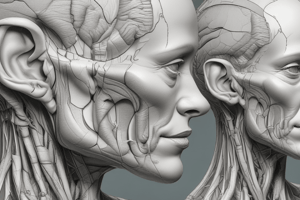
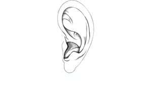
The Anatomy of the Ear
- External Ear
- Elastic cartilage
- Auricle
- External acoustic meatus
- Middle Ear
- Auditory ossicles
- Tympanic cavity
- Tympanic membrane
- Internal Ear
- Semicircular canals
- Petrous part of temporal bone
- Facial nerve (VII)
- Vestibulocochlear nerve (VIII)
- Bony labyrinth
- Auditory tube
- To nasopharynx
Module 15.16: The Ear is Divided into the External Ear, the Middle Ear, and the Internal Ear
- External ear—collects/directs sound waves toward middle ear
- Auricle—elastic cartilage
- External acoustic meatus—passageway in temporal bone
- Ceruminous glands—secrete waxy cerumen (earwax); keeps foreign objects out; slows growth of microorganisms
- Hairs—trap debris
Module 15.16: Anatomy of the Ear
- Middle ear (tympanic cavity) = air-filled chamber from tympanic membrane to auditory ossicles; connects to pharynx by auditory tube
- Tympanic membrane (tympanum, eardrum) = thin, semitransparent sheet that separates external ear and middle ear
- Auditory ossicles = three tiny bones; connect tympanic membrane and inner ear
Module 15.16: Anatomy of the Ear (2 of 7)
- Internal ear
- Contains sensory organs for hearing and equilibrium
- Receives amplified sound waves from middle ear
- Superficial contours established by layer of dense bone = bony labyrinth
Module 15.16: Anatomy of the Ear (3 of 7)
- Middle ear
- Auditory tube (pharyngotympanic tube, eustachian tube)
- Connects middle ear to nasopharynx
- Allows pressure equalization across tympanic membrane
- Can allow microorganisms into middle ear, causing infection (otitis media)—can impair hearing, may invade internal ear
- Auditory tube (pharyngotympanic tube, eustachian tube)
Module 15.16: Anatomy of the Ear (4 of 7)
- Auditory ossicles
- Malleus (malleus, hammer)—attaches to tympanic membrane
- Incus (incus, anvil)—attaches malleus to stapes
- Stapes (stapes, stirrup)—attached to oval window
Module 15.16: Anatomy of the Ear (5 of 7)
- Middle ear muscles
- Tensor tympani muscle connects malleus to temporal bone
- Contraction stiffens tympanic membrane, reduces vibration
- Innervated by mandibular branch of trigeminal nerve (V)
Module 15.16: Anatomy of the Ear (6 of 7)
- Middle ear muscles (continued)
- Stapedius muscle
- Connects stapes to posterior wall of middle ear
- Reduces stapes movement at oval window
- Stapedius muscle
Module 15.16: Anatomy of the Ear (7 of 7)
- Amplification and protection
- Sound waves vibrate tympanic membrane; convert sound into mechanical movement
- Auditory ossicles conduct vibrations to internal ear
- Focuses sound on oval window and amplifies it
- Contractions of tensor tympani and stapedius muscles protect tympanic membrane and ossicles from violent movement under very noisy conditions
The Anatomy of the Internal Ear
- Bony labyrinth
- Shell of dense bone surrounding/protecting membranous labyrinth
- Filled with perilymph—liquid similar to CSF; Between bony labyrinth and membranous labyrinth
- Three parts
- Semicircular canals, vestibule, cochlea
- Membranous Labyrinth
- Collection of fluid-filled tubes/chambers
- Houses receptors for hearing and equilibrium
- Contains fluid called endolymph
Module 15.17: Labyrinths of the Internal Ear
- Three parts (semicircular canals, utricle, and saccule are part of the vestibular complex, which maintains equilibrium)
- Semicircular ducts (within semicircular canals)
- Receptors stimulated by rotation of head
- Within the vestibule-utricle and saccule
- Provide sensations of gravity and linear acceleration
- Cochlear duct (within cochlea)
- Sandwiched between pair of perilymph-filled chambers
- Resembles snail shell
- Receptors stimulated by sound
- Semicircular ducts (within semicircular canals)
Module 15.18: Receptors for Equilibrium
- Semicircular ducts (continuous with utricle and filled with endolymph)
- Ampulla = enlarged part of duct that houses receptors
- Ampullary crest = region in wall of ampulla with receptors
- Ampullary cupula = gelatinous structure extending through ampulla with kinocilia and stereocilia of hair cells embedded in it
Module 15.18: Receptors for Equilibrium (2 of 6)
- Head rotating in the plane of a duct moves endolymph; pushes ampullary cupula to side, distorting receptor processes
- Movement in one direction causes stimulation; opposite direction causes inhibition
- Ampullary cupula rebounds to normal position when endolymph stops moving
Module 15.18: Receptors for Equilibrium (3 of 6)
- Even complex angular movements can be analyzed by movement of the three rotational planes
- Horizontal rotation ("no") stimulates lateral duct receptors
- Nodding ("yes") stimulates anterior duct receptors
- Tilting head to side stimulates posterior duct receptors
Module 15.18: Receptors for Equilibrium (4 of 6)
- Utricle and saccule
- Provide equilibrium sensations, whether body is stationary or moving
- Connected by slender passageway continuous with endolymphatic duct that ends in endolymphatic sac
- Sac projects into subarachnoid space
- Endolymphatic duct continuously secretes endolymph; returns to general circulation at endolymphatic sac
Module 15.18: Receptors for Equilibrium (5 of 6)
- Utricle and saccule (continued)
- Utricle and saccule contain hair cells clustered in maculae
- Macula of utricle senses horizontal movement
- Macula of saccule senses vertical movement
- Hair cell processes embedded in gelatinous otolithic membrane
- Surface has densely packed calcium carbonate crystals (otoliths, or "ear stones")
Module 15.18: Receptors for Equilibrium (6 of 6)
- Utricle and saccule (continued)
- Change in head position causes distortion of hair cell processes in the maculae, sending signals to the brain
- Head in upright position—otoliths sit on top of otolithic membrane in utricle
- Head in tilted position or with linear movement—gravity pulls on otoliths, shifts them to side
- Movement distorts hair cell processes; stimulates macular receptors
Module 15.19: The Cochlear Duct Contains the Hair Cells of the Spiral Organ That Function in Hearing
- Cochlear duct (scala media)
- Filled with endolymph
- Between two chambers (with perilymph)
- Scala vestibuli (vestibular duct)
- Scala tympani (tympanic duct)
- Encased by bony labyrinth except at oval/round windows
- Interconnect at tip of cochlear, forming single long chamber from oval window to round window
Module 15.19: Receptors for Hearing
- Vestibular membrane separates cochlear duct/scala vestibule
- Basilar membrane separates cochlear duct from scala tympani
- Hair cells for hearing located in cochlear duct in the spiral organ (organ of Corti) on basilar membrane
Module 15.19: Receptors for Hearing (2 of 5)
- Cross-sectional anatomy of cochlea
- Scala vestibuli and scala tympani filled with perilymph
- Cochlear duct filled with endolymph and contains spiral organ (with receptors for hearing)
- Spiral ganglion-cell bodies of sensory neurons monitoring adjacent hair cells in spiral organ
- Axons from spiral ganglion in cochlear nerve of vestibulocochlear nerve (VIII)
Module 15.19: Receptors for Hearing (3 of 5)
- Spiral organ
- Hair cells lack kinocilia
- Stereocilia are in contact with overlying tectorial membrane
- Bulk of hair cell embedded in basilar membrane
Module 15.19: Receptors for Hearing (4 of 5)
- Spiral organ (continued)
- Sound waves create pressure waves in perilymph
- Pressure waves cause basilar membrane to vibrate up and down
- Vibrations of basilar membrane press stereocilia into tectorial membrane; distorting them
Module 15.19: Receptors for Hearing (5 of 5)
- Spiral organ (continued)
- Distortion triggers nerve impulse
- Sensory neurons relay signal through spiral ganglion to cochlear branch of vestibulocochlear nerve (VIII)
External and Cross-Sectional Views of the Cochlea
- Shows the components of the cochlea relative to each other and external views of the components
Anatomy of the Spiral Organ
- Shows the components of the spiral organ (tectorial membrane, basilar membrane, hair cells, and nerve fibers)
A Pressure Wave in the Perilymph Causes Movement of the Hair Cells and Basilar Membrane
- Shows the process at rest, and with pressure wave in perilymph
Module 15.20: Sound Waves Lead to Movement of the Basilar Membrane in the Process of Hearing
- Hearing = perception of sound; sound = waves of pressure
- In air, pressure wave causes alternating areas of compressed/separated molecules
- Wavelength of sound = distance between adjacent wave crests (peaks) or adjacent troughs
Module 15.20: Physiology of Hearing
- Sound waves travel at the same speed (speed of sound = 1235 km/h)
- If frequency increases, wavelength decreases
Module 15.20: Physiology of Hearing (2 of 6)
- Frequency = number of waves (cycles) passing fixed point in given time
- Measured as cycles per second (cps); units = hertz (Hz)
- Wavelength and frequency inversely related
- Pitch = our perception of frequency
- High frequency (short wavelength) = high pitch
Module 15.20: Physiology of Hearing (3 of 6)
- Intensity (loudness) = amount of energy in sound waves
- Amplitude of sound wave reflects amount of energy (intensity). Greater energy = larger amplitude = louder sound
- Measured in decibels (dB)
Intensity of Representative Sounds
- Table of Intensity of Representative Sounds, decibel levels, and examples
Module 15.20: Physiology of Hearing (4 of 6)
- Sound waves and vibration
- Energy of sound waves is physical pressure
- Sound waves strike flexible object (i.e., tympanic membrane); object responds
- At particular frequency and amplitude, object will vibrate at same frequency = resonance
- Tympanic membrane resonates with sound waves, generating movement of stapes at oval window
- Basilar membrane regions resonate at different frequencies
Module 15.20: Physiology of Hearing (5 of 6)
- For hearing
- Stapes pushes on the oval window
- Inward movement causes distortion of basilar membrane toward the round window
- Opposite action when stapes moves outward
- Flexibility of basilar membrane varies along its length
- Different sound frequencies affect different parts of the membrane
- Location of vibration interpreted as pitch
- Number of stimulated hair cells interpreted as volume
- Stapes pushes on the oval window
Module 15.20: Physiology of Hearing (6 of 6)
- For hearing: (continued)
- Flexibility of basilar membrane varies along its length
- Different sound frequencies affect different parts of the membrane
- Location of vibration interpreted as pitch
- Number of stimulated hair cells interpreted as volume
The Role of the Basilar Membrane in Hearing
- Shows the stapes at oval window relative to the cochlea, and basilar membrane at different Hz levels
Module 15.21: The Vestibulocochlear Nerve Carries Equilibrium and Hearing Sensations to the Brainstem
- Equilibrium (balance)
- Hair cells of the vestibule and semicircular ducts monitor body position and motion
- Information carried on the vestibular nerve of the vestibulocochlear nerve (VIII)
Neural Pathways for the Sense of Equilibrium
- Information carried on vestibular nerve of the vestibulocochlear nerve (VIII)
Module 15.21: Vestibulocochlear Nerve Function
- Hearing
- Nerve signals for hearing are carried on the cochlear nerve, which is part of the vestibulocochlear nerve
Module 15.21: Vestibulocochlear Nerve Function (2 of 2)
- Hearing
- Nerve signals for hearing are carried on the cochlear nerve, which is part of the vestibulocochlear nerve
Neural Pathways for the Sense of Hearing
- Stimulation of hair cells activates sensory neurons
- Sending information to the ipsilateral auditory cortex
- Low-frequency sounds go to vestibular nerve
- High-frequency sounds go to vestibulocochlear nerve
Module 15.22: Aging Is Associated With Many Disorders of the Special Senses
- Olfaction disorders
- Head injury—damage to olfactory nerve (I)
- Age-related changes
- Olfactory receptors are regularly replaced by stem cells; but number declines with age
- Remaining receptors become less sensitive
Module 15.22: Disorders of the Special Senses
- Gustation disorders
- Problems with olfactory receptors—decreased smell dulls taste
- Damaged taste buds—mouth infection, inflammation
- Damaged cranial nerves (VII, IX, X)—trauma or compression
- Natural age-related changes
Module 15.22: Disorders of the Special Senses (2 of 6)
- Vision disorders
- Senile cataract—lens loses transparency
- Natural consequence of aging; can be surgically corrected
- Progresses—person needs more light to read; acuity may decline to blindness
- Presbyopia—age-related farsightedness due to loss of lens elasticity (less accommodation possible for close vision)
- Senile cataract—lens loses transparency
Module 15.22: Disorders of the Special Senses (3 of 6)
- Equilibrium disorders
- Vertigo—false perception of spinning
- From conditions altering function of : Internal ear receptor complex, vestibular nerve (of vestibulocochlear nerve VIII), sensory nuclei and CNS pathways
- Can be due to vision problems or drug use (including alcohol)
- Vertigo—false perception of spinning
Module 15.22: Disorders of the Special Senses (4 of 6)
- Vertigo (continued)
- Stimulated by anything that sets endolymph in motion
- Motion sickness is most common cause
- Symptoms—headache, sweating, flushing of face, nausea, vomiting
Module 15.22: Disorders of the Special Senses (5 of 6)
- Hearing disorders
- Partial hearing deficit affects 37.5 million in United States
- Two types: conductive and sensorineural
- Conductive hearing loss—problem conducting sound waves
- Causes include impacted earwax, infection, perforated tympanic membrane
Module 15.22: Disorders of the Special Senses (6 of 6)
- Sensorineural hearing loss—damage to cochlea or nerve pathways from internal ear to brain
- Causes include exposure to loud noise, head trauma, and aging
- Age changes
- Tympanic membrane loses flexibility
- Articulations between auditory ossicles stiffen
- Round window may start to ossify
Studying That Suits You
Use AI to generate personalized quizzes and flashcards to suit your learning preferences.