Podcast
Questions and Answers
Which muscle attaches to the ischial tuberosity and medial tibial condyle?
Which muscle attaches to the ischial tuberosity and medial tibial condyle?
- Pectineus
- Biceps femoris (short head)
- Adductor magnus
- Semimembranosus (correct)
What is the main nerve responsible for the innervation of the biceps femoris (short head) muscle?
What is the main nerve responsible for the innervation of the biceps femoris (short head) muscle?
- Common fibular nerve (correct)
- Tibial nerve
- Obturator nerve
- Femoral nerve
Which muscle group is composed of the semimembranosus, biceps femoris (long head), and the other posterior thigh compartment muscle?
Which muscle group is composed of the semimembranosus, biceps femoris (long head), and the other posterior thigh compartment muscle?
- ADDUCTOR
- HAMSTRING
- QUADRICEPS
- HASMTRING (correct)
Which artery is responsible for supplying the medial compartment of the thigh and the femoral head?
Which artery is responsible for supplying the medial compartment of the thigh and the femoral head?
Which muscle is responsible for extending the hip joint and flexing and internally rotating the knee joint?
Which muscle is responsible for extending the hip joint and flexing and internally rotating the knee joint?
What is the order of the structures in the femoral triangle from lateral to medial?
What is the order of the structures in the femoral triangle from lateral to medial?
What is the function of the patellar ligament in the knee joint?
What is the function of the patellar ligament in the knee joint?
What is the stronger ligament in the knee joint?
What is the stronger ligament in the knee joint?
Which of the following muscles is NOT a border of the femoral triangle?
Which of the following muscles is NOT a border of the femoral triangle?
What is the purpose of the femoral sheath in the femoral triangle?
What is the purpose of the femoral sheath in the femoral triangle?
Which nerve innervates the pectineus muscle in the medial thigh compartment?
Which nerve innervates the pectineus muscle in the medial thigh compartment?
Which of the following muscles is NOT a part of the quadriceps femoris muscle group?
Which of the following muscles is NOT a part of the quadriceps femoris muscle group?
What is the function of the iliopsoas muscle in the anterior thigh compartment?
What is the function of the iliopsoas muscle in the anterior thigh compartment?
Which of the following muscles attaches to the medial border of the tibial tuberosity?
Which of the following muscles attaches to the medial border of the tibial tuberosity?
What is the common innervation of the anterior thigh compartment muscles?
What is the common innervation of the anterior thigh compartment muscles?
Which nerve is responsible for innervating the adductor magnus muscle?
Which nerve is responsible for innervating the adductor magnus muscle?
Which muscle is a hip flexor and knee extensor?
Which muscle is a hip flexor and knee extensor?
Which compartment of the thigh contains muscles that are primarily extensors of the hip or flexors of the knee?
Which compartment of the thigh contains muscles that are primarily extensors of the hip or flexors of the knee?
Which muscle attaches to the trochanteric fossa and externally rotates the hip joint?
Which muscle attaches to the trochanteric fossa and externally rotates the hip joint?
Which structure contains the femoral artery and vein?
Which structure contains the femoral artery and vein?
What is the function of the menisci in the knee joint?
What is the function of the menisci in the knee joint?
Which structure is NOT present in the popliteal fossa?
Which structure is NOT present in the popliteal fossa?
What is the division of the leg into three fascial compartments?
What is the division of the leg into three fascial compartments?
What is the termination point of the small saphenous vein?
What is the termination point of the small saphenous vein?
What is the branch of the sciatic nerve that is present in the popliteal fossa?
What is the branch of the sciatic nerve that is present in the popliteal fossa?
Which muscle is responsible for unlocking the knee joint?
Which muscle is responsible for unlocking the knee joint?
What is the function of the fibularis tertius muscle?
What is the function of the fibularis tertius muscle?
Which nerve innervates the muscles of the posterior compartment?
Which nerve innervates the muscles of the posterior compartment?
Which muscle is not present in all individuals?
Which muscle is not present in all individuals?
Which muscle attaches to the calcaneus bone via the common calcaneal tendon?
Which muscle attaches to the calcaneus bone via the common calcaneal tendon?
Which muscle group serves to plantarflex and evert the foot?
Which muscle group serves to plantarflex and evert the foot?
What is the function of the tibialis posterior muscle?
What is the function of the tibialis posterior muscle?
Which muscle is responsible for flexing the great toe?
Which muscle is responsible for flexing the great toe?
Which nerve innervates the muscles of the anterior compartment?
Which nerve innervates the muscles of the anterior compartment?
What is the function of the flexor digitorum longus muscle?
What is the function of the flexor digitorum longus muscle?
What is the primary area of the leg supplied by the anterior tibial artery?
What is the primary area of the leg supplied by the anterior tibial artery?
Which artery gives rise to the lateral and medial plantar arteries?
Which artery gives rise to the lateral and medial plantar arteries?
Which vein drains the lateral aspect of the leg?
Which vein drains the lateral aspect of the leg?
What is the primary nerve responsible for supplying sensation to the plantar surface of the foot?
What is the primary nerve responsible for supplying sensation to the plantar surface of the foot?
What is the structure that provides support to the plantar surface of the foot?
What is the structure that provides support to the plantar surface of the foot?
Flashcards are hidden until you start studying
Study Notes
Femoral Triangle
- Located in the inguinal region, contains femoral nerve, artery, vein, and lymphatics (NAVL) arranged from lateral to medial.
- Bordered by sartorius muscle, adductor longus muscle, and inguinal ligament.
- Surrounded by connective tissue femoral sheath which encloses vessels and lymphatics, excluding the femoral nerve.
Knee Joint
- Composed of tibiofemoral and patellofemoral joints, sharing a synovial capsule.
- Supported by multiple ligaments, including:
- Patellar ligament: Connects quadriceps to tibia.
- Medial collateral ligament: Resists valgus forces; connects femur, medial meniscus, and tibia.
- Lateral collateral ligament: Resists varus forces; connects femur to fibular head.
- Anterior cruciate ligament (ACL): Resists anterior translation of tibia on femur.
- Posterior cruciate ligament (PCL): Stronger than ACL; resists posterior translation of tibia.
Muscles Involved in Knee Movement
- Semimembranosus: Extends hip, flexes, and internally rotates knee; attaches to ischial tuberosity.
- Biceps Femoris (Long Head): Extends hip, flexes, and externally rotates knee; attaches to ischial tuberosity.
- Biceps Femoris (Short Head): Flexes and externally rotates knee; attaches to linea aspera.
Vascularization
- Primarily supplied by femoral artery and branches; also some contributions from the obturator artery.
- Obturator artery arises from internal iliac artery, supplies medial compartment and femoral head.
- Femoral artery gives rise to profunda femoris artery, which branches to supply the posterior thigh and hip joint.
Knee Joint Support Structures
- Menisci made of elastic fibrocartilage and providing support, reducing friction.
- Bursa around the knee aid in friction reduction.
Popliteal Fossa
- Diamond-shaped area behind the knee, protected by a layer of fat.
- Contains popliteal artery and vein, small saphenous vein, common fibular and tibial nerves, and lymph nodes.
- Popliteal artery gives rise to several genicular branches that supply the knee capsule.
Leg Anatomy
- Runs from knee to ankle, divided into anterior, lateral, and posterior fascial compartments.
- Anterior compartment: Dorsiflex foot; innervated by deep fibular nerve (e.g., tibialis anterior).
- Lateral compartment: Plantarflex and evert foot; innervated by superficial fibular nerve (e.g., fibularis longus).
- Posterior compartment: Plantarflex and invert foot; innervated by tibial nerve (e.g., gastrocnemius, soleus).
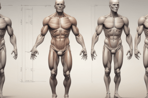
Thigh Anatomy
- Divided into anterior, medial, and posterior compartments.
- Anterior compartment: Innervated by femoral nerve; includes iliopsoas, sartorius, and quadriceps femoris.
- Quadriceps extend the knee; composed of rectus femoris, vastus lateralis, vastus intermedius, and vastus medialis.
- Medial compartment: Innervated by obturator nerve; includes adductor muscles (pectineus, adductor longus/magnus/brevis, gracilis).
- Posterior compartment: Primarily extensors of hip/flexors of knee; innervated by tibial nerve (e.g., semitendinosus).
Blood Supply and Drainage
- Anterior tibial artery: Supplies anterior and some lateral compartments; continues to form dorsalis pedis artery.
- Posterior tibial artery: Supplies posterior compartment; branches into lateral/medial plantar arteries for foot supply.
- Venous drainage via small saphenous (laterally) and great saphenous veins (medially).
Foot Anatomy
- Contains plantar aponeurosis for support; medial and lateral plantar nerves supply most muscles and sensation.
- Dorsum sensory supplied by dorsal lateral cutaneous nerve of the foot.
- Features complex muscular layers and compartments, aiding in foot function.
Studying That Suits You
Use AI to generate personalized quizzes and flashcards to suit your learning preferences.