Podcast
Questions and Answers
Which bone sits between the lunate and the hamate bone?
Which bone sits between the lunate and the hamate bone?
- Pisiform bone
- Trapezoid bone
- Trapezium bone
- Capitate bone (correct)
What is being shown with the blue star in this x-ray?
What is being shown with the blue star in this x-ray?
- Cortex (correct)
- Pedicle
- Articular pillar
- Trabecula
What is being shown with the pink star in this x-ray?
What is being shown with the pink star in this x-ray?
- Trabecula (correct)
- Cortex
- Pedicle
- Lamina
What is being shown as #5 in the picture of the cervical vertebrae
What is being shown as #5 in the picture of the cervical vertebrae
What is being shown as #6 in the picture of the cervical vertebrae
What is being shown as #6 in the picture of the cervical vertebrae
What is being shown as #2 in the picture of the cervical vertebrae
What is being shown as #2 in the picture of the cervical vertebrae
What vertebrae is ring-shaped, lacks a body, and has a facet for the dens?
What vertebrae is ring-shaped, lacks a body, and has a facet for the dens?
Bone #1
Bone #1
Bone #2
Bone #2
Bone #3
Bone #3
Bone #4
Bone #4
Bone #5
Bone #5
Bone #6
Bone #6
Bone #7
Bone #7
Bone #8
Bone #8
What abnormal vertebrae has a small body and a dens for an articular facet for articulation with the atlas?
What abnormal vertebrae has a small body and a dens for an articular facet for articulation with the atlas?
What 2 x-ray views are most commonly used for c-spine imaging?
What 2 x-ray views are most commonly used for c-spine imaging?
C-Spine XR is ALWAYS indicated when there is a high energy trauma
C-Spine XR is ALWAYS indicated when there is a high energy trauma
C-spine XR is indicated in low energy trauma WITH any of the following, EXCEPT:
C-spine XR is indicated in low energy trauma WITH any of the following, EXCEPT:
Clinical picture always considered in context of imaging and deciding if imaging is needed for a c-spine injury
Clinical picture always considered in context of imaging and deciding if imaging is needed for a c-spine injury
70 % of injuries are visible on the
70 % of injuries are visible on the
What is not part of the trauma series for c-spine injuries?
What is not part of the trauma series for c-spine injuries?
On a lateral view for a c-spine XR, what is considered adequate coverage?
On a lateral view for a c-spine XR, what is considered adequate coverage?
In a lateral view cervical x-ray, all bodies (not c1) should be same size.
In a lateral view cervical x-ray, all bodies (not c1) should be same size.
The AP View of a cervical XR should have spinous process in straight line with no space > 50% wider than the one above or below it.
The AP View of a cervical XR should have spinous process in straight line with no space > 50% wider than the one above or below it.
It is harder to see c-spine fractures on the AP view versus the lateral view.
It is harder to see c-spine fractures on the AP view versus the lateral view.
Thoracic vertebra are more stable than cervical due to attachment with ribs and musculature
Thoracic vertebra are more stable than cervical due to attachment with ribs and musculature
What is the most unstable, but most movable joint in the body?
What is the most unstable, but most movable joint in the body?
What is NOT a standard shoulder radiograph view?
What is NOT a standard shoulder radiograph view?
What shoulder XR view is best for dislocations?
What shoulder XR view is best for dislocations?
What x-ray view of the shoulder is best for looking at articular surface abnormalities?
What x-ray view of the shoulder is best for looking at articular surface abnormalities?
AP routine is the better view when looking for the joint space and joint injury.
AP routine is the better view when looking for the joint space and joint injury.
What view is NOT used in elbow radiographs?
What view is NOT used in elbow radiographs?
What special lines on an elbow XR is seen mid-shaft of the radius and should bisect capitulum? It is seen on ALL views.
What special lines on an elbow XR is seen mid-shaft of the radius and should bisect capitulum? It is seen on ALL views.
What elbow imaging should be used for supracondylar fractures in peds?
What elbow imaging should be used for supracondylar fractures in peds?
Finding on elbow radiography which suggests a fracture of one or more bones at the elbow and may indicate an occult fracture that is not directly visible, is called:
Finding on elbow radiography which suggests a fracture of one or more bones at the elbow and may indicate an occult fracture that is not directly visible, is called:
The "posterior fat pad sign" and is often the only visible marker of a fracture, particularly in the pediatrics population.
The "posterior fat pad sign" and is often the only visible marker of a fracture, particularly in the pediatrics population.
What wrist radiograph helps find fracture fragments?
What wrist radiograph helps find fracture fragments?
What is being shown in this image?
What is being shown in this image?
What is being shown in this image?
What is being shown in this image?
Flashcards are hidden until you start studying
Study Notes
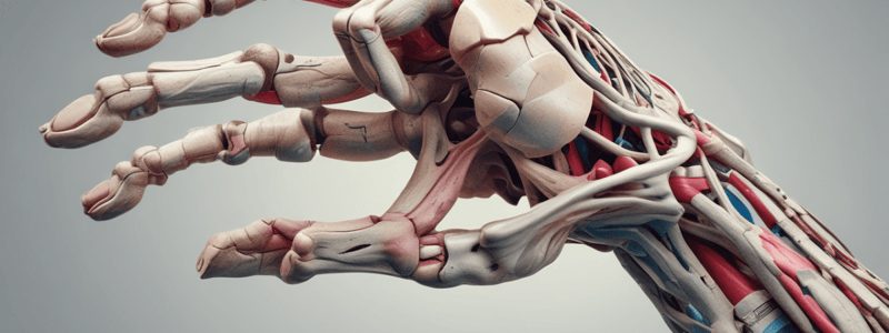
Anatomy of Hand and Wrist Bones
The hand and wrist are composed of several bones arranged in intricate ways, providing structure and stability while allowing for a great range of motion. Understanding these bones and their functions is essential for medical professionals, patients recovering from injuries, or anyone interested in human physiology.
Carpal Bones
Carpal bones are found between the forearm and the metacarpals. There are eight carpal bones in total, comprising two rows: the proximal row and the distal row. These small flat bones have varying shapes and sizes, and they interlock with each other to form a complex system of joints called the carpus. The carpus provides support for the base of the thumb and forms the wrist joint with the radius and ulna.
Proximal Row of Carpal Bones
Including the scaphoid, lunate, triquetral, pisiform, and trapezium and trapezoid bones, the proximal row is located closest to the forearm. The scaphoid bone, also known as the navicular bone, lies directly under the radius and is among the most commonly fractured carpal bones due to its location and thinness. The lunate bone fits into a groove on the radius and replaces the distal end of the ulna during supination. It has a concave surface which articulates with the radial styloid process. The triquetral bone, named after its horseshoe shape, is medial to the lunate. Its posterior part articulates with the hamulus of the ulna, forming the radio-ulnar joint. The pisiform bone overlies the triquetral bone and is one of the few sesamoid bones in the body, formed by the flexor carpi ulnaris tendon passing over it. The trapezium and trapezoid bones help form the first carpometacarpal joint at the base of the thumb.
Distal Row of Carpal Bones
This group includes the capitate, hamate, and lateral and medial uncinate bones. The capitate bone sits between the lunate and the hamate bone. The hamate bone's medial side connects with the hook of the hamulus of the ulna, creating another radioulnar joint. The lateral and medial uncinate bones are smaller bones situated on either side of the scapholunate ligament.
Metacarpal Bones
Metacarpal bones are the five long bones found between the wrist and the phalanges of the hand. They are numbered 1 through 5 from the radial side of the hand. The first metacarpal bone forms the base of the thumb, while the rest provide the foundation for the fingers. The metacarpal bones are connected to the wrist by the carpometacarpal joints. The first metacarpal bone is different in shape and size, giving the thumb its unique range of motion.
Phalanges
Phalanges are the bones found in the fingers and thumb of the hand. There are three phalanges in each finger, numbered 1-3 from proximal to distal. The proximal phalanges connect to the metacarpal bones, the middle phalanges are the longest of the three, and the distal phalanges are the smallest. The thumb has only two phalanges, the proximal and distal, due to a reduced middle phalanx.
Bones of the Hand
The bones of the hand include the bones mentioned above, such as the carpal bones, metacarpal bones, and phalanges, as well as the wrist and finger bones. These bones work together to provide the structure, stability, and flexibility necessary for the hand's function. Understanding their anatomy can help medical professionals in diagnosing and treating injuries, as well as providing insight into the overall health and function of the hand and wrist.
Studying That Suits You
Use AI to generate personalized quizzes and flashcards to suit your learning preferences.


