Podcast
Questions and Answers
What is the primary function of the connective tissue surrounding a bronchopulmonary segment?
What is the primary function of the connective tissue surrounding a bronchopulmonary segment?
- To provide a pathway for lymphatic vessels
- To separate the bronchopulmonary segments from each other (correct)
- To provide a site for gas exchange
- To support the segmental bronchus
What is the difference between the distribution of pulmonary arteries and pulmonary veins within a bronchopulmonary segment?
What is the difference between the distribution of pulmonary arteries and pulmonary veins within a bronchopulmonary segment?
- Pulmonary arteries and veins both accompany the segmental bronchus
- Pulmonary arteries accompany the segmental bronchus, while pulmonary veins run independently (correct)
- Pulmonary arteries and veins both run in the connective tissue
- Pulmonary arteries run in the connective tissue, while pulmonary veins accompany the segmental bronchus
What is the characteristic of the cartilage found in the smaller bronchi, compared to the trachea?
What is the characteristic of the cartilage found in the smaller bronchi, compared to the trachea?
- The number of cartilage plates increases in the smaller bronchi
- U-shaped bars of cartilage are replaced by irregular plates of cartilage (correct)
- Irregular plates of cartilage are replaced by U-shaped bars of cartilage
- The cartilage becomes more rigid in the smaller bronchi
What is the result of the repeated division of the segmental bronchus within a bronchopulmonary segment?
What is the result of the repeated division of the segmental bronchus within a bronchopulmonary segment?
What is the significance of each bronchopulmonary segment having its own lymphatic vessels and autonomic nerve supply?
What is the significance of each bronchopulmonary segment having its own lymphatic vessels and autonomic nerve supply?
What is the primary reason for the lungs becoming dark and mottled with age?
What is the primary reason for the lungs becoming dark and mottled with age?
What is the name of the surface of the lung that lies against the mediastinum anteriorly and the vertebral column posteriorly?
What is the name of the surface of the lung that lies against the mediastinum anteriorly and the vertebral column posteriorly?
What is the shape of each lung?
What is the shape of each lung?
What is the name of the border that separates the base from the costal surface of the lung?
What is the name of the border that separates the base from the costal surface of the lung?
Which of the following is a characteristic of the right lung?
Which of the following is a characteristic of the right lung?
What is the name of the area on the mediastinal surface of the pleura that provides entrance for the root of lung structures?
What is the name of the area on the mediastinal surface of the pleura that provides entrance for the root of lung structures?
What is the term for the thin vertical region that extends from the hilum downward to enable descent of the root of the lung on inspiration?
What is the term for the thin vertical region that extends from the hilum downward to enable descent of the root of the lung on inspiration?
During full inspiration, what happens to the lungs in relation to the pleural cavities?
During full inspiration, what happens to the lungs in relation to the pleural cavities?
What is the name of the space between the costal and diaphragmatic parietal pleurae that is separated only by a capillary layer of pleural fluid?
What is the name of the space between the costal and diaphragmatic parietal pleurae that is separated only by a capillary layer of pleural fluid?
During expiration, what happens to the lower margins of the lungs in relation to the costodiaphragmatic recesses?
During expiration, what happens to the lower margins of the lungs in relation to the costodiaphragmatic recesses?
What is the name of the partition that divides the chest cavity?
What is the name of the partition that divides the chest cavity?
How far does the chest cavity extend upward into the neck?
How far does the chest cavity extend upward into the neck?
What is the name of the layer that lines the thoracic wall and covers the thoracic surface of the diaphragm?
What is the name of the layer that lines the thoracic wall and covers the thoracic surface of the diaphragm?
What is the term for the structures that the pleura surrounds at the hilum of each lung?
What is the term for the structures that the pleura surrounds at the hilum of each lung?
What is the term for the type of cells that form the pleura?
What is the term for the type of cells that form the pleura?
What is the primary purpose of the pleural fluid in the pleural cavity?
What is the primary purpose of the pleural fluid in the pleural cavity?
What is the name of the loose fold of pleura that hangs down to allow for movement of the pulmonary vessels and large bronchi during respiration?
What is the name of the loose fold of pleura that hangs down to allow for movement of the pulmonary vessels and large bronchi during respiration?
Which part of the pleura covers the thoracic surface of the diaphragm?
Which part of the pleura covers the thoracic surface of the diaphragm?
Where does the mediastinal pleura become continuous with the visceral pleura?
Where does the mediastinal pleura become continuous with the visceral pleura?
What is the name of the space that separates the parietal and visceral layers of pleura?
What is the name of the space that separates the parietal and visceral layers of pleura?
What is the location of the spinous process of the vertebra at which the oblique fissure begins in the right lung?
What is the location of the spinous process of the vertebra at which the oblique fissure begins in the right lung?
What is the level of the costal cartilage at which the horizontal fissure runs across the costal surface in the right lung?
What is the level of the costal cartilage at which the horizontal fissure runs across the costal surface in the right lung?
What is the name of the tongue-like extension of the lower part of the superior lobe in the left lung?
What is the name of the tongue-like extension of the lower part of the superior lobe in the left lung?
What is the name of the bronchi that pass to a lobe of the lung?
What is the name of the bronchi that pass to a lobe of the lung?
What is the relationship between the oblique and horizontal fissures in the right lung?
What is the relationship between the oblique and horizontal fissures in the right lung?
What occurs when air enters the pleural cavity from the lungs or through the chest wall?
What occurs when air enters the pleural cavity from the lungs or through the chest wall?
What is the term for the condition when the gas within the pleural cavity accumulates to such an extent that the mediastinum is pushed to the opposite side?
What is the term for the condition when the gas within the pleural cavity accumulates to such an extent that the mediastinum is pushed to the opposite side?
What was the purpose of artificially injecting air into the pleural cavity in the old treatment of tuberculosis?
What was the purpose of artificially injecting air into the pleural cavity in the old treatment of tuberculosis?
What is the most common cause of pneumothorax?
What is the most common cause of pneumothorax?
What is a potential complication of pneumothorax in patients with cancer?
What is a potential complication of pneumothorax in patients with cancer?
What determines the symptoms of pneumothorax?
What determines the symptoms of pneumothorax?
What is a common symptom of pneumothorax?
What is a common symptom of pneumothorax?
What is a potential consequence of a severe pneumothorax?
What is a potential consequence of a severe pneumothorax?
What is a risk factor for spontaneous pneumothorax?
What is a risk factor for spontaneous pneumothorax?
What is the term for the intentional injection of air into the pleural cavity to collapse and rest the lung?
What is the term for the intentional injection of air into the pleural cavity to collapse and rest the lung?
Flashcards are hidden until you start studying
Study Notes
Bronchopulmonary Segments
- Each segmental bronchus connects to a bronchopulmonary segment, which is the smallest independent unit in a lung lobe.
- Surrounding connective tissue separates bronchopulmonary segments, allowing for functional independence.
- Segmental bronchi are accompanied by branches of the pulmonary artery; pulmonary veins run in connective tissue between segments.
- Each segment has its dedicated lymphatic vessels and autonomic nerve supply.
Anatomy of Lungs
- Lungs shrink to one-third of their volume upon opening the thoracic cavity.
- In children, lungs appear pink, darkening with age due to trapped dust particles.
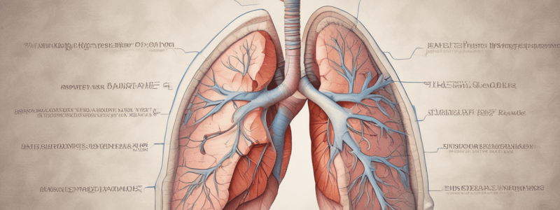
- Lungs are cone-shaped, covered with visceral pleura, and each is suspended in its pleural cavity connected by a root.
- The base of the lung rests on the diaphragm, while the apex extends above the first rib.
Surfaces and Borders
- The costal surface is adjacent to the thoracic wall; the mediastinal surface faces the heart and contains the hilum.
- The inferior border is sharp, separating the base from the costal surface; anterior and posterior borders separate the costal from the medial surface.
Lung Fissures and Lobes
- The right lung is shorter, wider, and larger than the left lung, comprising three lobes (upper, middle, lower) separated by oblique and horizontal fissures.
- The left lung has two lobes (upper and lower) divided by an oblique fissure, with a tongue-shaped extension called the lingula.
Pleurae Structure
- Pleura consists of a single layer of mesothelial cells and supporting connective tissue.
- Features two layers: parietal layer (lining thoracic wall and diaphragm) and visceral layer (covering lung surfaces).
- Pleural cavity contains pleural fluid, allowing smooth movement between the two pleura layers during respiration.
Thoracic Cavity
- Bounded anteriorly by the sternum, laterally by ribs, and posteriorly by thoracic vertebrae.
- Divided into the mediastinum and laterally placed pleurae and lungs.
Costodiaphragmatic Recesses
- Slit-like spaces between parietal pleurae allow lungs to expand during inspiration; they do not completely fill the pleural cavities.
Pneumothorax
- Traumatic pneumothorax occurs when air enters the pleural cavity due to injury, causing lung collapse.
- Tension pneumothorax refers to extensive air accumulation that shifts the mediastinum, requiring emergency intervention.
- Pneumothorax can arise spontaneously or due to trauma, inflammation, or smoking; symptoms include pain and shortness of breath.
Clinical Concepts
- Historical treatment for tuberculosis involved creating artificial pneumothorax by injecting air into the pleural cavity to rest the lung.
- Spontaneous pneumothorax can complicate cancer treatment, particularly in patients undergoing chemotherapy.
Studying That Suits You
Use AI to generate personalized quizzes and flashcards to suit your learning preferences.


